Introduction to Neuroanatomy: Difference between revisions
No edit summary |
No edit summary |
||
| (31 intermediate revisions by 5 users not shown) | |||
| Line 5: | Line 5: | ||
</div> | </div> | ||
== Overview == | == Overview == | ||
The nervous system is made up of vast neural networks; signalling within these circuits enables thinking, language, feeling, learning, memory, and all motor function and sensation. It is well-established that through [[Neuroplasticity|plasticity]] of existing cells our nervous systems can adapt to situations not previously encountered, but it also has been shown that neural stem cells are plastic and involved in creating new connections in adaptation and response to injury.<ref name=":0">Parker E. Ludwig; [https://www.ncbi.nlm.nih.gov/books/NBK442010/ Matthew Varacallo Neuroanatomy, Central Nervous System (CNS)] Feb 2019. | |||
The nervous system is made up of vast neural networks; signalling within these circuits enables thinking, language, feeling, learning, memory, and all function and sensation. It is well-established that through [[Neuroplasticity|plasticity]] of existing cells our nervous systems can adapt to situations not previously encountered, but it also has been shown | </ref><blockquote>'''Neuroplasticity''' or '''brain plasticity''' is defined as the ability of the nervous system to change its activity in response to intrinsic or extrinsic stimuli by reorganizing its structure, functions, or connections. </blockquote> | ||
[[File:Functions of nervous system.jpeg|border|right|400x400px]] | |||
'''The nervous system has three specific functions''': | |||
#'''Sensory Input''' - [[Sensation|Sensory receptors]] present in the [[skin]] and organs respond to external and internal stimuli by generating [[Introduction to Neurophysiology|nerve impulses]] that to the central nervous system | |||
#'''Integration''' - The [[Brain Anatomy|brain]] and [[Spinal cord anatomy|spinal cord]] of the central nervous system combine and sum up all the data received from the body and send out nerve impulses. | |||
#'''Motor Output''' - The nerve impulses from the central nervous system go to the effectors ([[Muscle|muscles]] and [[Hormones|glands]]). Muscle contractions and gland secretions are responses to stimuli received by sensory receptors. | |||
== Building Blocks of the Nervous System == | |||
=== Neurons and Glial Cells === | |||
[[Neurone|Neurons]] are cells of the nervous system<ref>Mercadante AA, Tadi P. Neuroanatomy, Gray Matter. StatPearls Publishing; 2024 Jan-. Available from: https://www.ncbi.nlm.nih.gov/books/NBK553239/</ref> and are responsible for all neurological functions of the brain<ref>Ludwig PE, Reddy V, Varacallo M. Neuroanatomy, Neurons. StatPearls Publishing; 2024 Jan-. Available [[from: https://www.ncbi.nlm.nih.gov/books/NBK441977/]]</ref>. | |||
The | |||
The basic components of a neuron include: | |||
[[File:Neuron Part 1.png|border|right|500x500px]] | |||
[[ | * '''Cell body''': contains the nucleus, mitochondria, and other organelles necessary for neuronal functioning. These structures support the chemical processes of the neuron, most notably the production of [[neurotransmitters]]. | ||
* '''Axons''': originate from the cell body at a specialized area called the axon hillock. Axons relay efferent information to neighboring neurons. | |||
* '''Dendrites''': radiate out from the cell body and receive afferent signals from other neurons. They react to neurotransmitters released by neighboring neurons and relay information via electrical signals through the cell body. A neuron may have a single dendrite or many dendrites depending on its function and location in the nervous system. | |||
The CNS | * '''Synapse''': microscopic gaps found between neurons, typically between the axon of one neuron and the dendrite of another. They are the site of chemical communication between neurons. The neuron sharing information on one side of the synapse is called the '''pre-synaptic neuron''', while the neuron on the receiving side of the synapse is called the '''post-synaptic neuron'''. | ||
Please see [[Neurone|this article]] to learn more about neuronal function and structure. | |||
* [[ | The CNS is made up of [[Grey and White Matter|grey matter and white matter]]: | ||
* [[Grey and White Matter|'''Grey matter''']]: named for its pinkish-gray color, is home to neural cell bodies, axon terminals, and dendrites, and nerve synapses. This nervous tissue is abundant in the [[cerebellum]], [[cerebrum]], and [[Brainstem|brain stem]]. It also forms a butterfly-shaped portion of the central [[Spinal cord anatomy|spinal cord]]. | |||
* [[Grey and White Matter|'''White matter''']]: composed of bundles of [[axons]]. These axons are coated with myelin, a mixture of proteins and lipids, that helps conduct [[Neurone|nerve]] signals and protect the axons. White matter conducts, processes, and send nerve signals up and down the spinal cord<span class="reference" id="cite_ref-:0_1-0"></span>.<blockquote>In this context, '''afferent''' describes information moving inward or into the neuron and '''efferent''' describes information moving outward or away from the neuron.</blockquote> | |||
Neurons can be classified structurally and functionally. | |||
Structurally: | |||
* '''Bipolar neurons''': which have one axon and one dendrite extending from the cell body. | |||
[[ | * '''Multipolar neurons''': which have one axon and multiple dendrites extending from the cell body. | ||
* '''Anaxonic neurons''': which have no identifiable axon that can be distinguished from the dendrites. | |||
* '''Pseudounipolar neurons''': which have a single axon that splits into two branches. The branches cover both functions of sending and receiving signals. | |||
[[File:Types of neuron structures.jpeg|border|center|500x500px]] | |||
Functionally: | |||
* '''Sensory neurons''': which respond to sensory inputs from the environment. | |||
* '''Motor neurons''': which send signals to muscles to control movement. | |||
* '''Interneurons''': which enable communication between sensory and motor neurons | |||
[[File:Types of neuron function.jpeg|border|center|500x500px]] | |||
=== Glial Cells === | |||
[[File:Glia.png|right|frameless|border]] | |||
[[Glial Cells|Glial cells]] (also known as neuroglia) are the most abundant cell in the CNS.<ref>Xu S, Lu J, Shao A, Zhang JH, Zhang J. Glial cells: role of the immune response in ischemic stroke. Frontiers in immunology. 2020 Feb 26;11:502688.</ref> However, they are present in both the CNS and the PNS. They are support cells to neurons and provide essential nervous system function and homeostasis.<ref name=":3">Ludwig P, Das J. Histology, Glial Cells [Internet]. 2023 [cited 22/05/2024]. Available from: https://www.ncbi.nlm.nih.gov/books/NBK441945/</ref> | |||
There are four main classes of glial cells within the CNS. | |||
#'''Astrocytes''': make up the majority of cells in CNS. They are important in maintaining cellular homeostasis. They perform tasks such as clearing excess neurotransmitters, maintaining the blood-brain barrier, and promoting synapse formation.<ref>Wei D, Morrison E. Histology, Astrocytes [Internet]. 2023 [cited 22/05/2024]. Available from: https://www.ncbi.nlm.nih.gov/books/NBK545142/</ref> | |||
#'''Oligodendrocytes''': myelin-forming cells found in the CNS<ref name=":3" /> | |||
#'''Ependymal cells''': are found lining the ventricle and other fluid filled spaces of the CNS. They are involved in the secretion of cerebral spinal fluid.<ref name=":3" /> | |||
#'''Microglial cells''': function as the macrophages of the CNS<ref name=":3" /> | |||
In the PNS: | |||
#'''Schwann Cell''': myelin-forming cells found in the PNS<ref name=":3" /> | |||
== | == Nervous System Organisation == | ||
[[ | The nervous system is divided into two main divisions:<ref name="Barker">Thau L, Reddy V, Singh P. Anatomy, Central Nervous System. StatPearls Publishing; 2024 Jan-. Available [[from: https://www.ncbi.nlm.nih.gov/books/NBK542179/]]</ref> | ||
# Central nervous system (CNS) | |||
# Peripheral nervous system (PNS) | |||
[[Image:Nervous System.jpg|Nervous System|frameless|781x781px|center]] | |||
The hypothalamus | === Central Nervous System (CNS) === | ||
[[File:Central Nervous System parts.jpeg|border|right|514x514px]] | |||
The CNS consists of two parts: the brain and spinal cord: | |||
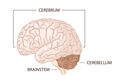
# The [[Brain Anatomy|brain]] is the central organ of the nervous system and the location of information processing and coordination. It is divided into four main parts<ref name=":0" />: | |||
* [[Brainstem|'''Brain stem''']]: consisting of the medulla, pons, and midbrain | |||
* '''[[Cerebellum]]''' | |||
* '''Diencephalon''': consisting of [[thalamus]], the subthalamus, the [[hypothalamus]], and the epithalamus | |||
* [[Cerebrum|'''Cerebral hemispheres''']] (comprised of the [[Cerebral Cortex|cerebral cortex]], [[Basal Ganglia|basal ganglia]], [[Hippocampus|hippocampi]] and [[Amygdala|amygdalae]]). The right and left hemisphere are connected by the [[Corpus Callosum|corpus callosum]] which facilitates communication between both sides of the brain. The hemispheres are then further divided into four lobes. | |||
2. The [[Spinal cord anatomy|'''spinal cord''']] is the caudal extension of the brain. It functions to relay information between the brain and the PNS. | |||
=== CNS coverings: the Bones and Meninges === | |||
== Meninges == | The CNS is enclosed within the [[Bones of the skull|'''skull''']] and '''vertebral column'''. These structures are separated by a series of membranes known as the [[meninges]]<ref>Sehgal I, M Das J. Anatomy, Back, Spinal Meninges. StatPearls Publishing; 2024 Jan-. Available from: https://www.ncbi.nlm.nih.gov/books/NBK547755/</ref>. The pia mater is separated from the delicate arachnoid membrane by the subarachnoid space, which is then in turn separated from the dura mater by the sub-dural space. | ||
The CNS is enclosed within the [[Bones of the skull|skull]] and vertebral column. These structures are separated by a series of membranes known as the [[ | [[File:Meninges of CNS.jpeg|border|right|500x500px]] | ||
* The outermost layer is the '''dura mater'''. It is composed of two fused layers: the outer layer that is directly attached to the skull is the periosteal layer and the inner layer is the meningeal layer. Only the meningeal layer continues down to surround the spinal cord. As the spinal dura mater is not directly attached to the vertebrae, there is an epidural space in the spine. In the cranial dura mater, there are a few places where the meningeal layer separates from the periosteal layer and folds down into the cranial cavity to create dural reflections. The falx cerebri runs in the longitudinal fissure to separate the left and right hemispheres of the cerebrum. The other main dural reflection is the tentorium cerebelli, which separates the occipital and temporal lobes of the cerebrum from the cerebellum and brainstem. | |||
* The middle layer of the meninges is the '''arachnoid mater''', a spider web-like layer closely attached to the meningeal layer of the dura mater. Arachnoid granulations which protrude into the dura mater allow drainage of cerebrospinal fluid into the systemic venous system. | |||
* The innermost layer is the '''pia mater'''. It lies right along the surface of the brain, following the ridges and grooves like tightly adhered plastic wrap. It also forms sheaths around blood vessels as they enter the brain. | |||
=== CNS Neural Structures === | |||
<gallery> | |||
File:Brain anatomy.jpeg|Sections of the brain | |||
File:Brain cross-section.jpeg|Cross section of the brain showing lobes of the brain | |||
File:Brain cross section with subcortical structures.jpeg|Cross section of the brain showing the basal ganglia, thalamus, and hypothalamus. | |||
File:Basal Ganglia and Related Structures.svg.png|Special focus on the basal ganglia | |||
File:Spinal cord and levels.jpeg|Spinal cord levels of innervation | |||
File:Dermatomes for Thoracic Spine Course.jpg|Dermatomes | |||
</gallery> | |||
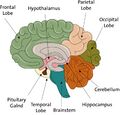
== | ==== Brainstem ==== | ||
[[ | The [[Brainstem|'''brainstem''']] lies at the base of the brain and the top of the spinal cord. It is the structure that connects the cerebrum of the brain to the spinal cord and cerebellum. It is composed of three sections in descending order: the midbrain, pons, and medulla oblongata. It is responsible for many vital functions of life, such as breathing, consciousness, blood pressure, heart rate, and sleep. | ||
==== Cerebrum ==== | |||
The [[cerebrum|'''cerebrum''']] consists of two cerebral hemispheres, the right and left. The two cerebral hemispheres are connected by the [[Corpus Callosum|'''corpus callosum''']] which facilitates communication between both sides of the brain. | |||
The hemispheres are then further divided into '''four lobes:''' | |||
#[[Occipital Lobe|'''Occipital''']] | |||
#[[Parietal Lobe|'''Parietal''']] | |||
#[[Temporal Lobe|'''Temporal''']] | |||
#[[Frontal Lobe Brain Injury|'''Frontal''']] | |||
===== Cerebral Cortex ===== | |||
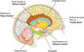
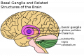
The outer layer of the cerebral hemisphere is termed the [[Cerebral Cortex|'''cerebral cortex''']]. This is interconnected via pathways that run sub-cortically. It is these connections as well as the connections from the cerebral cortex to the brainstem, spinal cord and [[Basal Ganglia|nuclei deep]] within the cerebral hemisphere that form the [[Grey and White Matter|white matter]] of the cerebral hemisphere. The deep nuclei include structures such as the [[Basal Ganglia|basal ganglia]] and the [[thalamus]]. | |||
== | ===== Basal Ganglia ===== | ||
The [[ | The [[Basal Ganglia|'''basal ganglia''']] are a group of subcortical nuclei within the brain responsible primarily for motor control, as well as other roles such as motor learning, executive functions, emotional behaviours, and play an important role in reward and reinforcement, addictive behaviours and habit formation. | ||
[[ | ==== Hypothalamus ==== | ||
The [[hypothalamus|'''hypothalamus''']] is an organ central to many autonomous functions of the human body, notably the regulation of homeostasis. It has a significantly large efferent output to the autonomic nervous system and has a highly significant role in the control of pituitary endocrine function. | |||
The hypothalamus lies on either side of the 3rd ventricle, below the [[thalamus]] and between the optic chiasm and the midbrain. It receives a large input from limbic structures. | |||
The [[ | |||
==== Limbic System ==== | |||
The [[Limbic System|'''limbic system''']] refers to a number of areas within the brain lying mainly on the medial side of the temporal lobe. | |||
* The limbic system provides high level processing of sensory information. The main outflow of the limbic system is to the prefrontal cortex and the hypothalamus as well as to cortical areas. It appears to have a role in attaching behavioural significance and response to a given stimulus. | |||
* Damage to this area has profound effects on emotional responses. | |||
* Long term potentiation is the increase in the strength of a synaptic transmission with repetitive use, it can be seen to be effected in the hippocampus (primarily is involved with memory) and is thought to be important for memory acquisition. | |||
== | === The Cerebellum === | ||
[[ | The [[cerebellum|'''cerebellum''']]<ref>Kebschull JM, Casoni F, Consalez GG, Goldowitz D, Hawkes R, Ruigrok TJ, Schilling K, Wingate R, Wu J, Yeung J, Uusisaari MY. Cerebellum lecture: the cerebellar nuclei—core of the cerebellum. The Cerebellum. 2023 Feb 13:1-58. | ||
</ref> is a vital component in the human brain as it plays a role in motor movement regulation and balance control. The cerebellum is neuron-rich, containing 80% of the brain’s neurons organized in a dense cellular layer, and it's surface area when unfolded is nearly 75% of the surface area of the cerebrum. | |||
=== Spinal Cord === | |||
The [[Spinal cord anatomy|'''spinal cord''']]<ref>Bess S, Line B. Embryology and anatomy: spine/spinal cord. InThe Growing Spine: Management of Spinal Disorders in Young Children 2022 Feb 3 (pp. 3-12). Cham: Springer International Publishing. | |||
</ref> is part of the central nervous system and consists of a tightly packed column of nerve tissue that extends downwards from the brainstem through the central column of the spine. It is a relatively small bundle of tissue (weighing 35g and just about 1cm in diameter) but is crucial in facilitating daily activities. | |||
The spinal cord carries nerve signals from the brain to other parts of the body and receives sensory input from the body, partially processes it, and then transmits that information to the brain. | |||
''' | [[Spinal Nerves|'''Spinal nerves''']] contain both motor and sensory nerve fibres, therefore are considered '''mixed nerves'''. The cell bodies of the sensory neurons are housed in an enlargement called the '''dorsal root ganglion'''. Structurally, these sensory neurons are pseudounipolar. The cell bodies of the motor neurons are housed in the grey matter of the spinal cord, specifically the anterior horn. The '''ventral (or anterior) root ganglion''' are were the nerve roots exit the spinal cord to the PNS. Sensory and motor neurons communicate at each spinal level through '''interneurons'''. | ||
The dorsal root of each spinal cord segment is responsible for innervating specific corresponding areas of skin that can be mapped onto the surface of the body, termed '''[[dermatomes]]'''. Similarly, [[myotomes|'''myotomes''']] are groups of muscles that are innervated by a single ventral nerve root. Dermatomes and myotomes allow for clinical assessment of specific nerve root injuries or the level of a spinal cord injury. | |||
''' | Spinal nerves C7 and above exit the vertebral column above their corresponding vertebrae. Spinal nerve C8 exits between the C7 and T1 vertebrae, and the rest of the spinal nerves exit below their corresponding vertebrae. Because the spinal cord is shorter than the vertebral column and ends at around the L1 or L2 vertebra, the spinal nerve roots below that level have to descend past the end of the spinal cord in the vertebral column before exiting below their corresponding vertebrae. This forms a bundle of spinal nerve roots below the spinal cord termed the [[Cauda Equina|'''cauda equina''']]. | ||
After the spinal nerves exit the vertebral column they split up into '''dorsal and ventral rami'''. Dorsal rami innervate the skin and muscles of the back, while the ventral rami innervate the skin and muscles of the ventral and lateral trunk and extremities. In areas other than the thoracic, the ventral rami intermingle with each other to form networks called '''nerve plexuses'''. These eventually give rise to peripheral nerves that provide sensorimotor innervation to the body. | |||
=== CNS Blood Supply === | |||
| | [[File:Circle of Willis en.svg.png|alt=|thumb|Circle of Willis]]The CNS vasculature provides the nutrients necessary for the correct functioning of the central nervous system<ref>Weerasuriya, A., Mizisin, A.P. The Blood-Nerve Barrier: Structure and Functional Significance. In: Nag, S. (eds) The Blood-Brain and Other Neural Barriers. Methods in Molecular Biology, 2011. vol 686. Humana Press. </ref>. | ||
| | |||
''' | ===== '''Brain''' ===== | ||
Arterial blood supply to the brain comes from four vessels; | |||
*Right and left [[Internal Carotid Artery|internal carotid ]] | |||
*Right and left [[Vertebral Artery|vertebral arteries]] | |||
The internal carotid arteries branch to form two major cerebral arteries, the '''anterior and middle cerebral arteries'''. The '''right and left vertebral arteries''' come together at the level of the pons on the ventral surface of the brainstem to form the '''midline basilar artery'''. | |||
The cerebral | '''Circle of Willis''': The basilar artery joins the blood supply from the internal carotids in an arterial ring at the base of the brain (in the vicinity of the hypothalamus and cerebral peduncles) called the circle of Willis. The '''posterior cerebral arteries''' arise at this confluence, as do two small bridging arteries, the '''anterior and posterior communicating arteries'''. Conjoining the two major sources of cerebral vascular supply via the Circle of Willis presumably improves the chances of any region of the brain continuing to receive blood if one of the major arteries becomes occluded<ref>Purves D, Augustine GJ, Fitzpatrick D, Katz LC, LaMantia AS, McNamara JO, Williams S. The blood supply of the brain and spinal cord. Neuroscience. 2001;2.Available: https://www.ncbi.nlm.nih.gov/books/NBK11042/<nowiki/>(accessed 6.5.2022)</ref>. | ||
===== '''Spinal Cord''' ===== | |||
The spinal cord is supplied by a single '''anterior spinal artery''' and paired '''posterior spinal arteries'''. | |||
* Anterior spinal artery: arises from the vertebral arteries and extends from the level of the lower brainstem to the tip of the conus medullaris. It supplies the ventral medial surface of the medulla and anterior 2/3 of the spinal cord. | |||
* The posterior spinal arteries supply the dorsal 1/3 of the cord. There are reinforcing branches from other arteries along the length of the cord. | |||
If occlusion occurs, it is normally of the anterior spinal artery, producing loss of power and spinothalamic sensory deficit, but dorsal column sensory capabilities are maintained. | |||
== | ===== '''Venous Drainage''' ===== | ||
[[File: | [[File:Brain Sinuses.jpeg|right|frameless]] | ||
The cerebrum, cerebellum and brainstem are drained by numerous veins, which empty into the '''dural venous sinuses'''<ref>Bayot ML, Reddy V, Zabel MK. Neuroanatomy, Dural Venous Sinuses. StatPearls Publishing; 2024 Jan-. Available from: https://www.ncbi.nlm.nih.gov/books/NBK482257/</ref>. The spinal cord is supplied by '''anterior and posterior spinal veins''', which drain into the '''internal and external vertebral plexuses'''. | |||
If occlusion of either of these venous systems then raised intracranial pressure can develop. | |||
=== Peripheral Nervous System (PNS) === | |||
== Peripheral Nervous System (PNS) == | [[File:Autonomic and Somatic Nervous System.png|right|frameless|527x527px]] | ||
[[File:Nervous | The peripheral nervous system<ref name=":1">Murtazina A, Adameyko I. The peripheral nervous system. Development. 2023 May 1;150(9):dev201164.</ref> includes the nerves and [[Ganglion|ganglia]] that are outside of the central nervous system. | ||
The peripheral nervous system is made up of two divisions: | |||
* autonomic system. | |||
* somatic nervous system | |||
Each part of this system plays a vital role in how information is communicated throughout the body. | |||
=== Autonomic Nervous System (ANS) === | === Autonomic Nervous System (ANS) === | ||
The | The [[Autonomic Nervous System|'''autonomic system''']]<ref>Jänig W. The integrative action of the autonomic nervous system: neurobiology of homeostasis. Cambridge University Press; 2022 Aug 4.</ref> is responsible for regulating involuntary body functions. Functions of the ANS include the regulation of “circulation, respiration, metabolism, secretion, body temperature, and reproduction.” | ||
The ANS | The ANS is divided into two divisions: | ||
#[[Sympathetic Nervous System|Sympathetic]]: Preganglionic neurons found in lateral horn of spinal cord from upper thoracic to mid-lumbar cord (T1-L3). Postganglionic cell bodies found in vertebral and prevertebral ganglia. Uses Noradrenalin as postganglionic transmitter. This system is responsible for the "fight or flight" response. | |||
#[[Parasympathetic System|Parasympathetic]]: Preganglionic neurons have cell bodies in the brainstem and sacrum. Postganglionic cell bodies are found adjacent to or within the walls of the organ they supply. Uses [[acetylcholine]] (ACh) as postganglionic transmitter. This system is responsible for the "rest and digest" response. | |||
=== Somatic Nervous System === | |||
[[File:Nervous system diagram.png|right|frameless|660x660px]] | |||
The '''somatic nervous system'''<ref name=":2">Michael-Titus AT, Shortland P. The Nervous System: The Nervous System, E-Book. Elsevier Health Sciences; 2022 Jun 11.</ref> is responsible for carrying sensory and motor information to and from the central nervous system. The somatic nervous system derives its name from the Greek word soma, which means "body." | |||
[[Cranial Nerves|'''Cranial''']] and [[Spinal Nerves|'''spinal nerves''']] contribute to the somatic nervous system. | |||
* Cranial nerves provide voluntary motor control and sensation to the head and face | |||
* Spinal nerves supply the trunk and limbs. | |||
The | The posterior rami travel backwards to supply the vertebral column, vertebral muscles and skin of the back whilst the anterior rami supply the limbs and anterior trunk. The majority of anterior rami combine to form nerve plexuses from which many major peripheral nerves stem. The exception to this is the anterior rami of the thoracic region which travel relatively independently from one another without forming plexuses, as the intercostal and subcostal nerves of the trunk. | ||
Nervous plexuses are as follows: | |||
* C1-C4 form the [[Cervical Plexus|cervical plexus]] | |||
[[ | * C5-T1 combine into the [[Brachial Plexus|brachial plexus]] | ||
* T12-L4 form the [[Lumbar Plexus|lumbar plexus]] | |||
* L4 - S4 combine into the [[Sacral Plexus|sacral plexus]] | |||
The sensory | ==== Sensory Systems ==== | ||
The sensory nervous system<ref name=":2" /> is a part of the nervous system responsible for processing sensory information being where information is transmitted to the spinal cord and brain from peripheral sensory receptors. The sensory receptors are specialised neurons or nerve endings. For more see [[Sensation]] | |||
There are five main (primary or special) senses: | |||
#touch/ | #touch/somatosensory | ||
#vision | #vision | ||
#hearing | #hearing | ||
#taste | #taste/gustatory | ||
#smell/olfaction | #smell/olfaction | ||
<blockquote> | |||
==== Clinical Pearl: Pain Sensations ==== | |||
[[Pain Behaviours|Pain]] is defined as an unpleasant sensory or emotional experience, associated with potential or actual tissue damage. Nociception defines the processing of information about damaging stimuli by the nervous system up to the level of the cortex. Potentially damaging mechanical, thermal, and chemical stimuli are detected by nerve endings called nociceptors, which are found in the skin, on internal surfaces such as the periosteum, joint surfaces, and in some internal organs. | |||
There are two types of nociceptor: ''A delta fibres'': activated by high threshold mechanoreceptors. thinly myelinated; ''Unmyelinated C-fibres'': activated by polymodal nociceptors(PMN) and respond to intense mechanical stimulation, high temperatures and irritant chemicals. | |||
A | |||
There are three main pathways that transmit nociceptive signals to the brain: | |||
#[[Spinothalamic tract|Spinothalamic]] tract | |||
#Spino reticular tract. | |||
#Spino mesencephalic | |||
</blockquote> | |||
==== Motor Systems ==== | |||
[[File:Neuromuscular junction illustration of action.jpeg|border|right|463x463px]] | |||
Motor systems are the areas of the nervous system responsible for controlling movement. Neural control of the somatic motor system involves complex feedback mechanisms between the brain, spinal cord, peripheral nerves, and musculoskeletal structures. Each component is functionally and structurally capable of adaptation and modulation to maintain as much efficiency as possible<ref>Seffinger MA, Hruby RJ. Evidence-based manual medicine: a problem-oriented approach. Elsevier Health Sciences; 2007. Available: https://www.sciencedirect.com/topics/neuroscience/somatic-motor-system (accessed 6.5.20220</ref>. | |||
In | ===== Neuromuscular Junction ===== | ||
Peripheral motor nerves<ref name=":1" /> consist of a '''neuromuscular junction''', which is the synaptic connection between the motor neuron and the muscle. Information in the form of electrical impulses reach the presynaptic membrane at the end of an axon and trigger the release of neurotransmitters. In skeletal muscles, the neurotransmitter [[acetylcholine]] is often used. The neurotransmitters then cross the gap between the motor neuron and the motor end plate on the muscle, a space termed the synaptic cleft. The motor end plate contains receptors which then receive the neurotransmitters, and this signal is then translated into muscle action. | |||
== Additional Resources == | |||
# | The following videos are optional additional resources which provide a deeper understanding and alternative mode of learning about the neuroanatomical structures discussed in this article. | ||
This video is 8:21 and provides an overview of the anatomy of the cerebral cortex.{{#ev:youtube|mGxomKWfJXs|400}}<ref>Neuroscientifically Challenged. 2-Minute Neuroscience: Hypothalamus & Pituitary Gland. Available from: http://www.youtube.com/watch?v=TVhm2rBGhB0 [last accessed 19/10/2019] </ref> | |||
This video is 2:01 and provides an overview of the physiology of the hypothalamus and the pituitary gland.{{#ev:youtube|TVhm2rBGhB0|400}}This video is 9:36 and is a discussion of the motor system and functional motor units.{{#ev:youtube|vXb0ZvkFkS8|400}}<ref>khanacademymedicine. Motor unit. Available from: http://www.youtube.com/watch?v=vXb0ZvkFkS8 [last accessed 19/10/2019] </ref> | |||
This video is 9:05 and provides an overview of pain physiology and the different types of pain the body can experience. | |||
{{#ev:youtube|i5V_q7XqQN8|400}}<ref>UCL Centre for Anaesthesia. An Introduction to Pain Pathways and Mechanisms. Available from: http://www.youtube.com/watch?v=i5V_q7XqQN8 [last accessed 19/10/2019] </ref> | {{#ev:youtube|i5V_q7XqQN8|400}}<ref>UCL Centre for Anaesthesia. An Introduction to Pain Pathways and Mechanisms. Available from: http://www.youtube.com/watch?v=i5V_q7XqQN8 [last accessed 19/10/2019] </ref> | ||
These last two videos, 10:22 and 4:33 respectively, are continuations of each other and provide a review of cerebral anatomy and a discussion of cerebral blood vessels and supply. | |||
{| width="100%" cellspacing="1" cellpadding="1" | |||
|- | |||
|{{#ev:youtube|hfG8J_X1D5Q|350}}<ref>khanacademymedicine. Cerebral blood supply - Part 1. Available from: http://www.youtube.com/watch?v=hfG8J_X1D5Q [last accessed 19/10/2019] </ref> | |||
|{{#ev:youtube|kVulo3qDcUo|350}}<ref>khanacademymedicine. Cerebral blood supply - Part 2. Available from: http://www.youtube.com/watch?v=kVulo3qDcUo [last accessed 19/10/2019] </ref> | |||
|} | |||
{{#ev:youtube| | |||
== References == | == References == | ||
| Line 238: | Line 260: | ||
[[Category:Anatomy]] | [[Category:Anatomy]] | ||
[[Category:Course Pages]] | [[Category:Course Pages]] | ||
[[Category: | [[Category:Plus Content]] | ||
Revision as of 04:38, 26 May 2024
Original Editor - Joanne Garvey and Naomi O'Reilly
Top Contributors - Joanne Garvey, Lucinda hampton, Naomi O'Reilly, Laura Ritchie, Stacy Schiurring, Kim Jackson, Jess Bell, Kate Sampson, Rachael Lowe, Tarina van der Stockt, Admin, WikiSysop, Simisola Ajeyalemi, Adam Vallely Farrell, Rucha Gadgil, Rewan Elsayed Elkanafany, George Prudden, Mande Jooste and Scott Buxton
Overview[edit | edit source]
The nervous system is made up of vast neural networks; signalling within these circuits enables thinking, language, feeling, learning, memory, and all motor function and sensation. It is well-established that through plasticity of existing cells our nervous systems can adapt to situations not previously encountered, but it also has been shown that neural stem cells are plastic and involved in creating new connections in adaptation and response to injury.[1]
Neuroplasticity or brain plasticity is defined as the ability of the nervous system to change its activity in response to intrinsic or extrinsic stimuli by reorganizing its structure, functions, or connections.

The nervous system has three specific functions:
- Sensory Input - Sensory receptors present in the skin and organs respond to external and internal stimuli by generating nerve impulses that to the central nervous system
- Integration - The brain and spinal cord of the central nervous system combine and sum up all the data received from the body and send out nerve impulses.
- Motor Output - The nerve impulses from the central nervous system go to the effectors (muscles and glands). Muscle contractions and gland secretions are responses to stimuli received by sensory receptors.
Building Blocks of the Nervous System[edit | edit source]
Neurons and Glial Cells[edit | edit source]
Neurons are cells of the nervous system[2] and are responsible for all neurological functions of the brain[3].
The basic components of a neuron include:

- Cell body: contains the nucleus, mitochondria, and other organelles necessary for neuronal functioning. These structures support the chemical processes of the neuron, most notably the production of neurotransmitters.
- Axons: originate from the cell body at a specialized area called the axon hillock. Axons relay efferent information to neighboring neurons.
- Dendrites: radiate out from the cell body and receive afferent signals from other neurons. They react to neurotransmitters released by neighboring neurons and relay information via electrical signals through the cell body. A neuron may have a single dendrite or many dendrites depending on its function and location in the nervous system.
- Synapse: microscopic gaps found between neurons, typically between the axon of one neuron and the dendrite of another. They are the site of chemical communication between neurons. The neuron sharing information on one side of the synapse is called the pre-synaptic neuron, while the neuron on the receiving side of the synapse is called the post-synaptic neuron.
Please see this article to learn more about neuronal function and structure.
The CNS is made up of grey matter and white matter:
- Grey matter: named for its pinkish-gray color, is home to neural cell bodies, axon terminals, and dendrites, and nerve synapses. This nervous tissue is abundant in the cerebellum, cerebrum, and brain stem. It also forms a butterfly-shaped portion of the central spinal cord.
- White matter: composed of bundles of axons. These axons are coated with myelin, a mixture of proteins and lipids, that helps conduct nerve signals and protect the axons. White matter conducts, processes, and send nerve signals up and down the spinal cord.
In this context, afferent describes information moving inward or into the neuron and efferent describes information moving outward or away from the neuron.
Neurons can be classified structurally and functionally.
Structurally:
- Bipolar neurons: which have one axon and one dendrite extending from the cell body.
- Multipolar neurons: which have one axon and multiple dendrites extending from the cell body.
- Anaxonic neurons: which have no identifiable axon that can be distinguished from the dendrites.
- Pseudounipolar neurons: which have a single axon that splits into two branches. The branches cover both functions of sending and receiving signals.

Functionally:
- Sensory neurons: which respond to sensory inputs from the environment.
- Motor neurons: which send signals to muscles to control movement.
- Interneurons: which enable communication between sensory and motor neurons

Glial Cells[edit | edit source]

Glial cells (also known as neuroglia) are the most abundant cell in the CNS.[4] However, they are present in both the CNS and the PNS. They are support cells to neurons and provide essential nervous system function and homeostasis.[5]
There are four main classes of glial cells within the CNS.
- Astrocytes: make up the majority of cells in CNS. They are important in maintaining cellular homeostasis. They perform tasks such as clearing excess neurotransmitters, maintaining the blood-brain barrier, and promoting synapse formation.[6]
- Oligodendrocytes: myelin-forming cells found in the CNS[5]
- Ependymal cells: are found lining the ventricle and other fluid filled spaces of the CNS. They are involved in the secretion of cerebral spinal fluid.[5]
- Microglial cells: function as the macrophages of the CNS[5]
In the PNS:
- Schwann Cell: myelin-forming cells found in the PNS[5]
Nervous System Organisation[edit | edit source]
The nervous system is divided into two main divisions:[7]
- Central nervous system (CNS)
- Peripheral nervous system (PNS)

Central Nervous System (CNS)[edit | edit source]

The CNS consists of two parts: the brain and spinal cord:
- The brain is the central organ of the nervous system and the location of information processing and coordination. It is divided into four main parts[1]:
- Brain stem: consisting of the medulla, pons, and midbrain
- Cerebellum
- Diencephalon: consisting of thalamus, the subthalamus, the hypothalamus, and the epithalamus
- Cerebral hemispheres (comprised of the cerebral cortex, basal ganglia, hippocampi and amygdalae). The right and left hemisphere are connected by the corpus callosum which facilitates communication between both sides of the brain. The hemispheres are then further divided into four lobes.
2. The spinal cord is the caudal extension of the brain. It functions to relay information between the brain and the PNS.
CNS coverings: the Bones and Meninges[edit | edit source]
The CNS is enclosed within the skull and vertebral column. These structures are separated by a series of membranes known as the meninges[8]. The pia mater is separated from the delicate arachnoid membrane by the subarachnoid space, which is then in turn separated from the dura mater by the sub-dural space.

- The outermost layer is the dura mater. It is composed of two fused layers: the outer layer that is directly attached to the skull is the periosteal layer and the inner layer is the meningeal layer. Only the meningeal layer continues down to surround the spinal cord. As the spinal dura mater is not directly attached to the vertebrae, there is an epidural space in the spine. In the cranial dura mater, there are a few places where the meningeal layer separates from the periosteal layer and folds down into the cranial cavity to create dural reflections. The falx cerebri runs in the longitudinal fissure to separate the left and right hemispheres of the cerebrum. The other main dural reflection is the tentorium cerebelli, which separates the occipital and temporal lobes of the cerebrum from the cerebellum and brainstem.
- The middle layer of the meninges is the arachnoid mater, a spider web-like layer closely attached to the meningeal layer of the dura mater. Arachnoid granulations which protrude into the dura mater allow drainage of cerebrospinal fluid into the systemic venous system.
- The innermost layer is the pia mater. It lies right along the surface of the brain, following the ridges and grooves like tightly adhered plastic wrap. It also forms sheaths around blood vessels as they enter the brain.
CNS Neural Structures[edit | edit source]
Sections of the brain
Cross section of the brain showing lobes of the brain
Cross section of the brain showing the basal ganglia, thalamus, and hypothalamus.
Special focus on the basal ganglia
Spinal cord levels of innervation
Dermatomes


Brainstem[edit | edit source]
The brainstem lies at the base of the brain and the top of the spinal cord. It is the structure that connects the cerebrum of the brain to the spinal cord and cerebellum. It is composed of three sections in descending order: the midbrain, pons, and medulla oblongata. It is responsible for many vital functions of life, such as breathing, consciousness, blood pressure, heart rate, and sleep.
Cerebrum[edit | edit source]
The cerebrum consists of two cerebral hemispheres, the right and left. The two cerebral hemispheres are connected by the corpus callosum which facilitates communication between both sides of the brain.
The hemispheres are then further divided into four lobes:
Cerebral Cortex[edit | edit source]
The outer layer of the cerebral hemisphere is termed the cerebral cortex. This is interconnected via pathways that run sub-cortically. It is these connections as well as the connections from the cerebral cortex to the brainstem, spinal cord and nuclei deep within the cerebral hemisphere that form the white matter of the cerebral hemisphere. The deep nuclei include structures such as the basal ganglia and the thalamus.
Basal Ganglia[edit | edit source]
The basal ganglia are a group of subcortical nuclei within the brain responsible primarily for motor control, as well as other roles such as motor learning, executive functions, emotional behaviours, and play an important role in reward and reinforcement, addictive behaviours and habit formation.
Hypothalamus[edit | edit source]
The hypothalamus is an organ central to many autonomous functions of the human body, notably the regulation of homeostasis. It has a significantly large efferent output to the autonomic nervous system and has a highly significant role in the control of pituitary endocrine function.
The hypothalamus lies on either side of the 3rd ventricle, below the thalamus and between the optic chiasm and the midbrain. It receives a large input from limbic structures.
Limbic System[edit | edit source]
The limbic system refers to a number of areas within the brain lying mainly on the medial side of the temporal lobe.
- The limbic system provides high level processing of sensory information. The main outflow of the limbic system is to the prefrontal cortex and the hypothalamus as well as to cortical areas. It appears to have a role in attaching behavioural significance and response to a given stimulus.
- Damage to this area has profound effects on emotional responses.
- Long term potentiation is the increase in the strength of a synaptic transmission with repetitive use, it can be seen to be effected in the hippocampus (primarily is involved with memory) and is thought to be important for memory acquisition.
The Cerebellum[edit | edit source]
The cerebellum[9] is a vital component in the human brain as it plays a role in motor movement regulation and balance control. The cerebellum is neuron-rich, containing 80% of the brain’s neurons organized in a dense cellular layer, and it's surface area when unfolded is nearly 75% of the surface area of the cerebrum.
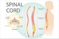
Spinal Cord[edit | edit source]
The spinal cord[10] is part of the central nervous system and consists of a tightly packed column of nerve tissue that extends downwards from the brainstem through the central column of the spine. It is a relatively small bundle of tissue (weighing 35g and just about 1cm in diameter) but is crucial in facilitating daily activities.
The spinal cord carries nerve signals from the brain to other parts of the body and receives sensory input from the body, partially processes it, and then transmits that information to the brain.
Spinal nerves contain both motor and sensory nerve fibres, therefore are considered mixed nerves. The cell bodies of the sensory neurons are housed in an enlargement called the dorsal root ganglion. Structurally, these sensory neurons are pseudounipolar. The cell bodies of the motor neurons are housed in the grey matter of the spinal cord, specifically the anterior horn. The ventral (or anterior) root ganglion are were the nerve roots exit the spinal cord to the PNS. Sensory and motor neurons communicate at each spinal level through interneurons.
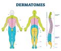
The dorsal root of each spinal cord segment is responsible for innervating specific corresponding areas of skin that can be mapped onto the surface of the body, termed dermatomes. Similarly, myotomes are groups of muscles that are innervated by a single ventral nerve root. Dermatomes and myotomes allow for clinical assessment of specific nerve root injuries or the level of a spinal cord injury.
Spinal nerves C7 and above exit the vertebral column above their corresponding vertebrae. Spinal nerve C8 exits between the C7 and T1 vertebrae, and the rest of the spinal nerves exit below their corresponding vertebrae. Because the spinal cord is shorter than the vertebral column and ends at around the L1 or L2 vertebra, the spinal nerve roots below that level have to descend past the end of the spinal cord in the vertebral column before exiting below their corresponding vertebrae. This forms a bundle of spinal nerve roots below the spinal cord termed the cauda equina.
After the spinal nerves exit the vertebral column they split up into dorsal and ventral rami. Dorsal rami innervate the skin and muscles of the back, while the ventral rami innervate the skin and muscles of the ventral and lateral trunk and extremities. In areas other than the thoracic, the ventral rami intermingle with each other to form networks called nerve plexuses. These eventually give rise to peripheral nerves that provide sensorimotor innervation to the body.
CNS Blood Supply[edit | edit source]

The CNS vasculature provides the nutrients necessary for the correct functioning of the central nervous system[11].
Brain[edit | edit source]
Arterial blood supply to the brain comes from four vessels;
- Right and left internal carotid
- Right and left vertebral arteries
The internal carotid arteries branch to form two major cerebral arteries, the anterior and middle cerebral arteries. The right and left vertebral arteries come together at the level of the pons on the ventral surface of the brainstem to form the midline basilar artery.
Circle of Willis: The basilar artery joins the blood supply from the internal carotids in an arterial ring at the base of the brain (in the vicinity of the hypothalamus and cerebral peduncles) called the circle of Willis. The posterior cerebral arteries arise at this confluence, as do two small bridging arteries, the anterior and posterior communicating arteries. Conjoining the two major sources of cerebral vascular supply via the Circle of Willis presumably improves the chances of any region of the brain continuing to receive blood if one of the major arteries becomes occluded[12].
Spinal Cord[edit | edit source]
The spinal cord is supplied by a single anterior spinal artery and paired posterior spinal arteries.
- Anterior spinal artery: arises from the vertebral arteries and extends from the level of the lower brainstem to the tip of the conus medullaris. It supplies the ventral medial surface of the medulla and anterior 2/3 of the spinal cord.
- The posterior spinal arteries supply the dorsal 1/3 of the cord. There are reinforcing branches from other arteries along the length of the cord.
If occlusion occurs, it is normally of the anterior spinal artery, producing loss of power and spinothalamic sensory deficit, but dorsal column sensory capabilities are maintained.
Venous Drainage[edit | edit source]

The cerebrum, cerebellum and brainstem are drained by numerous veins, which empty into the dural venous sinuses[13]. The spinal cord is supplied by anterior and posterior spinal veins, which drain into the internal and external vertebral plexuses.
If occlusion of either of these venous systems then raised intracranial pressure can develop.
Peripheral Nervous System (PNS)[edit | edit source]

The peripheral nervous system[14] includes the nerves and ganglia that are outside of the central nervous system.
The peripheral nervous system is made up of two divisions:
- autonomic system.
- somatic nervous system
Each part of this system plays a vital role in how information is communicated throughout the body.
Autonomic Nervous System (ANS)[edit | edit source]
The autonomic system[15] is responsible for regulating involuntary body functions. Functions of the ANS include the regulation of “circulation, respiration, metabolism, secretion, body temperature, and reproduction.”
The ANS is divided into two divisions:
- Sympathetic: Preganglionic neurons found in lateral horn of spinal cord from upper thoracic to mid-lumbar cord (T1-L3). Postganglionic cell bodies found in vertebral and prevertebral ganglia. Uses Noradrenalin as postganglionic transmitter. This system is responsible for the "fight or flight" response.
- Parasympathetic: Preganglionic neurons have cell bodies in the brainstem and sacrum. Postganglionic cell bodies are found adjacent to or within the walls of the organ they supply. Uses acetylcholine (ACh) as postganglionic transmitter. This system is responsible for the "rest and digest" response.
Somatic Nervous System[edit | edit source]

The somatic nervous system[16] is responsible for carrying sensory and motor information to and from the central nervous system. The somatic nervous system derives its name from the Greek word soma, which means "body."
Cranial and spinal nerves contribute to the somatic nervous system.
- Cranial nerves provide voluntary motor control and sensation to the head and face
- Spinal nerves supply the trunk and limbs.
The posterior rami travel backwards to supply the vertebral column, vertebral muscles and skin of the back whilst the anterior rami supply the limbs and anterior trunk. The majority of anterior rami combine to form nerve plexuses from which many major peripheral nerves stem. The exception to this is the anterior rami of the thoracic region which travel relatively independently from one another without forming plexuses, as the intercostal and subcostal nerves of the trunk.
Nervous plexuses are as follows:
- C1-C4 form the cervical plexus
- C5-T1 combine into the brachial plexus
- T12-L4 form the lumbar plexus
- L4 - S4 combine into the sacral plexus
Sensory Systems[edit | edit source]
The sensory nervous system[16] is a part of the nervous system responsible for processing sensory information being where information is transmitted to the spinal cord and brain from peripheral sensory receptors. The sensory receptors are specialised neurons or nerve endings. For more see Sensation
There are five main (primary or special) senses:
- touch/somatosensory
- vision
- hearing
- taste/gustatory
- smell/olfaction
Clinical Pearl: Pain Sensations[edit | edit source]
Pain is defined as an unpleasant sensory or emotional experience, associated with potential or actual tissue damage. Nociception defines the processing of information about damaging stimuli by the nervous system up to the level of the cortex. Potentially damaging mechanical, thermal, and chemical stimuli are detected by nerve endings called nociceptors, which are found in the skin, on internal surfaces such as the periosteum, joint surfaces, and in some internal organs.
There are two types of nociceptor: A delta fibres: activated by high threshold mechanoreceptors. thinly myelinated; Unmyelinated C-fibres: activated by polymodal nociceptors(PMN) and respond to intense mechanical stimulation, high temperatures and irritant chemicals.
There are three main pathways that transmit nociceptive signals to the brain:
- Spinothalamic tract
- Spino reticular tract.
- Spino mesencephalic
Motor Systems[edit | edit source]

Motor systems are the areas of the nervous system responsible for controlling movement. Neural control of the somatic motor system involves complex feedback mechanisms between the brain, spinal cord, peripheral nerves, and musculoskeletal structures. Each component is functionally and structurally capable of adaptation and modulation to maintain as much efficiency as possible[17].
Neuromuscular Junction[edit | edit source]
Peripheral motor nerves[14] consist of a neuromuscular junction, which is the synaptic connection between the motor neuron and the muscle. Information in the form of electrical impulses reach the presynaptic membrane at the end of an axon and trigger the release of neurotransmitters. In skeletal muscles, the neurotransmitter acetylcholine is often used. The neurotransmitters then cross the gap between the motor neuron and the motor end plate on the muscle, a space termed the synaptic cleft. The motor end plate contains receptors which then receive the neurotransmitters, and this signal is then translated into muscle action.
Additional Resources[edit | edit source]
The following videos are optional additional resources which provide a deeper understanding and alternative mode of learning about the neuroanatomical structures discussed in this article.
This video is 8:21 and provides an overview of the anatomy of the cerebral cortex.
[18] This video is 2:01 and provides an overview of the physiology of the hypothalamus and the pituitary gland.
This video is 9:36 and is a discussion of the motor system and functional motor units.
This video is 9:05 and provides an overview of pain physiology and the different types of pain the body can experience.
These last two videos, 10:22 and 4:33 respectively, are continuations of each other and provide a review of cerebral anatomy and a discussion of cerebral blood vessels and supply.
| [21] | [22] |
References[edit | edit source]
- ↑ 1.0 1.1 Parker E. Ludwig; Matthew Varacallo Neuroanatomy, Central Nervous System (CNS) Feb 2019.
- ↑ Mercadante AA, Tadi P. Neuroanatomy, Gray Matter. StatPearls Publishing; 2024 Jan-. Available from: https://www.ncbi.nlm.nih.gov/books/NBK553239/
- ↑ Ludwig PE, Reddy V, Varacallo M. Neuroanatomy, Neurons. StatPearls Publishing; 2024 Jan-. Available from: https://www.ncbi.nlm.nih.gov/books/NBK441977/
- ↑ Xu S, Lu J, Shao A, Zhang JH, Zhang J. Glial cells: role of the immune response in ischemic stroke. Frontiers in immunology. 2020 Feb 26;11:502688.
- ↑ 5.0 5.1 5.2 5.3 5.4 Ludwig P, Das J. Histology, Glial Cells [Internet]. 2023 [cited 22/05/2024]. Available from: https://www.ncbi.nlm.nih.gov/books/NBK441945/
- ↑ Wei D, Morrison E. Histology, Astrocytes [Internet]. 2023 [cited 22/05/2024]. Available from: https://www.ncbi.nlm.nih.gov/books/NBK545142/
- ↑ Thau L, Reddy V, Singh P. Anatomy, Central Nervous System. StatPearls Publishing; 2024 Jan-. Available from: https://www.ncbi.nlm.nih.gov/books/NBK542179/
- ↑ Sehgal I, M Das J. Anatomy, Back, Spinal Meninges. StatPearls Publishing; 2024 Jan-. Available from: https://www.ncbi.nlm.nih.gov/books/NBK547755/
- ↑ Kebschull JM, Casoni F, Consalez GG, Goldowitz D, Hawkes R, Ruigrok TJ, Schilling K, Wingate R, Wu J, Yeung J, Uusisaari MY. Cerebellum lecture: the cerebellar nuclei—core of the cerebellum. The Cerebellum. 2023 Feb 13:1-58.
- ↑ Bess S, Line B. Embryology and anatomy: spine/spinal cord. InThe Growing Spine: Management of Spinal Disorders in Young Children 2022 Feb 3 (pp. 3-12). Cham: Springer International Publishing.
- ↑ Weerasuriya, A., Mizisin, A.P. The Blood-Nerve Barrier: Structure and Functional Significance. In: Nag, S. (eds) The Blood-Brain and Other Neural Barriers. Methods in Molecular Biology, 2011. vol 686. Humana Press.
- ↑ Purves D, Augustine GJ, Fitzpatrick D, Katz LC, LaMantia AS, McNamara JO, Williams S. The blood supply of the brain and spinal cord. Neuroscience. 2001;2.Available: https://www.ncbi.nlm.nih.gov/books/NBK11042/(accessed 6.5.2022)
- ↑ Bayot ML, Reddy V, Zabel MK. Neuroanatomy, Dural Venous Sinuses. StatPearls Publishing; 2024 Jan-. Available from: https://www.ncbi.nlm.nih.gov/books/NBK482257/
- ↑ 14.0 14.1 Murtazina A, Adameyko I. The peripheral nervous system. Development. 2023 May 1;150(9):dev201164.
- ↑ Jänig W. The integrative action of the autonomic nervous system: neurobiology of homeostasis. Cambridge University Press; 2022 Aug 4.
- ↑ 16.0 16.1 Michael-Titus AT, Shortland P. The Nervous System: The Nervous System, E-Book. Elsevier Health Sciences; 2022 Jun 11.
- ↑ Seffinger MA, Hruby RJ. Evidence-based manual medicine: a problem-oriented approach. Elsevier Health Sciences; 2007. Available: https://www.sciencedirect.com/topics/neuroscience/somatic-motor-system (accessed 6.5.20220
- ↑ Neuroscientifically Challenged. 2-Minute Neuroscience: Hypothalamus & Pituitary Gland. Available from: http://www.youtube.com/watch?v=TVhm2rBGhB0 [last accessed 19/10/2019]
- ↑ khanacademymedicine. Motor unit. Available from: http://www.youtube.com/watch?v=vXb0ZvkFkS8 [last accessed 19/10/2019]
- ↑ UCL Centre for Anaesthesia. An Introduction to Pain Pathways and Mechanisms. Available from: http://www.youtube.com/watch?v=i5V_q7XqQN8 [last accessed 19/10/2019]
- ↑ khanacademymedicine. Cerebral blood supply - Part 1. Available from: http://www.youtube.com/watch?v=hfG8J_X1D5Q [last accessed 19/10/2019]
- ↑ khanacademymedicine. Cerebral blood supply - Part 2. Available from: http://www.youtube.com/watch?v=kVulo3qDcUo [last accessed 19/10/2019]

