
Cerebral Cortex
Original Editor - Lucinda hampton
Top Contributors - Lucinda hampton, Jess Bell, Stacy Schiurring, Rucha Gadgil, Kim Jackson, Tarina van der Stockt and Joao Costa
Introduction

The cerebrum is the largest anatomical area of the brain. It is a highly developed structure, with the cerebral cortex containing between 14 billion and 16 billion neurons. The major functions of the cerebrum include, but aren't limited to, controlling voluntary movements of the body, sensation, memory, emotions, and executive functioning.
The cerebrum is made up of both grey and white matter. The surface of the cerebrum is the cerebral cortex. This sheet of neural tissue has up to six layers of nerve cells. It is covered by the meninges and is often referred to as grey matter.[1][2] The cerebral cortex covers the internal white matter.
Cerebral Anatomy

The cerebrum is composed of two cerebral hemispheres (i.e. the right and left hemispheres). The two hemispheres are connected via subcortical pathways, including the corpus callosum. The corpus callosum is a thick tract of nerve fibres that facilitates communication between each side of the brain. These connections, as well as the connections from the cerebral cortex to the brainstem, spinal cord and subcortical nuclei deep within the cerebral hemisphere, form the white matter of the cerebral hemisphere. The deep nuclei include structures such as the basal ganglia and the thalamus.
The external surface of the brain is highly convoluted, with many folds. This distinct shape evolved as the volume of our cortex increased more rapidly than our cranial volume.
The grooves of the cortex are called sulci. The sulci separate the elevated regions or ridges, which are called gyri. There are three main sulci in each hemisphere: (1) central sulcus, (2) parieto-occipital sulcus and (3) lateral fissure. Each hemisphere is divided into four lobes: frontal, parietal, temporal and occipital lobes.[3][4] They are named after the overlying cranial bones.
-
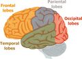 Lobes of the brain
 -
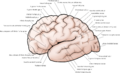 Cerebral cortex surface anatomy
 -
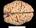 Note the raised gyri and depressed sulci forming the surface of the cerebrum
 -
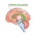 The area in green is the corpus callosum


Functions and Regions of the Cerebral Cortex
The cerebral cortex is the outer layer of the cerebral hemispheres. It is involved in many body functions including personality, intelligence,[5] memory, motor function/movement control, planning, processing sensory information, and language.[6]
The cerebral cortex is the site of data collection, processing, and sharing of information gathered from the different regions of the nervous system. This neuronal communication takes place via tracts or fasciculi which are organised as commissural fibres (between hemispheres), association fibres (within the hemispheres), and projection fibres (cortex to subcortical structures).
The cortex can be divided into primary areas and association areas:[3]
- primary areas: receive and send information
- association areas: process and interpret information
The cerebral cortex mainly contains sensory and motor areas.

Sensory areas: somatosensory information is sent to the thalamus. The thalamus then transfers this information to the primary somatosensory cortex in the parietal lobe. Other primary cortical sensory areas include the auditory cortex in the temporal lobe and the visual cortex in the occipital lobe. Association areas within these sensory regions give meaning to specific sensations.[4]
Motor areas: include the primary motor cortex and the premotor cortex. They are primarily located in the frontal lobe and are involved in regulating voluntary movement. Motor output from the brain to the body travels along upper and lower motor neurons. The upper motor neuron originates in the cortex or brainstem and synapses with the lower motor neuron in the brainstem or spinal cord, which then travels down to the target muscle.[7]
Neocortex

The neocortex is the "most recently evolved part of the brain".[8] The six-layer neocortex is present in all mammals, but there are differences in neocortex size between species[9]:
- neurons in various layers connect vertically to form small microcircuits, called 'columns'
- 90% of the cerebral cortex and 76% of the human brain volume is neocortex,[10] which makes it the largest area of the brain
- two distinct streams of information converge in the neocortex:
- "bottom-up" stream (i.e. signals from the environment)
- "top-down" stream (i.e. internally generated information is transmitted)
Allocortex
The allocortex (or the heterogenetic cortex) is phylogenetically older than the neocortex.[11] It has fewer layers than the neocortex and makes up around 10% of the cerebral cortex. Due to its simpler cellular organisation, the allocortex is unable to form as many complex microcircuits as the neocortex.[12]
The allocortex is divided into three cortical areas:[11]
- archicortex: confined to the hippocampus and the subiculum
- palaeocortex: olfactory cortex
- periarchicortex: entorhinal area, retrosplenial, and cingulate regions
Cerebral Lobes
Traditionally, the cerebrum is thought to contain four lobes: the (1) frontal, (2) parietal, (3) occipital, and (4) temporal lobes. There are also two "hidden lobes" in the cerebrum: the (1) insular and (2) limbic lobes.[13]
1. Frontal Lobe

The frontal lobe is anterior to the central sulcus and superior to the lateral fissure.[14] It is located under the frontal bone in the skull. It is divided into four main regions typically demarcated by convolutions in the brain's surface anatomy:
- precentral gyrus
- superior frontal gyrus
- middle frontal gyrus
- inferior frontal gyrus
The frontal lobe is further divided into the primary motor cortex, premotor cortex, frontal eye field, prefrontal cortex, and Broca's area.[15]
Primary motor cortex: found within the precentral gyrus. It controls voluntary movements on the contralateral, or opposite, side of the body. It is organised somatotopically, so the medial part controls the lower extremities, the intermediate part controls the trunk and upper extremities, and the lateral part controls the facial muscles. Damage to the primary motor cortex affects the upper motor neuron, resulting in symptoms consistent with upper motor neuron syndrome. This includes contralateral weakness; hypertonia, or increased muscle tone; and spasticity.
Premotor cortex[16]: lies anterior to the primary motor cortex. It communicates with the primary motor cortex, as well as other areas of the brain and spinal cord to influence movement functions, particularly in the selection of movement based on internal and external cues.
Frontal eye field: a small area anterior to the premotor cortex involved in voluntary control of certain types of eye movements, such as active visual search.
Prefrontal cortex: responsible for high-level human behaviours, such as executive functions (like planning and meeting goals), decision making, self-control, memory, and personality.
Broca's area: a small area within the inferior frontal gyrus that is responsible for speech output. It is present in the dominant hemisphere, which is the left hemisphere for most individuals. Damage in this region is associated with Broca's aphasia.
Clinical pearl: Broca's aphasia
Also known as expressive or non-fluent aphasia. Aphasia is normally considered a cortical sign and its presence suggests dysfunction or damage of the dominant cerebral cortex. Although the syndrome is named after Broca's area, current evidence indicates it arises from damage across a broader network rather than from a single discrete lesion site.[17][18]
Signs and symptoms:
- Spontaneous speech output is markedly diminished; there is a loss of normal grammatical structure: small linking words, conjunctions and the use of prepositions are lost
- Patients can exhibit interjectional speech when given enough time, but the words are expressed with much effort
- The ability to repeat heard phrases is impaired
- Despite impairments, produced words are often intelligible and contextually correct, and comprehension remains intact
- Patients may become frustrated with their difficulty in communicating clearly, causing some to slide into depression
- May often present with right hemiparesis/hemiplegia as the frontal lobe is also important for motor movements
2. Parietal Lobe

The parietal lobe lies posterior to the central sulcus, anterior to the parieto-occipital sulcus, and above the lateral fissure.[19] This lobe primarily integrates perception and sensation.
The following structures are found in the parietal lobe:
- postcentral gyrus between the central sulcus and postcentral sulcus. The primary somatosensory cortex is found here. This area is responsible for contralateral touch, temperature and pain perception. Like the primary motor cortex, it is also arranged somatotopically.
- superior and inferior parietal lobule, divided by the intraparietal sulcus, reside in the somatosensory association cortex and secondary somatosensory cortex. Both the somatosensory association cortex and the secondary somatosensory cortex communicate with the primary somatosensory cortex and other areas of the brain to integrate and process the sensory information received.[19]
3. Occipital Lobe

The occipital lobe is the smallest lobe in the cerebrum, and it lies posterior to the parieto-occipital sulcus. It receives and processes visual information. Each occipital lobe receives input from both eyes, processing the contralateral half of the visual field.
The occipital lobe consists of:
- primary visual cortex: located around the calcarine sulcus on the medial side of the occipital lobe
- secondary visual cortex
4. Temporal Lobe

The temporal lobe lies inferior to the lateral fissure and is responsible for memory, hearing, and language. It consists of the:
- primary auditory cortex, which lies in the superior temporal gyrus and receives input from the ears, both ipsilaterally and contralaterally
- auditory association area, which interprets auditory input
- Wernicke’s area, a small area in the superior temporal gyrus that is responsible for language comprehension. It is found in the dominant hemisphere, which is the left for most individuals. Broca’s area and Wernicke’s area are connected by a fibre tract called the arcuate fasciculus.
Clinical Pearl: Wernicke's aphasia
Also known as receptive or fluent aphasia. The most common cause of Wernicke’s aphasia is an ischaemic stroke that affects the dominant hemisphere temporal lobe.[20]
Signs and symptoms:[21]
- Impaired language comprehension
- Speech may have a normal rate, rhythm, and grammar: the individual is able to use complete sentences, but they are nonsensical and difficult to understand
"Hidden Lobes" of the Cerebral Cortex
When considering the hidden lobes, it can be helpful to think of the lobes as continuous neural networks rather than distinct anatomical structures. All six lobes of the cerebrum are either physically continuous and/or interconnected by neural pathways which allow them to function together to process and synthesise information.[13]
1. Insular Lobe (Insula)
The insular lobe is located within the lateral sulcus and has extensive interconnections with other regions of the brain.[22] It makes up about 2% of the total cortical area and is still poorly understood. It is involved in a variety of functions including consciousness, emotion, salience, mental imagery, cognitive functions, sensorimotor processing, taste, auditory and vestibular functioning, as well as pain pathways.[13][22][23] Isolated insular lesions, such as insular strokes, are uncommon; however, when they occur, they have wide-ranging effects.
2. Limbic Lobe
The limbic lobe is not a discrete lobe, as it crosses portions of the frontal, parietal, and temporal lobes on the medial side of each hemisphere. It is involved in motivationally driven and emotional behaviours, memory, homeostasis, and sexual behaviour.[13]
Blood supply

The circle of Willis plays an important role in the blood supply of the cerebral cortex - mainly the posterior cerebral artery, middle cerebral artery and the anterior cerebral artery:[24][25]
The posterior cerebral artery supplies the occipital lobe and parts of the temporal lobe through the temporal branch, the occipital branch, and the parieto-occipital branch. The middle cerebral artery supplies the insular cortex and parts of the frontal, parietal, and temporal lobes through the frontal branch, parietal branch, and temporal branch. Finally, the anterior cerebral artery supplies the frontal and parietal lobes through the frontal branch, orbital branch, and parietal branch.
References
- ↑ Britannica, The Editors of Encyclopaedia. "cerebrum". Encyclopedia Britannica, 13 Mar. 2024.
- ↑ Bui T, M Das J. Neuroanatomy, Cerebral Hemisphere. [Updated 2023 Jul 24]. In: StatPearls [Internet]. Treasure Island (FL): StatPearls Publishing; 2024 Jan-. Available from: https://www.ncbi.nlm.nih.gov/books/NBK549789/
- ↑ 3.0 3.1 Jawabri KH, Sharma S. Physiology, Cerebral Cortex Functions. StatPearls Publishing; 2024 Jan. https://www.ncbi.nlm.nih.gov/books/NBK538496/
- ↑ 4.0 4.1 Javed K, Reddy V, Lui F. Neuroanatomy, Cerebral Cortex. StatPearls Publishing; 2024 Jan-. Available from: https://www.ncbi.nlm.nih.gov/books/NBK537247/
- ↑ Dicke U, Roth G. Neuronal factors determining high intelligence. Philos Trans R Soc Lond B Biol Sci. 2016 Jan 5;371(1685):20150180.
- ↑ Jawabri KH, Sharma S. Physiology, Cerebral Cortex Functions. [Updated 2023 Apr 24]. In: StatPearls [Internet]. Treasure Island (FL): StatPearls Publishing; 2024 Jan-. Available from: https://www.ncbi.nlm.nih.gov/books/NBK538496/
- ↑ Zayia LC, Tadi P. Neuroanatomy, Motor Neuron. StatPearls Publishing; 2024 Jan-. Available from: https://www.ncbi.nlm.nih.gov/books/NBK554616/
- ↑ Science Daily. Not unique to humans but uniquely human: researchers identify factor involved in brain expansion in humans. Available from: https://www.sciencedaily.com/releases/2024/03/240327124641.htm (last accessed 17 June 2024).
- ↑ Cubillos P, Ditzer N, Kolodziejczyk A, Schwenk G, Hoffmann J, Schütze TM, et al. The growth factor EPIREGULIN promotes basal progenitor cell proliferation in the developing neocortex. EMBO J. 2024 Apr;43(8):1388-1419.
- ↑ Lui JH, Hansen DV, Kriegstein AR. Development and evolution of the human neocortex. Cell. 2011;146(1):18-36.
- ↑ 11.0 11.1 Creutzfeldt OD. The allocortex and limbic system. In Creutzfeldt OD (editor). Cortex cerebri: performance, structural and functional organisation of the cortex. Oxford: Oxford Academic, 1995; online edn, Oxford Academic, 22 Mar. 2012.
- ↑ Naidich TP, Nimchinsky EA, Pasik P. Chapter 10 - Cerebral cortex. In: Naidich TP, Castillo M, Cha S, Smirniotopoulos JG (editors).Imaging of the brain. W.B. Saunders, 2013.p154-173.
- ↑ 13.0 13.1 13.2 13.3 Kenhub. Lobes of the brain. Available from: https://www.kenhub.com/en/library/anatomy/lobes-of-the-brain (accessed 26 June 2024).
- ↑ El-Baba RM, Schury MP. Neuroanatomy, Frontal Cortex. StatPearls Publishing; 2024 Jan-. Available from: https://www.ncbi.nlm.nih.gov/books/NBK554483/
- ↑ El-Baba RM, Schury MP. Neuroanatomy, Frontal Cortex. [Updated 2023 May 29]. In: StatPearls [Internet]. Treasure Island (FL): StatPearls Publishing; 2024 Jan-. Available from: https://www.ncbi.nlm.nih.gov/books/NBK554483/
- ↑ Purves D, Augustine GJ, Fitzpatrick D, et al., editors. Neuroscience. 2nd edition. Sunderland (MA): Sinauer Associates; 2001. The Premotor Cortex. Available from: https://www.ncbi.nlm.nih.gov/books/NBK10796/
- ↑ Pracar AL, Biondo N, Dronkers NF and Ivanova MV. The neuroanatomy of Broca's aphasia. Front. Lang. Sci. 2025;4:1496209.
- ↑ Kumagae Y, Yamaguchi A. A consistent white-matter lesion is associated with network-level structural and functional disconnection in chronic Broca's aphasia. Neuroscience. 2026 Mar 27;598:140-156.
- ↑ 19.0 19.1 Dziedzic TA, Bala A, Marchel A. Cortical and subcortical anatomy of the parietal lobe from the neurosurgical perspective. Frontiers in Neurology. 2021 Aug 26;12:727055.
- ↑ Acharya A, Wroten M. Wernicke Aphasia [Internet]. 2023 [cited 28/May/2024]. Available from:https://www.ncbi.nlm.nih.gov/books/NBK441951/
- ↑ Williams S, Bell F, Northcott S, Beeke S. Speech and language therapists' views and experiences of working with people with Wernicke's aphasia: a qualitative interview study. Int J Lang Commun Disord. 2026 Sep-Oct;61(5):e70299.
- ↑ 22.0 22.1 Zhang Y, Becker B, Kendrick KM, Zhang Q, Yao S. Self-navigating the "Island of Reil": a systematic review of real-time fMRI neurofeedback training of insula activity. Transl Psychiatry. 2025 May 16;15(1):170.
- ↑ Silvanto J, Nagai Y. How interoception and the insula shape mental imagery and phantasia. Brain Topogr. 2025 Feb 6;38(2):27.
- ↑ Purves D, Augustine GJ, Fitzpatrick D, et al., editors. Neuroscience. 2nd edition. Sunderland (MA): Sinauer Associates; 2001. The Blood Supply of the Brain and Spinal Cord. Available from: https://www.ncbi.nlm.nih.gov/books/NBK11042/
- ↑ Musio F, Juchler N, Yang K, Shit S, Prabhakar C, Menze B, Hirsch S. Circle of Willis centerline graphs: A dataset and baseline algorithm. Neuroscience Informatics. 2026;6(1):100265.