When refering to evidence in academic writing, you should always try to reference the primary (original) source. That is usually the journal article where the information was first stated. In most cases Physiopedia articles are a secondary source and so should not be used as references. Physiopedia articles are best used to find the original sources of information (see the references list at the bottom of the article).
If you believe that this Physiopedia article is the primary source for the information you are refering to, you can use the button below to access a related citation statement.
The spinal cord consists of a densely packed column of nerve tissue, which runs from the brainstem to around L1-2 in adults.[1] The terminal end of the spinal cord is the conus medullaris. The cauda equina extends from the conus medullaris.[2] The spinal cord plays a vital role in facilitating functional activities as it sends information to and from the brain and periphery.[1]
The spinal cord sits inside the spinal or vertebral canal.[1] This canal also contains the meninges (membranes that protect the spinal cord), blood vessels, spinal nerve roots, and surrounding fatty and connective tissues.[4][5]
The spinal canal is formed by the vertebral foramina of the vertebral bodies. The anterior border of the canal is made up of the vertebral bodies, while the laminae (or vertebral arches) form the posterior border. The anterior longitudinal ligament prevents hyperextension of the vertebral column while the posterior longitudinal ligament prevents hyperflexion of the vertebral column.[6][7] The diameter of the spinal canal varies, ranging from 12 to 22 mm in the cervical region and 22 to 25 mm in the lumbar region.
five sacral vertebrae (fused in adults to form the sacrum)
four coccygeal vertebrae (fused to form the coccyx)[5]
An intervertebral disc sits between each vertebra. This provides a level of flexibility to the vertebral column. The cervical region is the most flexible area while the thoracic region is the least flexible.[5]
The spinal cord extends from the foramen magnum to the lowest border of the first lumbar vertebra. It is enlarged at two sites: C3-T1 and L1-S2.
Spinal Cord
The spinal cord is the major pathway and reflex centre between the peripheral nerves and the brain:
it transmits motor information from the brain to the muscles, tissues and organs
it sends sensory information from these areas back to the brain[8]
In adults, the spinal cord is around 45 cm long. It is cylindrical and slightly flattened anteriorly and posteriorly.[4]
Structure of the Spinal Cord
The upper end of the spinal cord is continuous with the medulla, beginning just above the first pair of cervical nerves.
The spinal cord structure includes both fissures and sulci.
Anterior structures:
the anterior median fissure is found centrally
the anterior nerve roots exit from the anterolateral sulcus
Posterior structures:
the posterior median sulcus is found posteriorly with the posterolateral sulcus present on either side
the posterior nerve roots exit from the posterolateral sulcus[9]
The lower end (i.e. the conus medullaris) ends at the L1-2 intervertebral disc in adults and at the L3 vertebra in newborns. Below this point, it becomes the cauda equina.[4]
The spinal cord has grey matter and white matter:[12]
the mass of butterfly-shaped grey matter is made up of the cell bodies of motor and sensory neurons, interneurons and neuropils.[1] The central canal runs through its centre. This central canal is filled with cerebrospinal fluid:[1]
the grey matter is organised into regions called horns:
anterior horns: location of motor neurons
posterior horns: location of sensory neurons
lateral horns: location of autonomic neurons
the lateral horns are only present in the thoracic, upper lumbar and sacral regions of the spinal cord
there is more grey matter in the cervical and lumbar regions
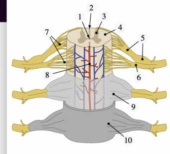
Spinal cord regions: 1. Central canal, 2. Posterior median sulcus, 3. Grey matter, 4. White matter, 5. Dorsal root and dorsal root ganglion, 6. Ventral root, 7. Fascicles, 8. Anterior spinal artery, 9. Arachnoid mater, 10. Dura mater.white matter surrounds the grey matter:[1]
divided into the posterior (dorsal) column, anterior (ventral) column and the lateral column
fibres decussate through the anterior white commissure
these tracts are largely made up of myelinated sensory and motor axons
ascending tracts: transmit information from the sensory receptors in the periphery for processing in the central nervous system
descending tracts: transmit information to the periphery from the central nervous system
Spinal Nerves
Spinal nerve.
Spinal nerves are "mixed nerves that interact directly with the spinal cord to modulate motor and sensory information from the body's periphery."[13]
Humans have 31 pairs of spinal nerves:
eight cervical
twelve thoracic
five lumbar
five sacral
one coccygeal
Each pair roughly corresponds to a segment of the vertebral column. While there are seven cervical vertebrae, there are eight pairs of cervical spinal nerves:[4]
the first pair of spinal nerves exits above the first cervical vertebra
the eighth pair exits below the last cervical vertebra
Spinal cord and spinal nerves
From the thoracic region, each spinal nerve exits below its corresponding vertebra.[4]
The spinal nerves are relatively large. They form from nerve fibres (fila radicularia) that extend from the posterior (dorsal) root and anterior (ventral) root of the spinal cord:
cell bodies of motor fibres are situated in the anterior horn of the spinal cord (anterior and lateral grey columns)
exits the spinal cord from the anterolateral sulcus[14]
The anterior and posterior roots join to form two spinal nerves, one on either side of the spine, which exit the vertebral canal through the intervertebral foramina. Once outside the intervertebral foramina, they form peripheral nerves.[5]
Alpha and Gamma motor neurons are both found in the anterior (ventral) horn.
Alpha motor neurons are the largest motor neurons in the nervous system and innervate skeletal muscle
Gamma motor neurons innervate intrafusal muscle fibres of the muscle spindle
Motor neurons are arranged somatotopically across the anterior horn. The more medially placed motor neurons innervate proximal muscles while laterally placed motor neurons innervate distal muscles.[4][5]
Associated Pathways
Brain and spinal cord tracts
The central nervous system uses ascending and descending pathways to communicate with the external environment. The tracts and pathways complete the circuit in the spinal cord.
Spinal reflexes[16] are involuntary actions to stimuli involving simple circuits at the level of the spinal cord. They allow for near-instantaneous responses because they can bypass processing from the brain. Although spinal reflexes can bypass the cortical circuit, the descending circuit of the brainstem and cortex can still modulate the intensity of the response.
These include:
myotatic or stretch reflex (also known as the jerk reflex): occurs when muscle spindle stretch receptors are activated. Information is sent along the afferent neuron to the spinal cord where it synapses on the efferent neuron to the same muscle, causing that muscle to contract in response. At the same time, reciprocal inhibition occurs. The afferent neurons also synapse with inhibitory interneurons in the spinal cord that prevent contraction of the antagonistic muscles.
flexor withdrawal reflex: occurs in response to noxious stimuli. For example: when you touch a hot object, a pain receptor in your hand picks up the noxious stimulus and the afferent neuron carries the impulse to the spinal cord where it synapses on an interneuron. The interneuron then synapses with the efferent neuron, which sends a signal to the muscle to withdraw your hand.
Blood Supply
The blood supply to the spinal cord "is highly complex and anastomotic to ensure adequate supply to the entire structure. Variability in the vasculature is not uncommon."[17]
Spinal arteries: The spinal arteries arise from the vertebral artery and run longitudinally down the spinal cord. Three major arteries supply the spinal cord:[18]
unpaired anterior spinal artery lies in the anterior median fissure of the spinal cord and extends from the level of the lower brainstem to the tip of the conus medullaris. It supplies the ventral medial surface of the medulla and anterior two-thirds of the spinal cord. Its diameter is narrower through the thoracic spine when compared to the cervical and lumbar spine. Therefore, much of the blood flow to the thoracic spine comes from segmental arteries that branch off from the dorsal aorta.[18]
paired posterior spinal arteries supply the posterior one-third of the spinal cord, the posterior (dorsal) columns and much of the dorsal horns (apart from their base).
the anterior spinal artery and posterior spinal arteries join circumferentially through the pial plexus (also called the vasocorona) and supply the spinal cord's periphery.[17]
Segmental arteries: the blood supply for the spinal cord is reinforced by segmental, medullary, and radicular arteries. These come from spinal branches of the cervical, posterior intercostal, and lumbar arteries. The segmental medullary arteries supply the anterior or posterior spinal arteries, while radicular arteries mainly supply the spinal nerve roots.
Occlusion of the anterior spinal artery can lead to a loss of power or paralysis and spinothalamic sensory deficit (i.e. pain and temperature sensation are affected).[19] Fine touch, two-point discrimination, vibration and proprioception, which are transmitted by the posterior (dorsal) columns, are maintained.[20][4][5] Occlusion of the posterior spinal artery affects the posterior (dorsal) column, leading to posterior cord syndrome. This presents with a loss of proprioception and vibration, lack of deep tendon reflexes, hypotonia, and ataxic gait. Anterior spinal artery occlusion is more common than posterior spinal artery blockage.
Posterior spinal cord, great posterior radiculomedullary artery
Spinal veins: blood from within the spinal cord travels through the intramedullary veins, to the anterior and posterior spinal veins. They form a reticulated network in the pia mater around the circumference of spinal cord:
the anterior spinal vein drains a significant portion of the gray matter of the spinal cord.
the posterior and lateral spinal veins run the length of the entire spinal cord and primarily drain the white matter and portions of the grey matter not drained by the anterior spinal vein. These vessels empty through the radicular veins into the external and internal vertebral venous plexuses, groups of valveless veins that extend from the coccyx to the base of the skull.
the vertebral and deep cervical veins drain venous blood from the cervical spine into the superior vena cava; the posterior intercostal and lumbar veins drain venous supply from the thoracic and lumbar spine into the azygos and hemiazygos veins; and the median and lateral sacral veins drain venous supply from the sacrum into the common iliac vein.[4][5]
References
↑ 1.01.11.21.31.41.5Harrow-Mortelliti M, Reddy V, Jimsheleishvili G. Physiology, Spinal Cord. [Updated 2023 Mar 17]. In: StatPearls [Internet]. Treasure Island (FL): StatPearls Publishing; 2024 Jan-. Available from: https://www.ncbi.nlm.nih.gov/books/NBK544267/
↑DeSai C, Reddy V, Agarwal A. Anatomy, Back, Vertebral Column. [Updated 2023 Aug 8]. In: StatPearls [Internet]. Treasure Island (FL): StatPearls Publishing; 2024 Jan-. Available from: https://www.ncbi.nlm.nih.gov/books/NBK525969/
↑ 13.013.113.2Kaiser JT, Lugo-Pico JG. Neuroanatomy, Spinal Nerves. [Updated 2023 Aug 14]. In: StatPearls [Internet]. Treasure Island (FL): StatPearls Publishing; 2024 Jan-. Available from:
↑ 14.014.1Khan YS, Lui F. Neuroanatomy, Spinal Cord. [Updated 2023 Jul 24]. In: StatPearls [Internet]. Treasure Island (FL): StatPearls Publishing; 2024 Jan-. Available from: https://www.ncbi.nlm.nih.gov/books/NBK559056/
↑ 15.015.1Xuan D. Spinal Cord Anatomy and Function Course. Plus, 2024.
↑Hultborn H. Spinal reflexes, mechanisms and concepts: from Eccles to Lundberg and beyond. Progress in neurobiology. 2006 Feb 1;78(3-5):215-32.
↑ 17.017.1Kaiser JT, Reddy V, Lugo-Pico JG. Anatomy, Back, Spinal Cord Arteries. [Updated 2023 Aug 14]. In: StatPearls [Internet]. Treasure Island (FL): StatPearls Publishing; 2024 Jan-. Available from: https://www.ncbi.nlm.nih.gov/books/NBK537219/
↑ 18.018.1Hasan S, Arain A. Neuroanatomy, Spinal Cord Arteries. [Updated 2023 Aug 8]. In: StatPearls [Internet]. Treasure Island (FL): StatPearls Publishing; 2024 Jan-. Available from: https://www.ncbi.nlm.nih.gov/books/NBK539889/
↑Sandoval JI, De Jesus O. Anterior Spinal Artery Syndrome. [Updated 2024 Jun 7]. In: StatPearls [Internet]. Treasure Island (FL): StatPearls Publishing; 2024 Jan-. Available from: https://www.ncbi.nlm.nih.gov/books/NBK560731/
↑l-Chalabi M, Reddy V, Alsalman I. Neuroanatomy, Posterior Column (Dorsal Column) [Updated 2023 Apr 8]. In: StatPearls [Internet]. Treasure Island (FL): StatPearls Publishing; 2024 Jan-. Available from: https://www.ncbi.nlm.nih.gov/books/NBK507888/