When refering to evidence in academic writing, you should always try to reference the primary (original) source. That is usually the journal article where the information was first stated. In most cases Physiopedia articles are a secondary source and so should not be used as references. Physiopedia articles are best used to find the original sources of information (see the references list at the bottom of the article).
If you believe that this Physiopedia article is the primary source for the information you are refering to, you can use the button below to access a related citation statement.
A ganglion is a collection of neuronal bodies found in the voluntary and autonomic branches of the peripheral nervous system (PNS).
Ganglia can be thought of as synaptic relay stations between neurons. The information enters the ganglia, excites the neuron in the ganglia and then exits[1][2].
Among vertebrate animals, there are three major groups of ganglia. These include:
Dorsal root ganglia or spinal ganglia where the cell bodies of sensory or afferent nerves are located See Image 1
Cranial nerve ganglia that contain the neurons of the selected cranial nerves See image 2 Trigeminal ganglion highlighted in green.
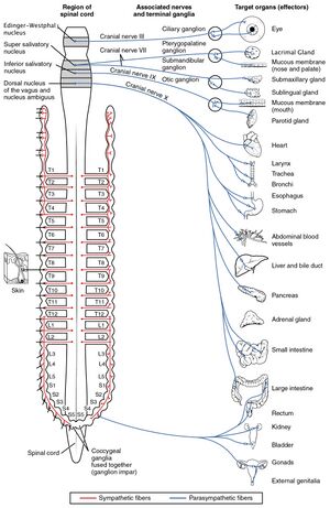
Autonomic ganglia, which contain the cell bodies of the autonomic nervous system. See image 3
Image 1: Shows the Autonomic Ganglia (red SNS, blue PNS)
Besides the ganglion of the peripheral nervous system, there are also parts of the brain that contains a cluster of interconnected nuclei called the basal ganglia[3][4]
Structure
Ganglia are oval in structure and contain
Neuronal cell bodies (somata)
Satellite glial cells, surround neurons in the sensory, sympathetic and parasympathetic ganglia and help regulate the chemical environment. They may contribute to chronic pain.[5]
A dense connective tissue capsule covers the ganglion, with a single layer of flat-shaped satellite cells surrounding each neuronal cell body.
A basement membrane covers the outer region of the satellite cells.
Three Major Groups Of Ganglia
A spinal ganglion (dorsal root ganglion) is a cluster of nerve bodies positioned along the spinal cord at the dorsal and ventral roots of a spinal nerve. The dorsal root ganglia contain the cell bodies of afferent nerve fibres (those carrying impulses toward the central nervous system); efferent neurons (carrying motor impulses away from the central nervous system) are present in the ventral root ganglia.See image 1.
Cranial Nerve Ganglion (not all CN have) is analogous to the dorsal root ganglion, except that it is associated with a cranial nerve, instead of a spinal nerve (associated with the spinal cord). The roots of cranial nerves are within the skull, whereas the ganglia are outside the skull. Eg, The vagus nerve has two sensory ganglia, the superior and the inferior ganglia both outside the skull; the trigeminal ganglion is exterior to the temporal bone. Like the sensory neurons associated with the spinal cord, the sensory neurons of cranial nerve ganglia are unipolar with associated satellite glial cells[6].
There are two types of Autonomic Ganglia: the sympathetic and the parasympathetic based on their functions. The former is located close to the spinal cord whereas the latter lie near or within the viscera of the peripheral organs that they innervate.[6]. See image 3 Shows the Autonomic Ganglia (red SNS, blue PNS).
Sensory ganglia
The somatic sensory and visceral neuron cell bodies are found in the dorsal root ganglia of spinal nerves, and on the ganglia of selected cranial nerves. Hence known as sensory ganglia.
1. Dorsal root ganglia:
most common type of sensory ganglia. They are found in the posterior (dorsal) root of spinal nerves, following the emergence of the dorsal root, that emerges from the intervertebral neural foramina
contain clusters of sensory neuron cell bodies which transmit messages relating to pain, touch, and temperature from the PNS, towards the CNS. Satellite glial cells separate and inhibit the interaction between cell bodies in the ganglion.
Most of the body’s sensory neurons are contained here.
2. Sensory ganglia of the cranial nerves
Another type of sensory ganglia are the ones that are found in the cranial nerves. Those are ganglia with special sensory functions and like the dorsal root ganglia except that they are associated with the cranial nerves and not the spinal nerves[1].
Key Facts
Ganglion: Collection of neuron cell bodies in the peripheral nervous system (PNS).
Types:
Sensory ganglia: Dorsal root ganglia of spinal nerves and the ganglia of selected cranial nerves.
Autonomic ganglia: Sympathetic (close to the spinal cord), Parasympathetic (near on in the viscera)[1].
With invertebrates, ganglia often do the work of a brain.
Eg The earthworm has a ganglion above the gut at the front. Nerve fibres running down each side of the gut link this to another under the gut. The rest of the central nervous system runs under the gut. This type of arrangement is found in several invertebrate phyla and contrasts with the vertebrates, who have their spinal cord above (dorsal to) their gut[8].
The brain and the spinal cord are the primary organs of the central nervous system. The nerves and ganglia are the primary components of the peripheral nervous system.[9][10]
↑ 6.06.1Barral J-P, Croibier A. Chapter 3 - Anatomical organization of cranial nerves. In Barral J-P, Croibier A. editors: Manual Therapy for the Cranial Nerves. Churchill Livingstone, 2009. p15-18. https://doi.org/10.1016/B978-0-7020-3100-7.50006-9.



.jpg)

