When refering to evidence in academic writing, you should always try to reference the primary (original) source. That is usually the journal article where the information was first stated. In most cases Physiopedia articles are a secondary source and so should not be used as references. Physiopedia articles are best used to find the original sources of information (see the references list at the bottom of the article).
If you believe that this Physiopedia article is the primary source for the information you are refering to, you can use the button below to access a related citation statement.
The human neuronal network is a complex electrochemical communication system that sends information at near instantaneous speeds. An understanding of the role and function of each level of this network can be a powerful tool for rehabilitation professionals, helping them identify appropriate treatment interventions and guide care plans and referrals.
Below is a summary of the components of the human neuronal network. Please see the linked reading pages for a more in-depth review of each topic.
Neurons
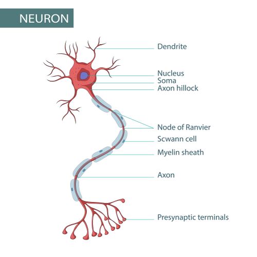
The neuron, or nerve cell, is the building block of the nervous system. Neurons come in many shapes and sizes, which dictates their function, but all neurons have the same basic structure.[1][2]
Cell body, also known as the soma: contains the nucleus, mitochondria, and other organelles necessary for neuronal functioning. These structures support the chemical processes of the neuron, most notably the production of neurotransmitters.
Dendrites: radiate out from the cell body and receive afferent signals from other neurons. They react to neurotransmitters released by neighbouring neurons and relay information via electrical signals through the cell body. A neuron may have a single dendrite or many dendrites depending on its function and location in the nervous system.
Axons: also originate from the cell body at a specialised area called the axon hillock. Axons relay efferent information to neighbouring neurons. Neuron communication speed is increased by a myelin sheath which surrounds and "insulates" axons. It should be noted that not all nerves are myelinated. Myelin sheaths are formed from concentric wraps of the plasma membrane of neuroglia cells around the axon. In the PNS, neuroglia cells are called Schwann cells, and in the CNS they are called oligodendrocytes.[3] The myelin sheath has regular gaps, called nodes of Ranvier, along the axon. The nodes of Ranvier are essential for the rapid propagation of action potentials (see below) along the axon.[4]
Communicating neurons interact at synapses. Synapses are microscopic gaps found between neurons, typically between the axon of one neuron and the dendrite of another. They are the site of chemical communication between neurons. The neuron that shares information on the transmitting side of the synapse is called the pre-synaptic neuron, while the neuron on the receiving side of the synapse is called the post-synaptic neuron.[1][2]
Action potentials and neurotransmitters: the language of nerves
The nervous system communicates in an electrochemical language. Action potentials serve as the electrical signals that propagate along neurons, while neurotransmitters facilitate the chemical communication between neurons at synapses. This intricate interplay allows the nervous system to transmit information rapidly and efficiently, thus enabling complex behaviours, reflexes, and cognitive processes. Understanding this communication is fundamental to neuroscience.
Action potentials are rapid electrical impulses that neurons use to convey information.[5][6]
Resting membrane potential: neurons maintain a resting membrane potential of about -70 mV (millivolts). This resting potential is primarily due to the distribution of ions across the cell membrane, with sodium (Na⁺) ions outside the membrane and potassium (K⁺) ions inside. The neuron is considered polarised at this stage.
Depolarisation: when a neuron receives a stimulus (from another neuron or an external signal), sodium channels in the membrane open, allowing sodium to rush into the cell. The influx of positive ions causes the membrane potential to become less negative. When it reaches a threshold, typically of around -55 mV, an action potential is triggered.
Propagation of the action potential: once the threshold is reached, a rapid depolarisation occurs as more sodium channels open, resulting in a spike in membrane potential. This is followed by the opening of potassium channels, allowing potassium to exit the neuron. This repolarises the membrane back to its resting state.
Refractory period: after action potential propagation, there is a brief period during which the neuron cannot fire another action potential (i.e., the absolute refractory period) and a relative refractory period where it is more difficult to fire. This ensures unidirectional signal propagation along the axon and prevents the neuron from becoming overstimulated.
Neurotransmitters are chemical messengers that transmit signals between neurons and other cells in the nervous system.[7][8]
Synaptic transmission: once the action potential reaches the axon terminal (the end of the neuron), it triggers voltage-gated calcium (Ca²⁺) channels to open. This allows calcium to enter the neuron. The influx of intracellular calcium causes synaptic vesicles (which are special organelles that store and release neurotransmitters) to fuse with the presynaptic membrane in preparation for the release neurotransmitters into the synapse.
Release of neurotransmitters: the fusion with the presynaptic cell membrane leads to the release of neurotransmitters into the synaptic cleft (the gap between the two neurons).
Binding to receptors: the released neurotransmitters diffuse across the synaptic cleft and bind to specific receptors on the postsynaptic neuron’s membrane. This binding can cause various effects, depending on the type of neurotransmitter and the receptor it interacts with.
Postsynaptic potential: the binding of neurotransmitters can generate a postsynaptic potential, which may be excitatory (depolarising the postsynaptic neuron) or inhibitory (hyperpolarising it):
excitatory postsynaptic potential (EPSP): if the neurotransmitter causes the postsynaptic membrane to become less negative (depolarisation), it may bring the neuron closer to its firing threshold, increasing the likelihood of an action potential.
inhibitory postsynaptic potential (IPSP): conversely, if the neurotransmitter makes the postsynaptic membrane more negative (hyperpolarisation), it decreases the likelihood of an action potential.
Termination of neurotransmitter action: after neurotransmitters have acted on the postsynaptic receptors, their effects are terminated through various mechanisms:
reuptake: many neurotransmitters are taken back into the presynaptic neuron through transporter proteins. This process allows the neuron to reuse the neurotransmitter and is crucial for terminating the signal.
enzymatic breakdown: some neurotransmitters are broken down by enzymes in the synaptic cleft, rendering them inactive.
diffusion: neurotransmitters may also diffuse away from the synaptic cleft, reducing their concentration and effect.
The following optional video includes a visual explanation of action potentials:
The size and shape of neurons give information about their function and location in the nervous system. Neurons are generally divided into four basic types.[2]
Unipolar: this neuron type is not found in vertebrates. They are characterised by a single structure extending away from the cell body.
Bipolar: these neurons have one axon and one dendrite extending away from the cell body (e.g., a retinal bipolar cell).
Multipolar: these neurons contain one axon and multiple dendrites. Multipolar neurons are found in the central nervous system (e.g., Purkinje cells in the cerebellum). Typically, most multipolar neurons are motor neurons. In fact, all motor neurons controlling skeletal muscles are multipolar neurons.
Pseudounipolar: these neurons share features of both unipolar and bipolar neurons. Most sensory neurons in the human body are pseudounipolar. They have a single process extending from the soma, which later branches into two distinct extensions. One extension connects to dendrites that receive afferent sensory information while the other transmits this information to the spinal cord.
There are three categories of neurons.
Motor neurons: located within the CNS. They control muscular or motor outputs in the periphery. Motor neurons can be classified into upper motor neurons (UMN) and lower motor neurons.[10] Please see Table 1.0 for more details on UMN and LMN.
Sensory neurons: relay sensory information from the PNS to the CNS.[11]
Interneurons: play varied and important roles in the CNS. They form the bridge between motor and sensory neurons. They also form complex communication networks among themselves. Interneurons allow the nervous system to coordinate complex motor movement patterns in response to environmental and sensory input, and play auxiliary roles in the organisation of brain activity and cognitive function.[12]
Table 1.0. Comparison of Upper and Lower Motor Neurons[10]
Cell body location
Neurotransmitters
Targeted Motor Control Location
Symptoms with Lesion
Clinical Examples
Upper Motor Neuron
Cortex
Glutamate
Within the CNS
Uncontrolled movement
Decreased sensitivity to superficial reflex stimulation
Spasticity
Stroke
Multiple sclerosis
Spinal cord injury
Lower Motor Neuron
Brainstem and spinal cord
Acetylcholine
Outside the CNS
Paralysis
Poliomyelitis
Bell's palsy
Spinal muscular atrophy
Amyotrophic lateral sclerosis (ALS)
If you would like to learn more about neurons in general, please see this article. If you would like to learn more about motor neurons, please review this article.
Nerves
A nerve is a group of neurons bundled together in the PNS. There are three classifications of nerves.
Motor nerves: contain only motor neurons. Motor nerves transmit efferent information from the CNS to the periphery to affect skeletal muscles and somatic tissues.[13]
Sensory nerves: contain only sensory neurons. Sensory nerves transmit afferent information from the PNS to the CNS. There are two sensory pathways within the PNS: (1) the somatosensory system, where spinal nerves transmit information about the external environment to the CNS, and (2) the autonomic nervous system, which transmits information about the internal environment via visceral sensory neurons.[14] Sensory nerves contain different nerve fibre types depending on their associated sensory receptors. Please see Table 2.0 for more information on the classification of sensory receptors, and Table 2.1 for differences between sensory nerve fibre types.
Mixed nerves: contain a combination of both motor and sensory neurons.
Table 2.0. Classification of sensory fibres and associated sensory receptors[14][15]
Sensory Receptor Type
Sensory Receptor Subtypes
Sensory Information and Location
Fibre Type
Chemoreceptors
Olfactory
Sense of smell
Location: olfactory epithelium in the nasal cavity
A-delta,
C-fibres
Gustatory
Sense of taste
Location: taste buds of the tongue, soft palate, pharynx, and upper part of the oesophagus
Internal chemoreceptors
Transmit information about blood gas levels to the central nervous system
Location: in the carotid and aortic bodies of the heart, and the brainstem, cerebellum, hypothalamus, and midbrain
Mechanoreceptors
Tactile receptors
Meissner’s corpuscles:
function: sensitive to light touch and changes in texture; respond quickly to stimuli
location: dermal papillae of the skin
Pacinian corpuscles:
function: detect deep pressure and vibration; respond rapidly
location: deep layers of the dermis and subcutaneous tissue
Merkel discs (tactile discs):
function: respond to sustained pressure and texture; provide fine spatial resolution
location: epidermis (especially in fingertips and hair follicles)
Ruffini endings (Ruffini corpuscles):
function: respond to skin stretch and sustained pressure; play a role in proprioception
location: deep in the dermis and joint capsules
A-beta, A-delta
Proprioceptors
Muscle spindles:
function: detect changes in muscle length (stretch) and contribute to the sense of body position and movement
location: within skeletal muscles
Golgi tendon organs:
function: monitor tension within muscles and tendons; help prevent muscle damage from excessive force
location: at the junction of muscles and tendons
Joint receptors:
function: respond to pressure, tension, and movement in joints, providing feedback about joint position and movement
location: within joint capsules
Baroreceptors
Detect changes in blood pressure
Location: arterial baroreceptors are found in the walls of the aorta of the heart and the carotid arteries. Low-pressure baroreceptors are located in the large veins, pulmonary vessels of the lungs, and the walls of the heart
Hearing receptors
Sense of hearing
Location: hair cells located in the inner ear respond to mechanical vibrations caused by sound waves
Nociceptors
Somatic nociceptors
Detect pain and potentially harmful stimuli
Located in the skin, subcutaneous tissues, fascia, other connective tissues, periosteum, endosteum, and joint capsules
A-delta (sharp, acute pain)
C-fibre (dull, throbbing pain)
Visceral nociceptors
Respond to pain in internal organs
Photoreceptors
Rods
Sensitive to low light levels and responsible for vision in dim conditions
A-fibres
Cones
Responsible for colour vision and function best in brighter light. There are three types of cones, each sensitive to different wavelengths of light corresponding to blue, green, and red
Thermoreceptors
Cold receptors
Respond to lower temperatures (e.g., Krause end bulbs)
A-delta (for cold)
C-fibres (for warmth)
Warm receptors
Respond to higher temperatures (e.g., Ruffini endings)
Table 2.1. Differences between sensory fibre types[14]
Structure
Conduction Velocity
Function
Sensation
A-beta
Myelinated
6-12 micrometres in diameter
Fastest (30-75 m/s)
Primarily responsible for transmitting touch, pressure, and vibration sensations
Associated with sensations of fine touch and proprioception, providing the ability to perceive texture and shape
A-delta
Myelinated
1-5 micrometres in diameter
Fast (5-30 m/s)
Transmit sharp, acute pain (nociceptive signals) and some temperature sensations (cold)
Associated with the initial, localised pain that occurs after a sharp stimulus, often referred to as "fast pain"
C-fibre
Unmyelinated
0.2-1.5 micrometres in diameter
Slow (0.5-2 m/s)
Transmit dull, aching pain, temperature sensations (warm), and some autonomic signals
Associated with the prolonged, diffuse pain that follows the initial sharp pain from A-delta fibres, often referred to as "slow pain"
Spinal Nerves
Spinal nerves branch directly from the spinal cord, but they are considered part of the PNS. They function as an intermediary between the CNS and the PNS. They are mixed nerves, that transmit sensory, motor, and autonomic information between the CNS and the body. There are 31 pairs of spinal nerves, grouped by spinal region: 8 cervical nerve pairs, 12 thoracic nerve pairs, 5 lumbar nerve pairs, 5 sacral nerve pairs, and one coccygeal nerve pair. Each spinal nerve is formed by nerve fibres, called fila radicularia, that extend from the dorsal (posterior) and ventral (anterior) roots of the spinal cord, where they connect through interneurons. These root fibres converge within the intervertebral foramina to create a spinal nerve.[16]
The dorsal root contains afferent sensory axons that relay visceral and somatic sensory information from peripheral receptors to the central nervous system. The areas of skin innervated by specific spinal nerves are organised into regions known as dermatomes. Sensory information from the dorsal root synapses in the dorsal horn of the spinal cord, while the cell bodies of these pseudounipolar neurons reside in the dorsal root ganglion. Dorsal root fibres can either synapse with interneurons in the grey matter for reflex actions or ascend through the spinal cord to reach the thalamus for further processing.[16]
Sensory dermatomes and their spinal levels of innervation
General idea of myotomes and their spinal levels of innervation
The ventral root is responsible for transmitting somatic motor signals from the brain and spinal cord to skeletal muscles. The cell bodies of the efferent motor fibres are located in the anterior horn of the spinal cord. The muscles innervated by a specific spinal nerve are referred to as the nerve’s myotomes. Notably, the spinal cord has enlargements in the cervical and lumbosacral regions, reflecting the higher density of muscle innervation in the upper and lower limbs.[16]
Spinal nerves exit the vertebral canal through the intervertebral foramina as mixed nerve fibre bundles. An exception occurs at the C1 level, where the C1 spinal nerve exits between the occiput and atlas (C1). Since the spinal cord does not extend the full length of the vertebral column, nerves in the cranial regions exit horizontally, while those below the L1 or L2 vertebrae travel downward before exiting, forming the cauda equina. Spinal nerves above C7 exit the column above their corresponding vertebrae. Spinal nerve C8 exits between C7 and T1. All other spinal nerves exit below their respective vertebrae.[16]
After exiting, the spinal nerve divides into ventral and dorsal rami. Both rami contain mixed fibres for sensory and motor innervation. The dorsal ramus is typically smaller than the ventral ramus. It divides into medial and lateral branches (and sometimes an intermediate branch). It is responsible for innervating the paraspinal muscles and associated skin regions. The functions of these branches vary by vertebral level, particularly above and below T6. In contrast, the larger ventral rami contribute to major neural plexuses, serving as the primary pathways for sensorimotor innervation throughout the body.[16] The major neural plexuses or spinal nerve pathways include: the (1) cervical plexus, (2) brachial plexus, (3) thoracic spinal nerves, (4) lumbar plexus, and (5) sacral plexus.
If you would like to learn more about these topics, please see:
Cranial nerves are excellent examples of different types of nerves: sensory, motor, and mixed function.
Cranial nerves (CN) provide innervation for the head and neck. They function similarly to spinal nerves. Like spinal nerves, cranial nerves are considered part of the PNS and function as either pure sensory, pure motor, or mixed nerves. Cranial nerves differ from spinal nerves in their neurological origins; spinal nerve roots originate from spinal cord grey matter while cranial nerve roots arise from neural processes associated with specific brainstem nuclei and cortical areas. The organisation of cranial nerve nuclei gives hints to their function - typically the more posterior and lateral nuclei tend to be sensory nerves, and the more anterior tend to be motor nerves.[17]
There are 12 paired cranial nerves that are numbered using Roman numerals.
If you would like to learn more about cranial nerves, please review this article.
Most cranial nerves (CN III-XII) arise from the brainstem (which consists of the midbrain, pons, and medulla). However, CN I and II originate from the forebrain.
Sensory cranial nerves connect directly to specific nuclei in the brainstem. Sensory ganglia contain the cell bodies of sensory neurons and function to relay and process sensory information between the periphery and the CNS. Sensory receptors transmit signals via cranial nerves to the sensory ganglia, then onto the appropriate brainstem nuclei and to higher brain centres.
Motor cranial nerves originate from nuclei in the brainstem.
Certain cranial nerves, like CN X (vagus nerve), have significant autonomic functions. The vagus nerve’s sensory and motor fibres connect to various organs, providing feedback to the CNS about visceral functions and enabling autonomic control over heart rate, digestion, and respiratory rate.
Cranial nerves exit the skull through specific foramina in the cranial bones. After exiting the CNS, cranial nerves travel to their target organs or muscles.
The Nervous System
The human nervous system has two divisions: the central nervous system and the peripheral nervous system.
Central Nervous System (CNS)
The CNS includes the brain and spinal cord. The brain and spinal cord are considered separate structures but are functional extensions of each other.
The brain is an organ comprised of nervous tissue. It performs countless functions continuously throughout life. It is responsible for integrating sensory input, controlling movement, facilitating cognitive functions and communication, regulating emotions, maintaining homeostasis, and supporting social interactions. The brain is protected from external forces by the skull, meninges, and cerebrospinal fluid (CSF). The brain has additional internal protection against harmful blood-circulating substances provided by the blood-brain barrier.[18]
The brain is divided into two hemispheres (left and right). Each hemisphere is responsible for different behaviours while remaining in constant communication with the other side - this is known as brain lateralisation. Every brain is unique, but typically, the left hemisphere is more dominant in language, logic, and math abilities, and the right hemisphere is dominant in artistic and musical situations and intuition. The cerebral cortex is the brain's outermost layer, consisting of grey matter that houses billions of neurons responsible for high-level executive functions. It is divided into four lobes: the (1) frontal, (2) parietal, (3) temporal, and (4) occipital. Each lobe plays a specific role in processing information and coordinating cognitive functions.[18]
Frontal lobe: responsible for voluntary motor functions, problem-solving, attention, memory, and language. It includes key areas like the motor cortex and Broca's area. The motor cortex enables precise voluntary movements of skeletal muscles, while Broca's area regulates the motor functions necessary for language production.
Parietal lobe: responsible for processing sensory information. It contains the somatosensory cortex, which processes sensory data and integrates this data with prior knowledge to create an understanding of tactile experiences.
Temporal lobe: responsible for processing auditory stimuli via the auditory cortex. Wernicke's area is also located in the temporal lobe, and is responsible for speech comprehension.
Occipital lobe: responsible for visual processing. The visual cortex is located here. It also receives input from the retina and interprets visual stimuli by drawing on past visual experiences to aid in recognition and understanding.
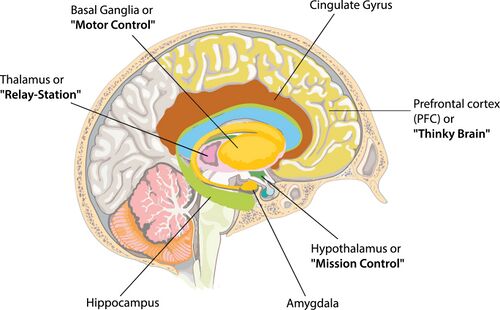
Basal ganglia: a group of interconnected structures in the brain that play a key role in regulating movement and coordinating voluntary motor control. They help facilitate desired movements while inhibiting unwanted ones, contributing to smooth and purposeful motion. Additionally, the basal ganglia are involved in various cognitive functions, including learning, habit formation, and emotional regulation. They work by integrating information from the cerebral cortex and sending outputs to other areas of the brain that manage movement and behaviour.
Thalamus: functions as a relay station in the brain, processing and transmitting sensory information to the appropriate areas of the cerebral cortex. It filters and integrates data from various sensory modalities, such as touch, pain, temperature, and visual and auditory signals, before sending this data to higher brain regions for further processing. Additionally, the thalamus plays a role in regulating sleep, alertness, and consciousness, contributing to the overall coordination of sensory and motor signals.
Hypothalamus: regulates essential bodily functions, including homeostasis, temperature control, hunger, thirst, and circadian rhythms. It plays a key role in maintaining the body's internal balance by influencing the autonomic nervous system and endocrine system. The hypothalamus also controls the release of hormones from the pituitary gland, which affects growth, metabolism, and stress responses. Additionally, it is involved in emotional responses and behaviours, linking physiological processes with emotional states.
Limbic system: a complex network of structures in the brain that plays a central role in regulating emotions, memory, and motivation. It is involved in processing and expressing feelings, such as fear, pleasure, and anger, and is crucial for forming and retrieving memories, particularly those related to emotional experiences. Key components of the limbic system include the amygdala, which processes emotions, the hippocampus, which is essential for memory formation, and the cingulate gyrus, which links emotional responses to behavioural outcomes. Overall, the limbic system integrates emotional experiences with higher cognitive functions, influencing behaviour and decision-making.
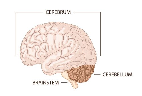
Cerebellum: responsible for coordinating voluntary movements and maintaining balance and posture. It fine-tunes motor activity by integrating sensory input from the body and the environment, ensuring smooth, accurate movements. Additionally, the cerebellum plays a role in learning motor skills and adapting movements based on experience. It also contributes to cognitive functions, such as attention and language processing, highlighting its importance beyond just motor control.
Brainstem: a vital structure that connects the brain to the spinal cord and regulates many essential life functions. It is responsible for controlling basic physiological processes, including heart rate, breathing, blood pressure, and digestion. The brainstem consists of three main parts: the midbrain, pons, and medulla. It also serves as a pathway for motor and sensory signals between the brain and the rest of the body. Additionally, the brainstem plays a role in regulating sleep and alertness, as well as coordinating reflexes and responses to sensory stimuli. It is crucial for maintaining basic bodily functions and enabling communication between different brain regions.
Reticular formation: a network of neurons located in the brainstem that plays an important role in regulating arousal, alertness, and attention. It helps control the sleep-wake cycle and is involved in filtering sensory information, allowing the brain to focus on relevant stimuli while ignoring distractions. Additionally, the reticular formation contributes to the coordination of motor functions and plays a part in reflexes and certain autonomic functions, such as heart rate and blood pressure regulation. It is essential for maintaining consciousness and facilitating responsiveness to the environment.
The spinal cord extends from the foramen magnum at the base of the skull to the first or second lumbar vertebrae, functioning as a two-way communication pathway between the brain and body. It is divided into four regions: (1) cervical, (2) thoracic, (3) lumbar, and (4) sacral, consisting of 31 segments and corresponding pairs of spinal nerves.
The spinal cord varies in width due to cervical and lumbar enlargements, which occur at C3-T1 and L1-S2, respectively. Structurally, the spinal cord contains white matter on the outside and grey matter in its core, with CSF within the central canal. The grey matter comprises the grey commissure, dorsal horns (containing interneurons), lateral horns, and ventral horns (housing somatic motor neurons). Afferent neurons in the dorsal roots transmit sensory information from the body to the spinal cord, where it begins processing. Efferent motor neurons in the ventral horns control voluntary and involuntary movements.
The spinal cord culminates in the conus medullaris, anchored to the coccyx by the filum terminale, with ligaments providing structural support throughout the spinal column.[18]
Ascending pathways to the brain: Sensory information travels from the body to the spinal cord and then ascends to the brain via first-, second-, and third-order neurons. First-order neurons receive sensory impulses and transmit them to the spinal cord. These first-order neurons synapse with second-order neurons located in the dorsal horn. Second-order neurons send signals to the thalamus and cerebellum. Third-order neurons in the thalamus relay this information to the somatosensory cortex, allowing for the perception of sensations such as pressure, pain, and temperature.[18] Ascending spinal tracts include:
lateral spinothalamic tract carries information about pain and temperature
anterior spinothalamic tract carries information about crude touch and pressure
spinocerebellar tract
located in the lateral column and carry proprioceptive information
Descending pathways to the brain: descending tracts carry motor signals from the brain to lower motor neurons, which then initiate muscle movement.[18] Descending spinal tracts include:
The PNS encompasses all nerves outside of the CNS, including the cranial nerves and spinal nerves. The PNS can be divided into two directional components: an afferent (sensory) division and an efferent (motor) division.[19]
The afferent division transmits impulses from peripheral sensory organs, such as tactile or pain receptors in the skin, taste buds on the tongue, visual input from the eyes, inward to the CNS.
The efferent division transmits impulses from the CNS out to the peripheral organs to cause an effect or action.
The PNS can also be functionally divided into the (1) somatic nervous system and the (2) autonomic nervous system.
Somatic Nervous System
The somatic nervous system plays a crucial role in enabling interaction with the environment through sensory perception and voluntary movement. The major functions of the somatic nervous system include:[20]
sensory innervation: the somatic nervous system transmits sensory information from the skin, muscles, and sensory organs to the CNS.
motor control and coordination of movement: the somatic nervous system innervates skeletal muscles, allowing for voluntary movements and conscious control of muscle activity. It also assists in the coordination of complex movements by integrating sensory feedback and motor responses, ensuring smooth and purposeful actions.
spinal reflexes: the somatic nervous system facilitates spinal reflexes, which are automatic responses to external stimuli. For example, the withdrawal reflex helps protect the body by allowing quick reactions, like pulling away from a hot surface.
Autonomic Nervous System
The autonomic nervous system is involved in the management of involuntary bodily functions, allowing the body to respond appropriately to varying internal and external stimuli. The autonomic nervous system has two main divisions:[21]
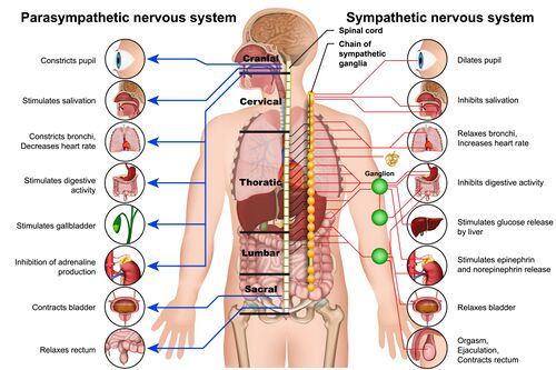
sympathetic nervous system: often referred to as the "fight or flight" system. It prepares the body for stressful or emergency situations by increasing heart rate, dilating airways, and redirecting blood flow to muscles. It primarily uses norepinephrine (noradrenaline) to communicate with target organs, affecting functions such as heart rate, sweat gland activity, and blood vessel constriction.
parasympathetic nervous system: also known as the "rest and digest" system. It promotes restorative processes when the body is at rest by decreasing heart rate, enhancing digestion, and conserving energy. Acetylcholine is the primary neurotransmitter used, acting on target organs to facilitate relaxation and energy conservation.
The major functions of the autonomic nervous system include:[22]
regulation of internal organs: the autonomic nervous system controls the function of internal organs, including the heart, lungs, digestive system, and glands, ensuring they operate efficiently without conscious effort:
heart and circulatory system: the autonomic nervous system can adjust heart rate and the strength of cardiac contractions. The sympathetic division increases heart rate and contractility, while the parasympathetic division (via the vagus nerve) decreases heart rate. The sympathetic division also acts to constrict blood vessels to increase blood pressure and redirect blood to essential organs and muscles during emergencies.
lungs: the sympathetic system dilates bronchioles, increasing airflow during stress, while the parasympathetic system constricts them during rest.
digestive system: the sympathetic system inhibits digestive secretions and peristalsis during stress, redirecting blood flow away from the gastrointestinal tract. In contrast, the parasympathetic system enhances digestive enzyme secretion and increases motility.
glands: the autonomic nervous system is involved in the regulation of glandular activity, such as sweating (sympathetic activation) and saliva production (parasympathetic activation).
maintaining homeostasis: the autonomic nervous system operates through feedback loops to maintain bodily homeostasis, which allows for constant monitoring of internal conditions via sensory receptors. This information is relayed to the CNS, which then adjusts autonomic output to maintain balance by adjusting physiological processes, such as temperature regulation, fluid balance, and energy usage.
reflex actions: the autonomic nervous system coordinates reflex actions that maintain homeostasis and facilitate rapid responses to stimuli. Through a network of receptors, neural pathways, and effectors, the autonomic nervous system ensures that the body can adapt to changes in the environment and internal conditions without requiring conscious effort. This involves two types of reflexes: (1) visceral reflexes, which involve internal organs and include responses that help control functions such as heart rate, digestion, and respiratory rate; and (2) homeostatic reflexes, which help maintain internal balance, such as regulating blood pressure, body temperature, and fluid balance.
↑ 1.01.1Vipula M, Atula M. Human Anatomy and Physiology: For Undergraduate Students of Pharmacy, Nursing, Physiotherapy and Other Paramedical Sciences. University Science Press; 2018.