
Motor Assessment
Original Editor - Stacy Schiurring
Top Contributors - Stacy Schiurring and Jess Bell
Motor System Introduction
Movement and movement control are vital to every aspect of life. The human motor system shapes all our movements, from balance to upright posture, walking, breathing, swallowing, and verbal and nonverbal communication. It is involved in autonomic functions, such as gut motility, and plays a role in the functioning of the sensory system. The motor and sensory systems are deeply interconnected—each system relies on the other to create coordinated, purposeful movements and enable perception of the environment.
Because the human nervous system is highly interconnected, it is difficult to isolate a discussion of one component from the rest of its neuronal network. While this article focuses on the motor assessment, aspects of sensory and cognition assessments are vital to a thorough and complete understanding of motor findings.
Please review the following articles on the sensory assessment and cognitive assessment for a holistic understanding of the rehabilitation assessment.
Motor Neuroanatomy Review
The motor system encompasses both voluntary and involuntary motor control, integrating pathways that originate in the brain and extend to the muscles via a complex hierarchy of neuroanatomical structures.

Voluntary movement begins in the primary motor cortex located in the frontal lobe, where upper motor neurons generate motor commands based on conscious intent. These impulses travel through the internal capsule and descend through the brainstem (passing sequentially through the midbrain, pons, and medulla). In the medulla, most corticospinal fibres cross over at the pyramidal decussation and descend in the lateral corticospinal tract of the spinal cord. The brainstem also houses critical descending motor pathways (like the vestibulospinal, reticulospinal, and rubrospinal tracts) that influence muscle tone, automatic postural adjustments, and locomotion—functions largely outside of conscious control. Additionally, cranial nerve motor nuclei in the brainstem control involuntary and voluntary movements of the head, neck, and face.[1]
The basal ganglia, a group of deep brain nuclei including the caudate, putamen, globus pallidus, subthalamic nucleus, and substantia nigra, play a vital role in initiating voluntary movements and suppressing unwanted or excessive motor activity. They form feedback loops with the motor cortex via the thalamus, fine-tuning movement initiation and amplitude. The cerebellum is responsible for motor coordination, precision, timing, and motor learning. It receives input from the motor cortex and sensory systems and sends corrective signals back to the cortex through the thalamus to ensure fluid, accurate movement.[2]
At the appropriate level, UMNs synapse with lower motor neurons in the anterior horn of the spinal cord, which then exit via spinal nerves to innervate skeletal muscles, producing voluntary contraction and movement.[1]
Upper Motor Neurons versus Lower Motor Neurons
Upper motor neurons (UMNs) and lower motor neurons (LMNs) are two components of the motor pathway that work together to produce voluntary movement, but they differ in location and function. Injuries of UMNs and LMNs also have different clinical presentations.
UMNs originate in the motor cortex of the brain and travel down through the brainstem and spinal cord, where they synapse with LMNs. In contrast, LMNs originate in the spinal cord or brainstem and extend out to innervate skeletal muscles.
UMN injuries typically result in weakness, increased muscle tone (spasticity), hyperreflexia, and the presence of pathological reflexes such as the Babinski sign. These symptoms occur because inhibitory control from the brain is lost. LMN injuries, on the other hand, lead to flaccid weakness, decreased or absent reflexes, muscle atrophy, and fasciculations due to direct disruption of the nerve supply to the muscle. Understanding the differences in these presentations is crucial for localising lesions and guiding appropriate rehabilitation and treatment strategies.[3]
Involuntary motor control, such as posture, balance, and reflexes, is coordinated primarily through subcortical structures, the brainstem, and spinal cord circuits.[2]

At the spinal level, reflex arcs enable rapid, automatic responses to sensory stimuli. In reflex arcs, sensory neurons synapse directly with motor neurons or interneurons in the spinal cord, bypassing higher brain centres for speed. There is continuous integration between voluntary and involuntary systems, allowing for real-time adjustments to maintain posture, balance, and smooth execution of movement.[1]
Please see the following articles for a more in-depth review of motor neuroanatomy:
Motor Examination
The motor assessment is a comprehensive evaluation used to examine the integrity and function of the neuromuscular system, guiding diagnosis and treatment planning in individuals with motor impairments. This assessment typically includes range of motion testing to evaluate joint flexibility and mobility; muscle strength testing to assess the force-generating capacity of individual muscles or muscle groups; and muscle tone testing to identify alterations in tone, such as spasticity or flaccidity. Reflex testing is used to evaluate the integrity of the peripheral and central nervous systems by eliciting involuntary responses. The motor assessment also includes testing a patient's balance and coordination; these assessments are significant because they reflect the integration of sensory input and motor output for postural control and smooth movement. Gross and fine motor assessments further examine the patient's ability to perform functional tasks requiring large body movements or precise manual dexterity. Together, these components provide a detailed understanding of motor function essential for designing effective rehabilitation interventions.[4]
Testing procedure: Prior to beginning a motor examination, the examiner should screen the patient's cognition to determine if they are able to fully and accurately participate in the assessment or if modifications are indicated. Observations of the patients movements and mobility can be performed prior to formal testing. This could include how a patient repositions in bed or moves from the waiting room to therapy area. The patient's general posture and ability to complete functional movements can provide insight into severity of symptoms, willingness to move, joint range of motion, and muscle strength. Please review this optional article if you would like to read more about posture and postural assessment.
The patient should be appropriately dressed and/or draped to allow the examiner to access the needed areas of their body while still maintaining privacy and dignity.
Rangle of Motion
Range of motion (ROM) refers to the extent of movement possible at a joint, measured in degrees. The ROM assessment includes: (1) active, (2) active assisted, and (3) passive ROM testing. Assessing ROM is a critical component of the motor assessment because it provides insight into joint integrity, muscle function, neurological status, and pain levels. Limitations in ROM can indicate specific impairments or compensatory patterns that may impact daily functioning. Evaluating all three types ROM allows clinicians to tailor interventions appropriately, monitor progress, and set realistic, individualised therapy goals. ROM can be assessed visually or using a goniometer for an objective measure.[4]
Active range of motion (AROM) is the unassisted voluntary osteokinematic movements of a joint. AROM can be assessed in straight planes of motion or using functional movement patterns. AROM is typically the first way joint motion is assessed during a motor examination, and can be limited by (1) pain, (2) the joint capsule, (3) ligament length, (4) muscle tightness or spasticity, (5) soft tissue tightness, (6) joint surface abnormalities, and/or (7) muscle weakness.
In active assisted range of motion (AAROM), the individual initiates the movement but has some help from either a therapist or a device, to complete the motion.
Passive range of motion (PROM) occurs when an external force, such as a therapist, moves the joint without the patient’s active muscle engagement. In addition to measuring the range of motion at a joint, PROM also assesses for (1) the impact of joint movement/muscle shortening or lengthening on symptoms, (2) joint end-feel, and (3) patterns of movement limitations. PROM can be limited by bone or joint abnormalities or tightness of the joint capsule, ligaments, muscles, tendons, and/or soft tissues.[4]
Please read the following articles if you would like to learn more about ROM:
Muscle Strength
Muscle strength is the amount of force exerted by a muscle (or group of muscles) to overcome resistance in one maximal effort.

Myotomes describe the pattern of muscle innervation by a single spinal nerve root. Each spinal nerve root sends motor signals to specific muscles. These patterns of innervation are consistent and predictable across individuals (see Table 1). Understanding myotomes is critical in a motor clinical assessment because muscle strength can be directly influenced by the health and function of the corresponding nerve root. When performing a strength assessment, identifying weakness in a specific muscle group can help the examiner localise a potential nerve root injury or compression. This targeted knowledge allows for more accurate diagnosis, effective treatment planning, and monitoring of neurological recovery or deterioration over time.[5]
| Spinal Nerve Level | Action to be Tested | Muscle |
|---|---|---|
| C5 | Shoulder ABDuction | Deltoid |
| C5, C6 | Elbow flexion | Biceps |
| C7 | Elbow extension | Tricpes |
| C8 | Ulnar deviation | Flexor carpi ulnaris
Extensor carpi ulnaris |
| L2, L3 | Hip flexion | Iliopsoas |
| L3, L4 | Knee extension | Quadraceps |
| L5 | Ankle dorsiflexion
Extension of great toe |
Anterior Tibialis
Extensor hallicus longus |
| S1 | Ankle plantarflexion | Gastrocnemius |
Manual muscle testing (MMT) is a clinical method used to assess and grade muscle strength based on gravity and manually applied resistance. Two primary techniques are used in MMT: the break test and the active resistance test. In the break test, the examiner positions the patient’s limb at the point of maximum contraction and applies resistance in the opposite direction while instructing the patient to "hold" the position. The test measures the muscle’s ability to resist this pressure before it "breaks" or yields to the resistance. In contrast, the active resistance test involves the patient actively moving through their full range of motion against the examiner's resistance, which is applied throughout the movement, rather than holding a static position.[4] MMT is typically graded on a 0-5 scale, acoording to the Medical Research Council Scale (also known as the Oxford Scale).[6]
| Grade | Description |
|---|---|
| 0 | No contraction |
| 1 | Flickering contraction |
| 2 | Full range of motion with gravity eliminated (more accurately called gravity minimised) |
| 3 | Full range of motion against gravity |
| 4 | Full range of motion against gravity with minimal resistance |
| 5 | Full range of motion against gravity with maximal resistance |
MMT is an essential part of the motor assessment because it helps identify muscle weakness, neurological deficits, and imbalances that can affect posture, mobility, and daily function. It also allows clinicians to evaluate the impact of injury, disease, or disuse on muscle performance.[4] By understanding the strength and capabilities of specific muscles, therapists can develop targeted intervention strategies, monitor progress over time, and adjust treatment plans to optimise recovery and functional outcomes.
Please see the following articles if you would like a more in-depth review of the strength assessment:
- Muscle Strength Testing
- Assessing Muscle Strength
- Performing Manual Muscle Tests (this link will take you to list of demonstrations for MMT of specific joints and movements)
- Myotomes
Muscle Tone
Tone refers to the continuous and passive partial contraction of a muscle, or a muscle’s resistance to passive stretch while at rest. It is a key indicator of the integrity of the motor system, particularly the neural pathways that control movement. Normal muscle tone allows for posture maintenance and readiness for voluntary movement. Tone can be influenced by many different factors, including: (1) volitional effect and movement, (2) stress and anxiety, (3) position and interaction of tonic reflexes, (4) medications, (5) overall health (including fever, infection, metabolic and /or electrolyte balance), (6) environmental temperature, and (7) CNS arousal or alertness.[4]
Assessing muscle tone involves evaluating a patient’s resistance to passive movement while the muscles are at rest in a supine position. The limb being examined must be fully supported to prevent any potential compensatory movements. The assessment begins with the joint in either a fully flexed or fully extended position (depending on the muscle group being tested). The examiner moves the patient’s limbs at a controlled speed through their range of motion while observing for the amount and quality of resistance. Tone should be assessed bilaterally for comparison, and findings are interpreted in the context of the patient’s neurological status to help localise and understand potential motor pathway dysfunction. In the Modified Ashworth Scale, tone is documented on a 0-4 scale.[7]
| Score | Clinical Findings |
|---|---|
| 0 | No increase in muscle tone |
| 1 | Slight increase in muscle tone, with a catch and release or minimal resistance at the end of the range of motion when an affected part(s) is moved in flexion or extension |
| 1+ | Slight increase in muscle tone, manifested as a catch, followed by minimal resistance through the remainder (less than half) of the range of motion |
| 2 | A marked increase in muscle tone throughout most of the range of motion, but affected part(s) are still easily moved |
| 3 | Considerable increase in muscle tone, passive movement difficult |
| 4 | Affected part(s) rigid in flexion or extension |
Hypertonia
Hypertonia is an increase in muscle tone that causes stiffness and resistance to passive movement. It is commonly associated with UMN lesions. Spasticity is a form of hypertonia characterised by a velocity-dependent increase in muscle tone, meaning that the faster a muscle is passively stretched, the greater the resistance it generates. It results from damage to UMNs, which disrupts the normal balance of excitatory and inhibitory signals in the spinal cord. A classic feature associated with spasticity is the clasp-knife response, where resistance to passive movement is initially high but then suddenly decreases, similar to the closing of a pocketknife. This phenomenon reflects exaggerated stretch reflexes and a release of inhibitory mechanisms. Clonus is another sign of spasticity. It is characterised by a series of involuntary, rhythmic, and rapid muscle contractions that are typically triggered by a sudden stretch. Clonus is most commonly seen at the ankle. It indicates heightened reflex activity due to UMN dysfunction. The Babinski sign is a pathological reflex also associated with UMN lesions. It is tested by firmly stroking the sole of the foot. A positive Babinski sign occurs when the big toe extends upward and the other toes fan out. This is an abnormal response in adults that suggests damage to the corticospinal tract.
Rigidity is a type of increased muscle tone that results in uniform resistance to passive movement, regardless of the speed or direction of the motion. Thus, unlike spasticity, rigidity is not velocity-dependent. It is often linked to extrapyramidal system disorders, particularly basal ganglia dysfunction, such as in Parkinson’s.
There are two main types of rigidity: (1) cogwheel rigidity and (2) lead pipe rigidity. Cogwheel rigidity is characterised by a ratchety resistance to passive movement, often felt as a series of brief catches or jerks during motion. It results from the combination of rigidity and tremor. Leadpipe rigidity involves a smooth, constant resistance throughout the entire range of passive movement, like bending a soft metal pipe. It is also associated with basal ganglia disorders and may occur without tremor.
Additional forms of abnormal rigidity include opisthotonus, decorticate rigidity and decerebrate rigidity. These types of rigidity are often seen in more severe neurological conditions include. Opisthotonus is a severe form of muscle rigidity where the back arches excessively, and the head and heels are pulled backward due to intense contraction of the back and neck muscles. It can occur in conditions such as severe meningitis, tetanus, or brain injury. Decorticate rigidity, which causes flexion of the arms, clenched fists, and extended legs, indicates damage to the brain above the level of the red nucleus, such as in the cerebral hemispheres or internal capsule. It suggests significant but potentially reversible brain injury. Decerebrate rigidity involves extension and pronation of the arms and legs, often with arching of the back. It reflects a more severe injury below the red nucleus, typically involving the brainstem. This posture generally indicates a worse prognosis than decorticate rigidity.[4]
-
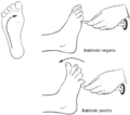 Babinski reflex and positive result (ie. Babinski sign)
 -
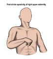 Flexor spasticity pattern of the upper extremity
 -
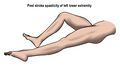 Extensor spasticity pattern of the lower extremity
 -
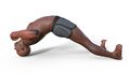 Opisthotonus posturing
 -
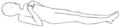 Decorticate posturing
 -
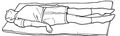 Decerebrate posturing


Hypotonia
Hypotonia is a decrease in muscle tone, meaning there is reduced resistance to passive movement of the muscles. It is often observed as muscle “floppiness,” especially in infants and children, but it can occur at any age depending on the underlying cause. Hypotonia is a symptom rather than a diagnosis and can be caused by dysfunction at any level of the motor pathway, including the central nervous system (brain and spinal cord), peripheral nerves, neuromuscular junction, or muscles themselves.
A severe form of hypotonia is flaccidity, where muscles offer no resistance to passive movement and may appear completely limp. Flaccid muscles also tend to exhibit decreased or absent reflexes, and over time, they may show signs of atrophy due to disuse or denervation.
Two important clinical scenarios involving acute hypotonia are spinal shock and cerebral shock. Spinal shock occurs immediately after an acute spinal cord injury. It is characterised by flaccid paralysis and loss of all reflexes below the level of the injury. This flaccid state is temporary and can last days to weeks; as spinal shock resolves, reflexes often return and may progress into spasticity. Cerebral shock refers to a transient phase of decreased muscle tone and reflexes following an acute injury to the brain, such as a stroke or traumatic brain injury. Like spinal shock, it can resemble lower motor neuron weakness early on, but over time may evolve into hypertonia as upper motor neuron pathways regain activity in a dysregulated way.[4]
Dystonia
Dystonia is a hyperkinetic movement disorder characterised by impaired or disordered tone that results in involuntary repetitive movements, twisting, or postures. These movements are typically patterned and may worsen with voluntary activity or stress, while often disappearing during sleep. Dystonia arises from dysfunction in the CNS, most commonly in the basal ganglia.[4]
Reflexes
A reflex is an involuntary and nearly instantaneous movement or response of a muscle or group of muscles to a specific stimulus, typically mediated by the nervous system. Reflexes are controlled by neural pathways called reflex arcs, which involve a sensory input to the spinal cord or brainstem and a corresponding motor output to the muscles, often without conscious involvement from the brain. Reflexes are used clinically to assess the integrity and function of the nervous system, particularly the spinal cord and peripheral nerves.[8]
Deep tendon reflex (DTR) testing, also known as muscle stretch reflexes (MSR), is a neurological assessment technique used to evaluate the integrity of the nervous system by eliciting involuntary muscle responses to specific stimuli, typically through the use of a reflex hammer. Each reflex corresponds to a particular spinal nerve root level; for example, the patellar tendon reflex assesses the L3-L4 spinal segments. A working knowledge of expected (normal) and abnormal reflex responses is essential in motor assessments because changes in reflexes can indicate underlying neurological dysfunction. Hyperreflexia may suggest an upper motor neuron lesion, while hyporeflexia or areflexia may point to lower motor neuron damage. Identifying these patterns helps examiners localise lesions, determine the severity of impairment, and track recovery or progression of neurological conditions, contributing to more precise and effective treatment strategies.[8]
| Spinal Nerve Root | Muscle Tested | Location to Strike to Elicit Response | Peripheral Nerve |
|---|---|---|---|
| C5-6 | Biceps | Just anterior to the elbow | Musculocutaneous nerve |
| C5-6 | Brachioradialis | Approximately 10 cm above the wrist on the radial aspect of the forearm | Radial nerve |
| C7 | Triceps | Just posterior to the elbow | Radial nerve |
| L3-4 | Quadriceps | Just inferior to the patella | Femoral nerve |
| S1 | Gastrocnemius | Posterior to the ankle joint on the Achilles tendon | Sciatic (tibial) nerve |
| Pons | Massester
(Jaw Jerk) |
Chin (mental protuberance) | Mandibular branch of the trigeminal nerve |
The degree of reflex activity is graded on a 0-4 scale, and can be highly subjective. Grades are assigned based on a predicted response and comparison between the two sides of the body.[4]
| Grade | Observed Response |
|---|---|
| 0 | No reflex response |
| 1 | Minimal response |
| 2 | Moderate response |
| 3 | Brisk, strong response |
| 4 | Clonus |

The Jendrassik maneuver is a technique used during reflex testing to enhance or elicit a stronger reflex response for the lower extremity and jaw reflexes. It involves the patient interlocking their fingers and forcefully pulling them apart while the examiner taps the reflex elicit point. This voluntary upper body activity helps distract the patient and increases general excitability of the spinal cord, thereby amplifying the reflex. The manoeuvre is especially useful when reflexes are difficult to observe or appear diminished, as it can help reveal a reflex that might otherwise seem absent.[8]
Primitive reflex testing in adults involves assessing for the presence of reflexes that are typically seen in infancy and should be inhibited by the developing brain within the first year of life. Examples include the Babinski reflex, grasp reflex, and rooting reflex. In a healthy adult nervous system, these reflexes are usually absent; their reappearance can indicate dysfunction in the central nervous system. A positive Babinski reflex is a sign of upper motor neuron damage or pyramidal tract lesion. A positive grasp reflex is associated with frontal lobe lesions and can be an early symptom of corticobasal syndrome, Lewy body dementia, and progressive supranuclear palsy. A positive rooting reflex can be a sign of diffuse cerebral atrophy.[9]
Please see the following articles if you would like to read more on reflex related topics:
- Reflexes (this link includes information about reflex assessment)
- Primitive reflexes list
- Babinski reflex
- Rooting reflex
Motor Cranial Nerve Testing

Cranial nerves (CN) are a set of twelve paired nerves that originate directly from the brain, primarily the brainstem, and are responsible for a wide range of autonomic, sensory, and motor functions. Cranial nerves with pure motor function include: CN III (Oculomotor nerve), CN IV (Trochlear nerve), CN VI (Abducens nerve), CN XI (Spinal Accessory nerve), and CN XII (Hypoglossal nerve). Cranial nerves with combined motor, sensory, and/or autonomic function include: CN V (Trigeminal nerve, CN VII (Facial nerve), CN IX (Glossopharyngeal nerve), and CN X (Vagus nerve).[4]
Below are concise summaries of how to perform the motor portions of CN testing, and normal versus abnormal results.[4]
CN III (Oculomotor nerve) is responsible for most eye movements, pupil constriction, and eyelid elevation. Testing CN III involves several steps to evaluate both motor and parasympathetic functions.
- First, the examiner assesses extraocular movements by asking the patient to follow a target in an "H" pattern, which tests the medial, superior, and inferior rectus muscles, as well as the inferior oblique. Expected results include smooth, coordinated movement of both eyes in all directions without double vision (diplopia) or any limitations in range. Abnormal findings may include strabismus (misalignment of the eyes), an eye that drifts "down and out" at rest (suggesting unopposed action of the lateral rectus and superior oblique muscles), or diplopia during certain gaze directions, all of which point to oculomotor nerve dysfunction.
- Next, the examiner checks for ptosis, or drooping of the upper eyelid. Normally, the eyelids should be symmetrical and fully elevated. Ptosis may indicate weakness of the levator palpebrae superioris, a muscle controlled by CN III, and can suggest nerve compression or a central lesion.
- Finally, pupillary function is assessed using the pupillary light reflex test. The examiner shines a light into each eye and observes for pupil constriction in both the eye being tested (direct response) and the opposite eye (consensual response). Expected results include brisk, equal constriction of both pupils. Abnormal results include a dilated pupil that does not constrict in response to light, which may indicate damage to the parasympathetic fibres of CN III. Anisocoria (unequal pupil sizes), especially if greater in bright light, can also be a sign of oculomotor nerve involvement.
CN IV (Trochlear nerve) innervates the superior oblique muscle, which primarily controls downward and inward movement of the eye, especially when the eye is adducted (looking toward the nose). Testing CN IV involves assessing the patient’s ability to move the eye in this specific direction. The examiner typically asks the patient to follow a target in an “H” pattern, paying close attention to the eye’s movement when looking down and in. Expected results include smooth and symmetrical eye movements without double vision (diplopia) or misalignment. Abnormal findings may include vertical diplopia (seeing double images one above the other), difficulty looking down when the eye is turned inward (such as when reading or walking down stairs), and a compensatory head tilt away from the affected side to minimise the double vision. These signs suggest a trochlear nerve palsy, which can result from trauma, microvascular disease, or compression.
Motor portion of CN V (Trigeminal nerve) testing involves testing the strength and symmetry of the muscles of mastication, including the masseter, temporalis, and pterygoid muscles. The examiner asks the patient to clench their teeth while palpating the masseter and temporalis muscles to assess for equal contraction on both sides. Additionally, the patient may be asked to open and move their jaw side-to-side against resistance. Expected results include strong, symmetrical muscle contraction and smooth, coordinated jaw movements. Abnormal findings might include weakness or atrophy on one side, jaw deviation toward the weak side when opening the mouth, or difficulty with chewing. Such deficits can indicate damage to the mandibular branch of CN V, potentially caused by trauma, brainstem lesions, or peripheral nerve disorders.
CN VI (Abducens nerve) controls the lateral rectus muscle, which is responsible for moving the eye outward (abduction). To test CN VI, the patient is asked to follow a target with their eyes in various directions, particularly observing for the ability to move each eye laterally. This is often done as part of the standard “H” pattern used in cranial nerve examination. Expected results include full, smooth, and symmetrical eye movements, with both eyes able to abduct equally without difficulty. Abnormal findings may include an inability to move one eye laterally, resulting in a medial deviation (esotropia) of the affected eye at rest, as well as horizontal diplopia that worsens when looking toward the side of the lesion. Such signs are indicative of CN VI palsy, which can result from increased intracranial pressure, diabetes, trauma, or lesions affecting the brainstem or the nerve along its long intracranial course.
The motor portion of CN VII (Facial nerve) controls the muscles of facial expression and plays a key role in motor function of the face. Motor testing of CN VII involves asking the patient to perform various facial movements, such as raising their eyebrows, closing their eyes tightly, smiling, frowning, and puffing out their cheeks. The examiner observes for symmetry and strength in these actions. Expected results include smooth, symmetrical movement on both sides of the face, with no weakness or drooping. Abnormal findings may include asymmetry, such as drooping of one side of the mouth, inability to fully close one eye, or loss of forehead movement. These signs suggest facial nerve weakness or paralysis. Importantly, the pattern of weakness can help localise the lesion: if both the upper and lower face are affected on one side, it suggests a lower motor neuron lesion (eg. Bell’s palsy), whereas sparing of the forehead often points to an upper motor neuron lesion (eg. stroke).
The motor portion of CN IX (Glossopharyngeal nerve) primarily innervates the stylopharyngeus muscle, which helps elevate the pharynx during swallowing and speech. Because direct testing of CN IX motor function is difficult to isolate, it is typically assessed together with CN X (Vagus nerve), as both contribute to palatal and pharyngeal movement. During testing, the examiner asks the patient to say “ah” while observing the soft palate and uvula. Expected results include symmetrical elevation of the soft palate and midline positioning of the uvula. Abnormal findings may include asymmetrical elevation or deviation of the uvula away from the affected side, which can indicate weakness in the muscles supplied by CN IX (and possibly CN X). Patients with motor deficits in CN IX may also have difficulty swallowing (dysphagia) or changes in voice quality. Additional signs of CN X dysfunction may include hoarseness, a weak or breathy voice, dysphagia, or nasal regurgitation of liquids during swallowing. In severe cases, aspiration can occur.
CN XI (Accessory or Spinal Accessory nerve) innervates the sternocleidomastoid and trapezius muscles, which are involved in head rotation and shoulder elevation. To test CN XI, the examiner assesses the strength of these muscles by asking the patient to turn their head against resistance (to evaluate the sternocleidomastoid) and to shrug their shoulders against resistance (to evaluate the trapezius). Expected results include strong, symmetrical movement on both sides, with no signs of muscle wasting or weakness. Abnormal findings may include weakness or paralysis on one side, such as difficulty turning the head away from the affected side or an inability to elevate the shoulder on that side. Muscle atrophy or shoulder droop may also be present. These signs suggest a lower motor neuron lesion of CN XI, which can occur due to surgical injury, trauma, or neurological disease.
CN XII (Hypoglossal nerve) is responsible for controlling the movements of the tongue, which are essential for speech, swallowing, and food manipulation. Testing CN XII involves asking the patient to stick out their tongue and move it side to side. Expected results include a tongue that protrudes straight and moves symmetrically in both directions without tremors or atrophy. Abnormal findings may include deviation of the tongue toward the side of the lesion, weakness when pushing the tongue against the inside of the cheek, visible fasciculations, or muscle atrophy. A lower motor neuron lesion, such as from trauma or nerve compression, typically causes deviation toward the affected side and atrophy, while upper motor neuron lesions may produce less obvious but still impaired movements.
Clinical Considerations
Balance
Balance testing is a critical component of the motor examination. It aims to evaluate a patient’s ability to maintain postural stability during static and dynamic activities. It assesses the integration of sensory input, motor responses, and central processing required to control the body’s position in space. Neuroanatomically, balance relies on the coordinated function of the vestibular system,[10] visual input processed by the occipital cortex, and somatosensory feedback transmitted via the dorsal columns to the parietal lobe. The cerebellum plays a central role in integrating these inputs and adjusting motor output to maintain equilibrium, while the basal ganglia contribute to postural adjustments and movement initiation. Descending pathways such as the vestibulospinal and reticulospinal tracts facilitate reflexive and voluntary postural control.[4]
Please see the following articles if you would like to read more about balance and balance treatment:
- Balance Assessment
- Balance Training
- Objective Vestibular Assessment
- Perturbation-Based Balance Training
- Postural Control System
- Reactive Balance: Intervention Strategies
Please see the following articles if you would like more information on balance testing options:
- Activities-Specific Balance Confidence Scale
- Balance Evaluation Systems Test
- Balance Outcome Measure for Elder Rehabilitation
- Berg Balance Scale
- Biodex Balance System
- Community Balance and Mobility Scale
- Dynamic Gait Index / 4-item Dynamic Gait Index
- Functional Reach Test
- Modified Clinical Test for Sensory Interaction on Balance
- Pediatric Balance Scale
- Romberg test
- Scale for the Assessment and Rating of Ataxia
- Single Leg Stance Test
- Star Excursion Balance Test
- Timed Up and Go Test
- Tinetti Test
- Y Balance Test
- 4-Stage Balance Test
Coordination
Coordination testing assess a patient’s ability to perform smooth, accurate, and controlled voluntary movements. This aspect of the motor examination primarily evaluates the integration of sensory input and motor output necessary for timing, force, and direction of movement. Neuroanatomically, coordination depends heavily on the cerebellum, which receives input from the motor cortex, proprioceptive pathways (especially the spinocerebellar tracts), and the vestibular system to fine-tune motor activity. The basal ganglia also contribute by regulating movement initiation and suppressing involuntary movements. Deficits in coordination may manifest as dysmetria, intention tremor, or ataxia.[4]
Please see the following articles if you would like to read more about coordination and coordination treatment:
Coordination testing is used to evaluate a patient’s ability to execute smooth, accurate, and purposeful movements. A standard element of coordination testing is the assessment of diadochokinesia, the ability to perform rapid, alternating movements such as flipping the hands back and forth on the thighs or quickly tapping the hands or feet. Impairment in diadochokinesia may indicate cerebellar dysfunction and is often observed as slowed, irregular, or uncoordinated movement.[11] By identifying such deficits, coordination testing provides valuable insight into neuromotor integration and guides targeted therapeutic interventions.
Please see the following articles if you would like to read more about coordination testing options:
- Fugl-Meyer Assessment
- Lower Extremity Motor Coordination Test
- Scale for the Assessment and Rating of Ataxia
| Test name | Description |
|---|---|
| Finger to nose | With the shoulder ABDucted to 90 degrees and the elbow extended, the patient is asked to bring the tip of their index finger to touch the tip of their nose. |
| Finger to examiner's finger | Patient sits opposite the examiner, whose finger is held in front of the patient. The patient is asked to touch the tip of their index finger to the examiner's finger. The examiner may change the position of their finger during testing. |
| Finger to finger | With bilateral shoulders ABDucted to 90 degrees, the patient is asked to bring both hands toward midline and touch the index finger tips from opposing hands. |
| Finger opposition | The patient touches the tip of the thumb to each of their other finger tips in sequence. Speed may be gradually increased. |
| Mass grasp | The patient alternates between opening and closing a fist (full finger flexion to full extension). Speed by be gradually increased. |
| Pronation/ supination | With elbows flexed to 90 degrees and maintained close to the body, the patient alternates turning the palms up and down in unison. Alternative testing position: shoulders flexed to 90 degrees and elbows extended. Speed may be gradually increased. |
| Rebound test | The patient maintains bilateral elbows flexed while the examiner applies enough manual resistance to produce an isometric contraction of the biceps. Resistance is suddenly removed. Normal response: the opposing muscle group (triceps) will contract and "check" the movement of the limb. This test can be performed on other muscle groups. |
| Tapping | (1) Hand: with the elbow flexed, forearm pronated, and palm resting on their knee, the patient is asked to "tap" their hand on their knee.
(2) Foot: the patient is asked to "tap" the ball of their foot on the floor without raising the knee, keeping the heel in contact with the floor. |
| Alternate heel to knee; heel to toe | In supine: the patient is asked to touch their knee and great toe alternately with the heel of the opposite side. |
| Heel on shin | In supine: the patient is asked to slide the heel of one foot up and down the shin of their opposite leg. |
| Drawing a circle | The patient is asked to draw an "imaginary" circle (alternatively a figure-eight pattern) with either the upper or lower extremity. A table or the floor may be used for a drawing surface. |
Fine Versus Gross Motor Skills
Within the broader context of the motor examination, fine motor and gross motor assessments serve distinct yet complementary roles in evaluating a patient’s motor function. The fine motor assessment focuses on small, precise movements (such as writing, buttoning a shirt, or manipulating small objects) which primarily involve the distal musculature of the hands and fingers. These tasks rely heavily on the integrity of the corticospinal tract, particularly the lateral corticospinal pathway, which originates in the primary motor cortex (precentral gyrus) and is responsible for voluntary, skilled movement. The premotor cortex and supplementary motor area also contribute to planning and coordinating these fine actions. Additionally, fine motor control depends on intact sensory feedback from the somatosensory cortex, basal ganglia, and integration through the cerebellum for coordination and timing.[12]
Please see the following articles if you would like to read more on fine motor testing options:
- Arm Motor Ability Test
- Arthritis Hand Function Test
- Box and Block Test
- Dexterity Tests
- Jebsen-Taylor Hand Function Test
- Minnesota Manual Dexterity test
- Nine-Hole Peg Test
- Outcome Measures for Aged Hand Function
- Sollerman Hand Function Test
The gross motor assessment evaluates large, whole-body movements such as walking, sitting, standing, and lifting—functions that depend more on proximal musculature and postural control. These movements are mediated not only by the corticospinal tract but also by extrapyramidal systems, such as the reticulospinal and vestibulospinal tracts, which originate in the brainstem and help regulate posture, balance, and automatic movements. The cerebellum and basal ganglia also play key roles in modulating gross motor activity, contributing to coordination, muscle tone, and initiation of movement.[13]
Please see the following articles if you would like to learn more about gross motor testing options. This list includes adult and paediatric tests:
- Beery-Buktenica Developmental Test of Visual-Motor Integration
- Dynamic Gait Index / 4-item Dynamic Gait Index
- Figure of 8 Walk Test
- Four Square Step Test
- Fugl-Meyer Assessment
- Functional Gait Assessment
- Functional Walking Test
- Gross Motor Function Classification System
- Gross Motor Function Measure
- Peabody Developmental Motor Scale
- Timed Up and Go Test
- 4 Metre Walk Test
- 6 Minute Walk Test
- 10 Metre Walk Test
- 30 Seconds Sit To Stand Test / Modified 30-Second Sit-to-Stand Test
Clinical Resources
The resources listed below include both written instructions and demonstration videos, and additional reference materials:
- How to Assess the Motor System (Merck Manuals)
- How to Assess Muscle Strength (Merck Manuals)
- How to Assess Reflexes (Merck Manuals)
- How to Assess the Cranial Nerves (Merck Manuals)
- How to Assess Gait, Stance, and Coordination (Merck Manuals)
Optional Reading
This website provides information on movement assessments, specifically in regards to fall education and training:
The following journal articles provide information on movement analysis
- Gill-Body KM, Hedman LD, Plummer L, Wolf L, Hanke T, Quinn L, Riley N, Kaufman R, Verma A, Quiben M, Scheets P. Movement system diagnoses for balance dysfunction: recommendations from the academy of neurologic physical Therapy’s movement system task force. Physical therapy. 2021 Sep 1;101(9):pzab153.
- Quinn L, Riley N, Tyrell CM, Judd DL, Gill-Body KM, Hedman LD, Packel A, Brown DA, Nabar N, Scheets P. A framework for movement analysis of tasks: recommendations from the Academy of Neurologic Physical Therapy’s Movement System Task Force. Physical therapy. 2021 Sep 1;101(9):pzab154.
References
- ↑ 1.0 1.1 1.2 Zayia L, Tadi P. Physiology, Neuroanatomy, Motor Neuron [Internet]. 2023 [cited 30/April/2025]. Available from: https://www.ncbi.nlm.nih.gov/books/NBK554616/
- ↑ 2.0 2.1 Thau L, Reddy V, Singh P. Anatomy, Central Nervous System [Internet]. 2022 [cited 30/April/2025]. Available from: https://www.ncbi.nlm.nih.gov/books/NBK542179/
- ↑ Stifani N. Motor neurons and the generation of spinal motor neuron diversity. Frontiers in cellular neuroscience. 2014 Oct 9;8:293
- ↑ 4.00 4.01 4.02 4.03 4.04 4.05 4.06 4.07 4.08 4.09 4.10 4.11 4.12 4.13 4.14 4.15 4.16 4.17 O'sullivan SB, Schmitz TJ. Physical rehabilitation: assessment and treatment. 1994.
- ↑ Kaiser J, Lugo-Pico J. Neuroanatomy, Spinal Nerves [Internet]. 2023 [cited 05/May/2025]. Available from: https://www.ncbi.nlm.nih.gov/books/NBK542218/
- ↑ 6.0 6.1 NaqviU, Sherman A. Muscle Strength Grading [Internet]. 2023 [cited 02/May/2025]. Available from: https://www.ncbi.nlm.nih.gov/books/NBK436008/
- ↑ 7.0 7.1 Harb A, Margetis K, Kishner. S. Modified Ashworth Scale [Internet]. 2024 [cited 05/May/2025]. Available from: https://www.ncbi.nlm.nih.gov/books/NBK554572/
- ↑ 8.0 8.1 8.2 8.3 Zimmerman B, Hubbard J. Deep Tendon Reflexes [Internet]. 2023 [cited 05/May/2025]. Available from: https://www.ncbi.nlm.nih.gov/books/NBK531502/
- ↑ Modrell A, Tadi P. Primitive Reflexes [Internet]. 2023 [cited 05/May/2025]. Available from: https://www.ncbi.nlm.nih.gov/books/NBK554606/
- ↑ Casale J, Browne T, Murray I, Gupta G. Physiology, Vestibular System [Internet]. 2023 [cited 06/May/2025]. Available from: https://www.ncbi.nlm.nih.gov/books/NBK532978/
- ↑ Cabrero FA, De Jesus O. Dysdiadochokinesia [Internet]. 2023 [cited 06/May/2025]. Available from: https://www.ncbi.nlm.nih.gov/books/NBK559262/
- ↑ Burr P, Choudhury P. Fine Motor Disability [Internet]. 2022 [cited 06/May/2025]. Available from: https://www.ncbi.nlm.nih.gov/books/NBK563266/
- ↑ Cleveland Clinic. Gross Motor Skills. Available from: https://my.clevelandclinic.org/health/articles/gross-motor-skills (accessed 06/May/2025).