
Sensory Assessment
Original Editor - Stacy Schiurring
Top Contributors - Stacy Schiurring and Jess Bell
Sensory System Introduction
Our sensory system provides input about the external world and internal environment of our bodies. This complex and interconnected system not only forms and frames our view of the world, but also influences voluntary and involuntary motor activity and how a person interacts with the environment.[1] A fully functioning sensory system allows us to safely negotiate our surroundings, be altered to danger, taste and smell our food, and experience all the world has to offer.
Sensorimotor integration is the ability to process sensory information and use it to generate appropriate motor responses. It involves the central nervous system integrating sensory input from various sources to produce coordinated movements.[2] This integration is crucial for tasks like walking, grasping, and balance, as well as learning new motor skills. Sensory information is also vital for cognitive functions as it forms the basis of our perception and interaction with the world. Sensory inputs, such as vision, hearing, and touch, fuel cognitive processes like memory, attention, and language. The brain integrates sensory input from multiple senses, enabling us to create a comprehensive understanding of our surroundings and guide our actions.[3]
The human nervous system is highly interconnected—it is difficult to isolate a discussion of one component from the rest of its neuronal network. This article focuses on the sensory assessment, but it's important to remember that aspects of motor and cognition assessments are vital to a thorough and complete understanding of sensory findings.
Please review the following articles on the motor assessment and cognitive assessment for a holistic understanding of rehabilitation assessment.
Sensory Neuroanatomy Review

Sensory input begins when specialised sensory receptors detect specific stimuli from the environment. These receptors are categorised by the type of stimulus they respond to: mechanoreceptors detect touch, pressure, vibration, and stretch; thermoreceptors sense temperature changes; nociceptors detect pain; photoreceptors, found in the retina, respond to light; chemoreceptors detect chemical changes involved in smell, taste, and blood chemistry; and proprioceptors provide information about body position and movement.[4]
Once a receptor is activated, the signal is transmitted by first-order neurons, whose cell bodies reside in the dorsal root ganglia (DRG, for body sensations) or the cranial nerve sensory ganglia (for the face and head). These neurons carry the signal to the spinal cord or brainstem, where they synapse with second-order neurons. Second-order neurons often decussate, or cross to the opposite side of the central nervous system. They then ascend through specific spinal tracts via the dorsal column–medial lemniscus pathway (fine touch and proprioception), or the spinothalamic tract (pain and temperature) towards the thalamus. In the thalamus, they synapse with third-order neurons, which project the signal to the cerebral cortex.[5]
The cerebral cortex contains specialised regions that process different types of sensory information. The parietal lobe, specifically the primary somatosensory cortex located in the postcentral gyrus, interprets sensations such as touch, pressure, pain, temperature, and proprioception. The occipital lobe processes visual information, while the temporal lobe handles auditory signals and aspects of olfaction. The insula, located deep within the lateral sulcus, is involved in gustatory (taste) perception and visceral sensation. Although primarily associated with motor functions, the frontal lobe contributes to higher-level sensory integration, helping to plan and execute responses based on sensory input.[5]
Please see the following articles for a more in-depth review of sensory neuroanatomy:
- The Human Neuronal Network
- Neurons
- Spinal cord
- Brainstem
- Thalamus
- Cranial nerves
- Parietal lobe
- Occipital lobe
- Temporal lobe
- Frontal lobe
Sensory Examination
The sensory examination is performed to localise the source of dysfunction along the sensory pathways. The examinations listed below are used to determine if the injury or lesion is located in the cerebral cortex, thalamus, sensory pathways in the brain or spinal cord, or peripheral nerves. For examination purposes, the sensory system can be organised into three divisions of sensory receptors. These divisions of sensory receptors include the (1) superficial, (2) deep, and (3) combined cortical sensations.[6]
Testing procedures: Prior to beginning a sensory assessment, the examiner should screen the patient's cognition to determine if they are able to fully and accurately participate in the assessment or if modifications are indicated. The patient should be appropriately dressed and / or draped to allow the examiner to access the needed areas of their body while maintaining patient privacy and dignity. Sensory assessments are typically performed in a distal to proximal direction. Write down findings on a sensory assessment form to highlight any potential patterns of sensory impairment. The person being assessed should not be able to see the areas being tested. During the assessment, sensory stimuli should be given randomly with variable timing—this makes it more difficult for the person to guess the correct response. The examiner should test the person bilaterally and look for symmetry of responses. Testing should begin in areas where normal sensation is expected to establish a baseline. Skin integrity is important as sensation testing over scar tissue or calluses will demonstrate a diminished response. Sensory assessments can be repeated as needed to check for patient comprehension versus sensory impairment.
Superficial (Exteroceptive) Sensations
Superficial or exteroceptive receptors receive sensory information about the external environment from the skin and subcutaneous tissues. They are responsible for the perception of (1) pain, (2) temperature, (3) light touch, and (4) pressure. A working knowledge of peripheral nerve innervation and dermatomal distribution patterns is important in narrowing the source of sensory impairments.[6]
Peripheral nerves are branches of spinal nerves that innervate specific regions of the body. Unlike dermatomes, peripheral nerve distributions may overlap and involve multiple spinal segments.
A dermatome is an area of skin primarily supplied by sensory fibres from a single spinal nerve root. Understanding dermatomes helps clinicians localise neurological deficits to specific spinal levels.
-
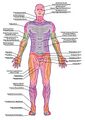 Anterior Cutaneous Innervation
 -
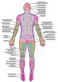 Posterior Cutaneous Innervation
 -
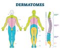 Dermatomal distribution


Pain (or pinprick) testing is used to evaluate the integrity of the sensory pathways responsible for transmitting pain signals, primarily through small A-delta and C nerve fibres. To perform this test, the examiner uses a sterile, sharp object such as a safety pin, reshaped paperclip or neurotip, and applies a light but firm pressure to the skin. The examiner alternates between sharp and dull stimuli and asks the patient to identify each sensation, ensuring that the patient can distinguish between them by stating "sharp," "dull," or "nothing." Abnormal findings, such as reduced / absent / exaggerated pain response (hyperalgesia), may indicate sensory nerve damage, spinal nerve root involvement, or central nervous system impairment.
Temperature discrimination testing is used to assess the function of small sensory nerve fibres (A-delta and C fibres) that transmit thermal sensations. To perform this test, the examiner uses two test tubes or metal instruments—one filled with warm water (around 40°C or 104°F) and the other with cold water (around 10°C or 50°F). The examiner alternately applies the warm and cold objects to the skin in a consistent manner and asks the patient to identify each as either "warm" or "cold." Abnormal responses, such as an inability to distinguish between temperatures or incorrect labelling of thermal stimuli, may suggest peripheral neuropathy, spinal cord lesions, or central sensory pathway dysfunction.
Light touch testing is used to evaluate the function of large myelinated A-beta fibres, which transmit non-painful tactile information. To perform this test, the examiner uses a soft object, such as a cotton wisp, and gently strokes the skin without pressing or causing deformation. The patient is asked to respond each time they feel the stimulus by saying “yes” or “now,” or by pointing to the location. Inconsistent responses, reduced sensitivity (hypoaesthesia), or absence of sensation (anaesthesia) may indicate a lesion in the peripheral nerves, nerve roots, spinal cord, or brain.[6]
If sensation impairments are found, the anatomical pattern suggests the general location of the injury or lesion:[7][8]
- distal peripheral nerves: stocking-glove distribution
- isolated nerves or nerve roots (radiculopathy): single dermatomal or nerve branch distribution
- brachial or pelvic plexus: patchy sensory, motor, and reflex deficits in a limb
- spinal cord: sensation reduced below a certain dermatomal level
- cauda equina: saddle area sensory loss
- brain stem: crossed face-body pattern
- thalamus: midline hemisensory loss
- brain: hemisensory loss
The location of the lesion can be further assessed by determining whether motor weakness and reflex changes follow a similar pattern.[7] For example, different spinal cord injury syndromes have different sensory and motor presentations. Knowledge of neuroanatomy and information gained from both the sensory and motor assessment can aid in syndrome identification.[9] The American Spinal Injury Association (ASIA) Impairment Scale is an example of a standardised assessment which uses both the sensory and motor assessment to determine the diagnosis.
Specific spinal cord injury syndromes and their symptom presentations are described below.
Complete spinal cord transection causes bilateral loss of motor function and all sensory modalities below the level of injury, including bowel, bladder, sexual dysfunction, and respiratory impairment depending on the level.
Central cord syndrome typically causes more weakness in the upper limbs than the lower limbs and possible loss of pain and temperature sensation below the level of injury.
Anterior cord syndrome results in bilateral motor paralysis and loss of pain and temperature sensation below the lesion, with preserved proprioception, vibration, and light touch.
Posterior cord syndrome causes loss of proprioception, vibration sense, and fine touch, while motor function and pain / temperature sensation remain intact.
Brown-Séquard syndrome results in ipsilateral loss of motor function, proprioception, and fine touch, and contralateral loss of pain and temperature sensation below the injury.
Conus medullaris syndrome manifests as bowel, bladder, and sexual dysfunction, along with absent Achilles reflexes and lower limb weakness.
Neurogenic shock causes hypotension and bradycardia due to loss of sympathetic tone following a high cervical spinal cord injury.
Deep (Proprioceptive) Sensations
Deep or proprioceptive receptors receive sensory information from the internal environment (i.e. structures such as muscles, tendons, ligaments, joints, and fascia). These receptors are responsible for proprioceptive awareness.[6] Proprioception includes the following subcategories of sensation: (1) position sense and awareness of joint location at rest, (2) kinesthesia, (3) sense of force (also known as sense of effort / heaviness / tension), and (4) sense of change in velocity, which also includes the ability to detect vibration.[10]
Proprioception or "joint position sense" testing evaluates a person’s awareness of joint position in space, relying on intact function of large-diameter sensory fibres and central proprioceptive pathways. To perform this test, the examiner stabilises the joint being tested, holding it lightly on the sides to avoid pressure cues. The joint is moved passively into a specific position, and the patient is asked to describe the joint position (e.g. “up,” “down,” “straight,” "bent") or to mirror the position with the contralateral limb. Impaired proprioception, demonstrated by incorrect or inconsistent responses, may suggest damage to peripheral nerves, the dorsal columns of the spinal cord, or areas of the brain involved in spatial awareness and coordination.
Kinesthesia or "awareness of movement" testing assesses a person’s ability to sense movement of a joint. It relies on the function of proprioceptive pathways, involving muscle spindles and large-diameter sensory fibres. To perform this test, the examiner lightly holds the sides of the joint being tested—such as a finger, toe, or limb segment, using a gentle grip that avoids giving pressure cues. The joint is moved slowly from mid-range through both end ranges. The examiner will let the patient feel the full range stating "this is up" while moving from midrange to one end range, and "this is down" while moving from midrange to the other end range. Next, the examiner moves the patient through the range of motion asking them to describe "which way their body is moving." The patient may also respond by duplicating the movement with the contralateral body part, though this is often impractical. Inability to correctly identify movement direction or inconsistent responses may indicate damage to peripheral nerves, dorsal columns of the spinal cord, or central pathways involved in proprioception.
Vibration sense testing is used to assess the integrity of large myelinated sensory fibres and the dorsal column–medial lemniscus pathway, which carry vibratory information to the brain. To perform this test, the examiner uses a 128 Hz tuning fork, striking it gently to initiate vibration. The vibrating fork is then placed on a bony prominence, such as the sternum, elbow, or malleolus. The patient is asked to report when they feel the vibration and when it stops. The examiner can dampen the tuning fork with their hand to stop the vibration without removing it, checking the patient’s accuracy in sensing the change. Diminished or absent vibration sense may indicate peripheral neuropathy, particularly in conditions like diabetes, or damage to the spinal cord’s posterior columns, such as in multiple sclerosis.[6]
Combined Cortical Sensations
This category of sensations requires information from both the superficial and deep sensory mechanisms AND intact functioning of higher cortical sensory association areas. Sensations at this level of processing include (1) stereognosis, (2) two-point discrimination, (3) barognosis (recognition of weight), (4) graphesthesia (traced figure identification), (5) tactile localisation, (6) recognition of texture, and (7) double simultaneous stimulation.[6]
Stereognosis or tactile object recognition testing evaluates a person’s ability to identify common objects using touch alone, relying on intact sensory pathways, cortical processing, and higher-level integration in the parietal lobe. To perform this test the examiner places a familiar or significant object (such as a key, coin, paperclip, or pen) in the patient’s hand and instructs them to manipulate and identify it using touch alone; the patient's eyes are kept closed throughout this assessment. The test is typically repeated with different objects in each hand to assess both sides of the body. It is important to ensure that the patient has sufficient motor function to explore the object and that primary tactile sensation (light touch, proprioception) is intact before conducting the test. An inability to recognise the object despite normal primary sensation (astereognosis) may suggest a lesion in the contralateral parietal cortex or issues with higher-order sensory integration, rather than peripheral nerve dysfunction.
Two-point discrimination testing assesses the ability to perceive two distinct points of contact on the skin, providing information about tactile spatial resolution and the functional integrity of the somatosensory cortex and peripheral nerves. To perform this test, the examiner uses a two-point discriminator, callipers, or a bent paperclip with two prongs set at a known distance. The clinician applies either one or two points simultaneously to the skin, typically on the fingertips, palm, or other areas of interest, and asks the patient to report whether they feel "one point or two." The distance between the two points is gradually decreased until the patient can no longer distinguish them as separate. Impaired two-point discrimination may indicate damage to peripheral nerves or lesions in the somatosensory cortex, especially in conditions such as stroke, neuropathy, or spinal cord injury.
Normal values for the two-point discrimination test vary across different body regions, reflecting the density of sensory receptors and the area of cortical representation. For example, on the fingertips, the typical threshold is between 2–3 mm, indicating high tactile acuity. The lips also exhibit high sensitivity, with thresholds ranging from 2–5 mm. In areas such as the palm, the threshold increases to approximately 8–10 mm.[11]
Barognosis or recognition of weight testing assesses the ability to perceive and compare the weight of objects using proprioceptive input and higher-level sensory processing in the parietal cortex. To perform this test, the examiner places objects of similar size but different weights (you can use calibrated test weights or everyday items) in one of the patient’s hands. The patient is asked to lift or hold each object and identify which is heavier or lighter, or to compare a series of objects by weight. This can be done sequentially in one hand or simultaneously with objects in both hands to compare sides. Adequate motor control is necessary to grasp and lift the objects effectively. A person with intact sensory integration should be able to distinguish between different weights. Difficulty performing this task, despite normal strength and tactile sensation, may indicate a lesion in the contralateral parietal lobe or disruption of proprioceptive pathways involved in cortical sensory interpretation.
Graphesthesia testing or traced figure identification evaluates the ability to recognise numbers, letters, or simple shapes that are drawn on the skin using only tactile input. This test assesses cortical sensory processing, particularly the function of the parietal lobe. To perform the test, the examiner traces a recognisable figure on the palm of the patient’s hand. After each tracing, the patient is asked to identify what was drawn. The test is repeated on both hands to compare sides. It is important that the patient has intact primary sensation (especially light touch) and a basic understanding of numbers or letters. Inability to correctly identify the figures, despite normal tactile sensation, may indicate a lesion in the contralateral parietal cortex or a disorder affecting higher-level somatosensory integration, such as after a stroke or traumatic brain injury.
Tactile localisation assesses the ability to identify the exact location on the skin where a light touch stimulus is applied, relying on intact primary sensory input and cortical processing in the parietal lobe. To perform this test, the examiner lightly touches a specific point on the skin using a fingertip, cotton wisp, or blunt probe. Immediately after the touch, the patient is asked to point to or verbally indicate the exact spot where they felt the stimulus. Inaccurate localisation, especially with preserved light touch sensation, may suggest a lesion in the contralateral parietal cortex or disruption in somatosensory integration pathways. This test is especially useful in identifying subtle cortical sensory deficits in conditions such as stroke or brain injury.
Recognition of texture testing evaluates the ability to identify different surface qualities (e.g. roughness, smoothness, or softness) using tactile input and higher-level sensory processing. To perform this test, the examiner places various textured materials (such as sandpaper, silk, velvet, or cotton) in the patient’s hand, one at a time. The patient is allowed to explore each texture using their fingers and is then asked to describe or identify it. Testing should be done on both hands to assess for sensory asymmetry, and materials should be distinctly different to ensure clear comparisons. This test relies on intact light touch sensation and cortical interpretation, particularly within the parietal lobe. Difficulty recognising textures, despite normal primary sensation, may indicate a lesion in the contralateral sensory cortex or impaired somatosensory integration, as can occur in stroke, brain injury, or certain neurodegenerative conditions.
Double simultaneous stimulation assesses cortical processing, particularly in the parietal lobes. To perform this test, the examiner applies a light tactile stimulus using a fingertip or soft object either to (1) identical locations on opposite sides of the body, (2) touches proximally and distally on opposite sides of the body, or (3) touches to proximal and distal locations on the same side of the body. The patient is asked to report where they felt the touch: left, right, or both. The stimuli should be identical in pressure and applied simultaneously during bilateral testing. If the patient can detect stimulation on each side individually but fails to perceive one side when both are touched at the same time, this is known as sensory extinction. This typically indicates a lesion in the contralateral parietal cortex, often associated with stroke or other cortical damage. Double simultaneous stimulation helps identify subtle deficits in attention and sensory integration that may not be apparent with unilateral testing.[6]
Sensory Cranial Nerve Testing

The cranial nerves (CN) are a set of twelve paired nerves that originate directly from the brain, primarily the brainstem, and are responsible for a wide range of autonomic, sensory, and motor functions. Cranial nerves with purely sensory function include: CN I (Olfactory nerve), CNII (Optic nerve), and CN VIII (Vestibulocochlear nerve). Cranial nerves with combined motor, sensory, and / or autonomic function include: CN V (Trigeminal nerve), CN VII (Facial nerve), CN IX (Glossopharyngeal nerve), and CN X (Vagus nerve).[6]
Below are concise summaries of how to perform the sensory portions of cranial nerve testing, and normal versus abnormal results.[6]
CN I (Olfactory nerve) testing evaluates the sense of smell, which can be affected by head trauma, neurodegenerative diseases, or nasal obstruction. To perform the test, the clinician first ensures that the patient’s nasal passages are clear. The patient is then asked to close their eyes and occlude one nostril. A familiar, non-irritating scent (e.g. coffee, vanilla, or peppermint) is presented to the open nostril using a scent-soaked cotton ball or vial held just beneath the nostril. The patient is asked to identify the smell. The process is then repeated with the other nostril. It's important not to use strong or noxious odours like alcohol or ammonia, as these can stimulate the trigeminal nerve instead. A normal response is the ability to detect and correctly identify the scent on both sides. Impaired or absent smell (anosmia or hyposmia) may indicate nasal pathology, olfactory nerve damage, or central nervous system disorders such as Parkinson’s or Alzheimer’s disease.
CN II (Optic nerve) testing assesses visual function, including visual acuity, visual fields, and the appearance of the optic nerve. To test visual acuity, the patient is asked to read a standard eye chart (such as a Snellen chart) from a specific distance, first with each eye separately and then with both eyes together, using corrective lenses if needed. To assess visual fields, the clinician performs confrontation testing by sitting facing the patient and asking them to cover one eye (covering the eye NOT being tested is the formal testing version performed by ophthalmologists, however it is not always covered in rehabilitation visuals screens). The clinician then moves a finger or object into the patient’s peripheral vision from various directions, and the patient signals when it becomes visible; this is repeated for both eyes. To inspect the optic disc, an ophthalmoscope is used to examine the retina and optic nerve through the pupil, checking for clarity, colour, and signs of swelling or atrophy. Abnormalities in any part of this testing may indicate issues such as optic neuritis, glaucoma, stroke, or intracranial pressure changes.
The sensory portion of the CN V (Trigeminal nerve) test evaluates sensation across the face in its three branches: ophthalmic (V1), maxillary (V2), and mandibular (V3). To perform this test, the patient is asked to close their eyes while the clinician lightly touches the skin of the forehead, cheeks, and jaw on both sides using a soft object such as a cotton wisp for light touch, and a blunt object or pin for pain (pinprick) sensation. The patient is asked to report when and where they feel the stimulus, ensuring symmetry and accuracy. Temperature discrimination can also be tested using warm and cool objects if needed. An abnormal response—such as reduced or absent sensation—may suggest a lesion affecting the trigeminal nerve or its central connections, possibly due to conditions like trigeminal neuralgia, stroke, or tumours. Sensory testing should be followed by motor testing if any abnormalities are found.
The sensory portion of the CN VII (Facial nerve) testing primarily evaluates taste sensation on the anterior two-thirds of the tongue. To perform this test, the examiner asks the patient to close their eyes and extend their tongue slightly. Using a cotton swab or small applicator, the clinician applies a taste stimulus (typically a sweet, salty, sour, or bitter solution) to the anterior part of the tongue. The patient is then asked to identify the taste. It's important to use substances that are easy to distinguish and not overly strong, such as sugar, salt, or lemon juice. Impaired taste sensation on the anterior tongue may indicate dysfunction of the facial nerve, often seen in conditions like Bell's palsy or lesions affecting the nerve’s peripheral branches.
CN VIII (Vestibulocochlear nerve) testing focuses on evaluating hearing, which involves the cochlear division of the nerve. To perform this test, the examiner begins with a basic hearing screening, such as the whisper test. In the whisper test, the patient covers one ear while the examiner stands behind and whispers a word or number into the opposite ear; the patient repeats what they hear. For more precise assessment, tuning fork tests are used.
- In the Rinne test, a vibrating 512 Hz tuning fork is placed on the patient’s mastoid bone (bone conduction); when the sound is no longer heard, it is moved next to the ear canal (air conduction). Normally, air conduction is greater than bone conduction.
- In the Weber test, the vibrating fork is placed in the middle of the forehead; the patient is asked if the sound is heard equally in both ears or louder on one side.
These tests help differentiate conductive from sensorineural hearing loss. Abnormal findings may indicate issues in the outer / middle ear (conductive loss) or damage to the cochlea or auditory nerve (sensorineural loss), which can result from noise exposure, infections, or neurological conditions.
The sensory portion of CN IX (Glossopharyngeal nerve) testing primarily evaluates taste sensation on the posterior third of the tongue and the sensory component of the gag reflex. Taste sensation is rarely tested in clinical practice. More commonly, the gag reflex is assessed by gently touching the posterior pharyngeal wall or soft palate with a tongue depressor or cotton swab while the patient’s mouth is open. A normal response is a brief contraction of the pharyngeal muscles and possibly a gag. Sensory input for this reflex comes from CN IX, while the motor response is mediated by CN X (Vagus nerve). An absent or asymmetrical gag reflex, along with impaired taste or swallowing, may suggest a lesion of the glossopharyngeal nerve, possibly due to stroke, tumour, or brainstem pathology.
The sensory portion of CN X (Vagus nerve) testing primarily involves assessing sensation in the pharynx, larynx, and external auditory canal, as well as its role in the gag reflex (explained above). While direct testing of vagal sensory function is limited in a clinical setting, the gag reflex is the most practical method.
Resources
Clinical Resources
The resources listed below include written instructions, demonstration videos and additional reference materials:
- How to Assess Mental Status (Merck Manuals)
- How to Assess Sensation (Merck Manuals)
- How to Assess the Cranial Nerves (Merck Manuals)
The resources below describes the items needed to create a cranial nerve testing kit:
- This short YouTube video quickly discusses and provides examples of items needed for cranial nerve testing.
References
- ↑ Gadhvi M, Moore M, Waseem M. Physiology, Sensory System [Internet]. 2023 [cited 24/04/2025]. Available from: https://www.ncbi.nlm.nih.gov/books/NBK547656/
- ↑ Guardado K, Sergent S. Sensory Integration [Internet]. 2023 [cited 24/04/2025]. Available from: https://www.ncbi.nlm.nih.gov/books/NBK559155/#
- ↑ Magosso E, Ursino M. The Sensory-Cognitive Interplay: Insights into Neural Mechanisms and Circuits. Journal of Integrative Neuroscience. 2023;22(1):1-3.
- ↑ Thau L, Reddy V, Singh P. Neuroanatomy, Central Nervous System. National Library of Medicine Stat Pearls. 10 October 2022.
- ↑ 5.0 5.1 Thau L, Reddy V, Singh P. Neuroanatomy, Central Nervous System. National Library of Medicine Stat Pearls. 10 October 2022.
- ↑ 6.0 6.1 6.2 6.3 6.4 6.5 6.6 6.7 6.8 O'sullivan SB, Schmitz TJ. Physical rehabilitation: assessment and treatment. 1994.
- ↑ 7.0 7.1 Chakraborty U, Mukherjee A, Pal J, Medicine Update 2021. Anatomic Localization of Classical Neurological Symptoms. Evangel: Edition 31, chapter 119.
- ↑ Merck Manual. How to Assess Sensation. Available from: https://www.merckmanuals.com/professional/neurologic-disorders/neurologic-examination/how-to-assess-sensation (accessed 28 April 2025).
- ↑ Bennett J, Das J, Emmady P. Spinal Cord Injuries [Internet]. 2024 [cited 30/04/2025]. Available from: https://www.ncbi.nlm.nih.gov/books/NBK560721/
- ↑ Proske U, Gandevia SC. The proprioceptive senses: their roles in signaling body shape, body position and movement, and muscle force. Physiological reviews. 2012 Oct 1.
- ↑ Thube S, Shah MR, Kothari PH, Shah V. Assessment of two point discrimination on hand in adult population: An observational study. Int J Health Sci Res. 2020;10(05):60.