Femoral Nerve: Difference between revisions
No edit summary |
No edit summary |
||
| Line 2: | Line 2: | ||
'''Top Contributors''' - {{Special:Contributors/{{FULLPAGENAME}}}}</div> | '''Top Contributors''' - {{Special:Contributors/{{FULLPAGENAME}}}}</div> | ||
'''This article or area is currently under review and may only be partially complete. Please come back soon to see the finished work!''' | |||
== Introduction == | == Introduction == | ||
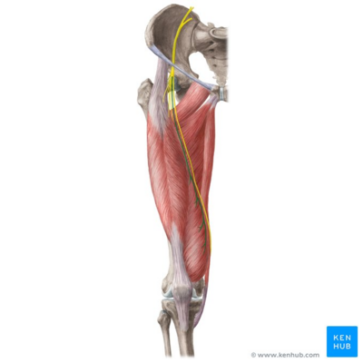
[[File:Muscular branches of femoral nerve - Kenhub.png|alt=Muscular branches of femoral nerve (highlighted in green) - anterior view|400x400px|'''Figure.1''' Muscular Branches of Femoral Nerve <ref>Muscular branches of femoral nerve (highlighted in green) - anterior view image - © Kenhub. Available from: https://www.kenhub.com/en/library/anatomy/femoral-nerve</ref>|thumb]] | [[File:Muscular branches of femoral nerve - Kenhub.png|alt=Muscular branches of femoral nerve (highlighted in green) - anterior view|400x400px|'''Figure.1''' Muscular Branches of Femoral Nerve <ref>Muscular branches of femoral nerve (highlighted in green) - anterior view image - © Kenhub. Available from: https://www.kenhub.com/en/library/anatomy/femoral-nerve</ref>|thumb]] | ||
The femoral nerve is the largest nerve of the [[Lumbar Plexus|lumbar plexus]]. It originates from the dorsal divisions of the L2-L4 ventral rami. It has a role in motor and sensory processing in the lower limbs. It controls: | The femoral nerve is the largest nerve of the [[Lumbar Plexus|lumbar plexus]]. It originates from the dorsal divisions of the L2-L4 ventral rami. It has a role in motor and sensory processing in the lower limbs. It controls: | ||
# The major [[Hip Flexors|hip flexor muscles]], as well as [[Quadriceps Muscle|knee extension]] muscles. | # The major [[Hip Flexors|hip flexor muscles]], as well as [[Quadriceps Muscle|knee extension]] muscles. | ||
# Sensation over the anterior and medial thigh, as well as medial leg down to the hallux (great toe). <ref>Refai NA, Tadi P. Anatomy, [https://www.ncbi.nlm.nih.gov/books/NBK556065/ Bony Pelvis and Lower Limb, Thigh Femoral Nerve.] StatPearls [Internet]. 2020 Oct 27. | # Sensation over the anterior and medial thigh, as well as medial leg down to the hallux (great toe). <ref>Refai NA, Tadi P. Anatomy, [https://www.ncbi.nlm.nih.gov/books/NBK556065/ Bony Pelvis and Lower Limb, Thigh Femoral Nerve.] StatPearls [Internet]. 2020 Oct 27.</ref> | ||
== Root == | |||
The femoral nerve originates from the dorsal divisions of the L2-L4 ventral rami, then it emerges from behind the [[Psoas Major|psoas muscle]] to run laterally, deep to the iliac [[fascia]] above the [[Iliacus|iliacus muscle]] in the pelvis. At the level of the thigh, it begins to pass lateral to the [[Femoral Artery|femoral artery]] (behind the inguinal ligament), dividing approximately 4 cm below the inguinal ligament into anterior and posterior divisions. | |||
=== Branches === | |||
''' In the [[Pelvis]]''' | |||
*Muscular branches are first given off to the psoas and then to the iliacus muscles (sometimes known together as the [[Iliopsoas Tendinopathy|iliopsoas]] muscle) before the nerve runs beneath the [[Inguinal Ligament|inguinal ligament]]. | |||
'''In the thigh'''[[File:1452198295 lateral-femoral-cutaneous-nerve.jpg|alt=Femoral and lateral-femoral-cutaneous-nerves|303x303px|thumb|Femoral and lateral-femoral-cutaneous-nerves]] | |||
*The anterior division gives rise to the medial and intermediate cutaneous nerves of the thigh and muscular branches to the [[sartorius]] and [[Pectineus Muscle|pectineus muscles]]. | |||
* The posterior division supplies the four heads of the [[Quadriceps Muscle|quadriceps femoris]] ([[Vastus Medialis|vastus medialis,]] [[Vastus Lateralis|vastus lateralis]], [[Vastus Intermedius|vastus intermedius]] and [[Rectus Femoris|rectus femoris]])<ref name=":0">Femoral Nerve. Available from: https://www.earthslab.com/anatomy/femoral-nerve/ (Accessed, 22/06/2018).</ref>and then continues along the medial border of the [[Calf Strain|calf]] as the saphenous nerve, that is considered as the largest and longest branch of the femoral nerve and supplies the [[skin]] over the medial side of the leg. | |||
* The | |||
<br> | <br> | ||
'''Articular Supply''' | '''Articular Supply''' | ||
Revision as of 01:57, 20 December 2023
This article or area is currently under review and may only be partially complete. Please come back soon to see the finished work!
Introduction[edit | edit source]

The femoral nerve is the largest nerve of the lumbar plexus. It originates from the dorsal divisions of the L2-L4 ventral rami. It has a role in motor and sensory processing in the lower limbs. It controls:
- The major hip flexor muscles, as well as knee extension muscles.
- Sensation over the anterior and medial thigh, as well as medial leg down to the hallux (great toe). [2]
Root[edit | edit source]
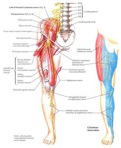
The femoral nerve originates from the dorsal divisions of the L2-L4 ventral rami, then it emerges from behind the psoas muscle to run laterally, deep to the iliac fascia above the iliacus muscle in the pelvis. At the level of the thigh, it begins to pass lateral to the femoral artery (behind the inguinal ligament), dividing approximately 4 cm below the inguinal ligament into anterior and posterior divisions.
Branches[edit | edit source]
In the Pelvis
- Muscular branches are first given off to the psoas and then to the iliacus muscles (sometimes known together as the iliopsoas muscle) before the nerve runs beneath the inguinal ligament.
In the thigh
- The anterior division gives rise to the medial and intermediate cutaneous nerves of the thigh and muscular branches to the sartorius and pectineus muscles.
- The posterior division supplies the four heads of the quadriceps femoris (vastus medialis, vastus lateralis, vastus intermedius and rectus femoris)[3]and then continues along the medial border of the calf as the saphenous nerve, that is considered as the largest and longest branch of the femoral nerve and supplies the skin over the medial side of the leg.
Articular Supply
- The femoral nerve also innervates the capsule of the hip joint and allows for proprioceptive feedback about the joint.[4]
- The knee joint is supplied by the nerves to the three vasti. The nerve to the vastus medialis contains numerous proprioceptive fibres from the knee joint, accounting for the thickness of the nerve.[5]
Note: The lateral thigh is not supplied by the femoral nerve but is innervated by the lateral femoral cutaneous nerve , which is derived directly from the lumbar plexus, receiving innervation from the L2–L3 nerve roots.[6]
Femoral Triangle[edit | edit source]
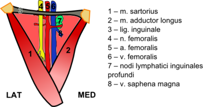
The following structures pass through the femoral triangle:
- Femoral nerve - Which innervates the anterior compartment of the thigh
- Femoral sheath containing: Femoral artery and branches - Arterial supply for majority of the lower limb; Femoral vein - The great saphenous vein drains into the femoral vein within the triangle; Femoral canal - Contains lymph nodes and vessels[7]
Physiotherapy Relevance[edit | edit source]
Femoral nerve damage (also referred to as femoral nerve dysfunction or neuropathy), can occur from an injury or prolonged compression. Typically, damage and dysfunction of the femoral nerve are associated with the leg weakness and sensation changes.
Injury
Injury of the femoral is uncommon but may be injured by a stab, gunshot wounds, or a pelvic fracture. The femoral nerve can be damaged during penetrating trauma to the thigh. It can also be damaged during hip replacement operations, particularly the anterior approach (not commonly used) where the nerve can be stretched and damaged. Listed here are the characteristic clinical features:
Motor Loss
- Poor flexion of the hip, because of paralysis of the iliacus, psoas and sartorius muscles.
- Inability to extend the knee, because of paralysis of the quadriceps femoris.
Sensory impairment
- Sensory decline over the anterior and medial aspects of the thigh, as a result of engagement of the intermediate and lateral cutaneous nerves of the thigh.
- Sensory loss on the medial side of the leg and foot up to the ball of the great toe (first metatarsophalangeal joint), because of engagement of the saphenous nerve.[8]
Apart from direct injury aside the femoral nerve damage can be caused by a number of other factors.
- Certain medical conditions eg diabetes, can damage this nerve due to impaired metabolic functioning, and is common.
- Other mediating factors include fracturing the pelvis, internal bleeding, or oxygen deprivation to the nerve due to becoming encased in a tumor or being subjected to pressure by the presence of a tumor.[9]
Other relevant issues
- Patellar Tendon Reflex: The femoral nerve is responsible for the patellar tendon reflex (tests L3-L4 spinal component
- Femoral nerve block: Femoral nerve block (in combination with sciatic nerve block) may be indicated in patients requiring lower limb surgery who cannot tolerate a general anaesthetic. A femoral nerve block can also be used as peri- and post-operative analgesia for patients with a fractured neck of femur who cannot tolerate particular analgesics.
- Femoral nerve tension test
Viewing[edit | edit source]
Below is a 6 minute video on the femoral nerve.[10]
References[edit | edit source]
- ↑ Muscular branches of femoral nerve (highlighted in green) - anterior view image - © Kenhub. Available from: https://www.kenhub.com/en/library/anatomy/femoral-nerve
- ↑ Refai NA, Tadi P. Anatomy, Bony Pelvis and Lower Limb, Thigh Femoral Nerve. StatPearls [Internet]. 2020 Oct 27.
- ↑ Femoral Nerve. Available from: https://www.earthslab.com/anatomy/femoral-nerve/ (Accessed, 22/06/2018).
- ↑ Femoral Nerve. Available from:https://www.kenhub.com/en/library/anatomy/femoral-nerve (Accessed, 24/06/2018).
- ↑ Chaurasia, B., 2013. Human Anatomy Volume 2 Regional and Applied Dissection and Clinical Lower Limb , Abdomen and Pelvis.. 6th ed. India CBS Publisher and Distributors Pvt Ltd.
- ↑ Musculoskeletal key femoral neuropathy Available: https://musculoskeletalkey.com/femoral-neuropathy/ (accessed 17.1.2022)
- ↑ Physiopedia The Femoral Triangle Available;https://www.physio-pedia.com/The_Femoral_Triangle?utm_source=physiopedia&utm_medium=search&utm_campaign=ongoing_internal (accessed 17.1.2022)
- ↑ Ellis, H., 2006. Clinical Anatomy A revision and applied anatomy for clinical students. 11th ed. Blackwell Publishing Ltd.
- ↑ The health board What can I do About Femoral Nerve Damage? Available: https://www.thehealthboard.com/what-can-i-do-about-femoral-nerve-damage.htm(accessed 17.1.2022)
- ↑ Femoral Nerve Anatomy - Everything You Need To Know - Dr. Nabil Ebraheim. Available from: https://www.youtube.com/watch?v=zdgJueAZaxU [last accessed 24/06/2018]


