Quadriceps Muscle
Original Editor -
Top Contributors - Lucinda hampton, Kim Jackson, Nikhil Benhur Abburi, Oyemi Sillo, Joao Costa, Wanda van Niekerk, Chrysolite Jyothi Kommu and Aminat Abolade
Description[edit | edit source]

The Quadriceps femoris is the most voluminous muscle of the human body.[1]
The quadriceps femoris is a hip flexor and a knee extensor. It consists of four individual muscles; three vastus muscles and the rectus femoris. They form the main bulk of the thigh, and collectively are one of the most powerful muscles in the body.[2]
It is located in the anterior compartment of the thigh.
The four 4 sub-components being:
- Rectus femoris
- Vastus lateralis
- Vastus medialis
- Vastus intermedius (see links for further elaboration)
Origin[edit | edit source]
The muscles that form the quadriceps femoris unite proximal to the knee and attach to the patella via the quadriceps tendon. In turn, the patella is attached to the tibia by the patella ligament.
Vastus Lateralis[edit | edit source]
- Proximal attachment: Originates from the greater trochanter and the lateral lip of linea Aspera.
- Actions: Extends the knee joint and stabilises the patella.
Vastus Intermedius[edit | edit source]
- Proximal attachment: Anterior and lateral surfaces of the femoral shaft.
- Actions: Extends the knee joint and stabilises the patella.
Vastus Medialis[edit | edit source]
- Proximal attachment: The intertrochanteric line and medial lip of the linea aspera.
- Actions: Extends the knee joint and stabilises the patella, particularly due to its horizontal fibres at the distal end.
Rectus Femoris[edit | edit source]
- Attachments: Originates from the ilium, just superior to the acetabulum. It runs straight down the leg (the Latin for straight is rectus), and attaches to the patella by the quadriceps femoris tendon.
- Actions: The only muscle of the quadriceps to cross both the hip and knee joints. It flexes the thigh at the hip joint, and extends at the knee joint.[2]
Nerve[edit | edit source]

Innervation of these muscles is by the femoral nerve. (L2, L3, L4)
Artery[edit | edit source]
The lateral femoral circumflex artery.
Function[edit | edit source]
The quadriceps all work to extend (straighten) the knee. The rectus femoris also flexes the hip, The vastus medialis adducts the thigh and also extends and externally rotates the thigh and stabilizes the kneecap
- The quadriceps are primarily active in kicking, jumping, cycling and running.[3] eg sports like basketball that requires jumps.
- In everyday life, they help you get up from a chair, walk, climb stairs and squat.
- They are used in walking and running at the onset of a stride and get used significantly when going downhill.
.
Clinical Relevance[edit | edit source]

From a sporting point of view, it is an extraordinarily important muscle, but due to the stress it receives, it is often subject to trauma. Injury to the quadriceps muscle group can be painful and debilitating. Strains, tears and contusions of the quadriceps are common in various sports, such as athletics, rugby, football, etc and result in lost time from training and competition.
The quadriceps is essential for daily activities, such as climbing stairs or getting up from the chair.
Myositis Ossificans(MO) is a complication associated with severe quadriceps contusions. This is a non-neoplastic proliferation of bone and cartilage in the area of the contusion injury. In contusions, the reported incidence is between 9% and 17%. Myositis Ossificans should be suspected if symptoms worsen after 2–3 weeks accompanied by a loss of knee flexion and persistent swelling.[3]
Adaptations of the Quadriceps Muscle in the Presence of Disease[edit | edit source]
Skeletal muscle adapts in the presence of systemic diseases. This means that the function of the muscle changes both metabolism and volumes, worsening the symptomatic picture eg
Ageing
The muscular adaptation of the quadriceps muscle with advancing age changes its morphology and function. The musculature loses mass and volume (sarcopenia) decreases strength and coordination. Motor units are lost (denervation processes), while the percentage of red fibers increases. This increases the fibrosis processes and intramuscular fat.
Chronic Obstructive Pulmonary Disease (COPD) and Chronic Heart Failure
The presence of chronic and ingravescent respiratory diseases such as COPD determines a decrease in muscle mass of the quadriceps femoris muscle, an increase of the connective tissue, and fibrosis phenomena. This leads to decreased contractile capacity, less strength, and less balance during walking or standing posture. The anaerobic fibers increase at the expense of the oxidative fibers; the muscles fatiguing more easily. An increase in intramuscular fat, with local and systemic metabolic alteration (increased cardiovascular risk) also occur. The females suffer more from the functional alteration of the muscle than the males.
Multiple Sclerosis
In multiple sclerosis, the quadriceps muscle loses muscle mass and strength, with a decrease in oxidative fibers but with an increase in anaerobic fibers. Although there is an increase in the number of white fibers, the latter suffer more atrophy. Increase intramuscular fat and fibrosis processes.[1]
Assessment[edit | edit source]
After obtaining a thorough history, a careful examination should ensue including observation, palpation, strength testing, and evaluation of motion. Strain injuries of the quadriceps may present with an obvious deformity such as a bulge or defect in the muscle belly.[4]
- Palpation of the anterior thigh should include the length of the injured muscle, locating the area of maximal tenderness and feeling for any defect in the muscle.
- Strength testing of the quadriceps should include the resistance of knee extension and hip flexion. Adequate strength testing of the rectus femoris must include resisted knee extension with the hip flexed and extended. Practically, this is best accomplished by evaluating the patient in both a sitting and prone-lying position. The patient is asked to extend the leg (at the knee) against resistance. If the femoral nerve is damaged, the contraction of the quadriceps femoris will be absent.[2] The prone-lying position also allows for optimum assessment of quadriceps motion and flexibility.
- Pain is typically felt by the patient with resisted muscle activation, passive stretching, and direct palpation over the muscle strain.
- Assessing tenderness, any palpable defect, and strength at the onset of muscle injury will determine the grading of the injury and provide direction for further diagnostic testing and treatment.[4]
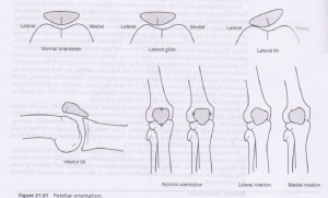
- Check for individual muscle head imbalances eg may lead to patella tracking problems.
- Check for muscle imbalances eg. Runners often develop an imbalance between strong hamstring muscles and less developed quadriceps.
- The quadriceps femoris muscle can be used to test the femoral nerve in cases of suspected nerve palsy.[2]
- Functional tests include: Timed Up and Go test (TUG) and a timed Stair Climbing Test (SCT)[5]

Treatment[edit | edit source]
See
Functional Coordination Between the Different Vasti[edit | edit source]
The myoelectric balance of the quadriceps is essential for a correct movement of the patella.
The proprioceptive afferents of the muscle contribute to maintaining adequate posture. Recent studies show that the activation of these afferents allows the contralateral quadriceps muscle to improve its coordination, increasing postural balance. The quadriceps allow an independent walk, helps with stair climbing and enables sit to stand.
The rectus femoris can activate its fibers in the longitudinal mode. It can activate the proximal fibers in the absence of contraction of the most distal fibers. If the action of the quadriceps continues, it will activate the most distal fibers, in the absence of the most proximal ones (probably a mechanism to delay the onset of fatigue).
The patellar tendon insertion of the vastus medialis is small and is not able to generate a force capable of medially stabilizing the patella during knee extension. The force expressed by the vastus medialis during the extension is modest. In reality, during its contraction, it pulls the aponeurosis of the vastus intermedius, counteracting the lateral forces on the patella of the vastus lateralis. The vastus medialis acts indirectly as a patellar stabilizer, placing its contractile force on the median axis of the femur.
The strength expressed by the vastus lateralis increases with the increase in knee flexion. This mechanism is due to the length of the fibers compared to the connective structure of the muscle. Longer fibers express greater strength and make better use of the elasticity or resistance of the connective tissue. When the knee is extended, the vastus lateralis places a small force that is useful for maintaining the position with minimal effort.[1]
Resources[edit | edit source]
Anatomy of the quadriceps femoris muscle [6]
Another video of the anatomy of the quadriceps muscle
References[edit | edit source]
- ↑ 1.0 1.1 1.2 Bordoni B, Varacallo M. Anatomy, Bony Pelvis and Lower Limb, Thigh Quadriceps Muscle. InStatPearls [Internet] 2018 Dec 15. StatPearls Publishing.Available from:https://www.ncbi.nlm.nih.gov/books/NBK513334/ (last accessed 7.2.2020)
- ↑ 2.0 2.1 2.2 2.3 Teach me anatomy. Muscles of the anterior thigh. Available from:https://teachmeanatomy.info/lower-limb/muscles/thigh/anterior-compartment/ (last accessed 8.2.2020)
- ↑ 3.0 3.1 Kary JM. Diagnosis and management of quadriceps strains and contusions. Current reviews in musculoskeletal medicine. 2010 Oct 1;3(1-4):26-31. Available from:https://www.ncbi.nlm.nih.gov/pmc/articles/PMC2941577/ (last accessed 7.2.2020)
- ↑ 4.0 4.1 Kary JM. Diagnosis and management of quadriceps strains and contusions. Current reviews in musculoskeletal medicine. 2010 Oct 1;3(1-4):26-31. Available from:https://www.ncbi.nlm.nih.gov/pmc/articles/PMC2941577/ (last accessed 8.2.2020)
- ↑ Mizner RL, Petterson SC, Stevens JE, Axe MJ, Snyder-Mackler L. Preoperative quadriceps strength predicts functional ability one year after total knee arthroplasty. The Journal of rheumatology. 2005 Aug 1;32(8):1533-9. Available from: http://www.jrheum.org/content/32/8/1533.short (last accessed 8.2.2020)
- ↑ Anatomy of the quadriceps femoris muscle video - © Kenhub https://www.kenhub.com/en/library/anatomy/the-quadriceps-femoris-muscle
- ↑ Bodyworks Prime. Quadriceps Anatomy: Origin, Insertion, Innervation & Action. Available from: https://www.youtube.com/watch?v=EoHyhIWfLig [last accessed 16/5/2023]

