Femoral Nerve
Introduction[edit | edit source]
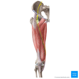
The femoral nerve is the largest nerve of the lumbar plexus. It originates from the dorsal divisions of the L2-L4 ventral rami. It has a role in motor and sensory processing in the lower limbs[1]. It controls:
- The major hip flexor muscles, as well as knee extension muscles.
- Sensation over the anterior and medial thigh, as well as medial leg down to the hallux (great toe). [2]
Root[edit | edit source]
The femoral nerve originates from the dorsal divisions of the L2-L4 ventral rami, then it emerges from behind the psoas muscle to run laterally, deep to the iliac fascia above the iliacus muscle in the pelvis. At the level of the thigh, it begins to pass lateral to the femoral artery (behind the inguinal ligament), dividing approximately 4 cm below the inguinal ligament into anterior and posterior divisions[3].
Branches[edit | edit source]
Motor[edit | edit source]
In the Pelvis
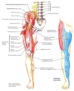
- Muscular branches are first given off to the psoas and then to the iliacus muscles (sometimes known together as the iliopsoas muscle) before the nerve runs beneath the inguinal ligament.
In the thigh
- The anterior division gives muscular branches to the sartorius and pectineus muscles.
- The posterior division supplies the four heads of the quadriceps femoris (vastus medialis, vastus lateralis, vastus intermedius and rectus femoris)[4].
Sensory[edit | edit source]
- The anterior division gives rise to the medial and intermediate cutaneous nerves of the thigh they give cutaneous innervation to the skin over the anterior and medial region of the thigh.
- The posterior division continues along the medial border of the calf as the saphenous nerve, that is considered as the largest and longest branch of the femoral nerve and supplies the skin over the medial side of the leg.
Articular Supply[edit | edit source]
- The femoral nerve also innervates the capsule of the hip joint and allows for proprioceptive feedback about the joint.
- The knee joint is supplied by the nerves to the three vasti. The nerve to the vastus medialis contains numerous proprioceptive fibres from the knee joint, accounting for the thickness of the nerve.[5]
Note: The lateral thigh is not supplied by the femoral nerve but is innervated by the lateral femoral cutaneous nerve , which is derived directly from the lumbar plexus, receiving innervation from the L2–L3 nerve roots[6].
Clinical Relevance[edit | edit source]
Femoral nerve damage (also referred to as femoral nerve dysfunction or neuropathy), can occur from an injury or prolonged compression. Typically, damage and dysfunction of the femoral nerve are associated with the leg weakness and sensation changes.
Injury of the femoral is uncommon but may be injured by a stab, gunshot wounds, or a pelvic fracture. The femoral nerve can be damaged during penetrating trauma to the thigh. It can also be damaged during hip replacement, abdominal, and pelvic surgeries.
There are several mechanisms for nerve damaged as a result from in direct trauma. Mechanical injury, such as from stretching or compression, that leads to neuropraxia, where nerve function is temporarily disrupted. Nerve can accidentally be damaged by sutures or staples. Ischaemic damage arises from restricted blood flow, often due to compression. Heat damage, from the heat released during hip prosthesis cementing, that can harm nearby nerves. Lastly, toxic damage that can caused by exposure to harmful substances or chemicals[7].
Assessment[edit | edit source]
The patient with femoral nerve injury may be presented with one or more of the following presentations[7]:
Motor Loss
- Poor flexion of the hip, because of paralysis of the iliacus, psoas and sartorius muscles.
- Inability to extend the knee, because of paralysis of the quadriceps femoris.
Sensory impairment
- Sensory decline over the anterior and medial aspects of the thigh, as a result of engagement of the intermediate and lateral cutaneous nerves of the thigh.
- Sensory loss on the medial side of the leg and foot up to the ball of the great toe (first metatarsophalangeal joint), because of engagement of the saphenous nerve.[8]
Other relevant issues
- Patellar Tendon Reflex: The femoral nerve is responsible for the patellar tendon reflex (tests L3-L4 spinal component)
- Femoral nerve block: Femoral nerve block (in combination with sciatic nerve block) may be indicated in patients requiring lower limb surgery who cannot tolerate a general anaesthetic. A femoral nerve block can also be used as peri- and post-operative analgesia for patients with a fractured neck of femur who cannot tolerate particular analgesics.
- Femoral nerve tension test, it is used to evaluate the mechanical sensitivity and the neural mobility of the femoral nerve. In addition, to detect lesions or irritations of the femoral nerve[9].
Treatment[edit | edit source]
Surgical Management[edit | edit source]
There are three surgical approaches for managements of injured femoral nerve:
- Sural nerve graft, is often the best first choice to repair the nerve directly at the site of damage, and leading to faster and better recovery outcomes
- Obturator nerve trunk transfer when nerve graft is not possible, obturator nerve transfer can be an alternative and save approach, nerve truck can be done for injury at the level of pelvis
- Mortal branch of obturator nerve transfer for injury at the thigh level[10].
Physical Therapy Role[edit | edit source]
After a nerve injury, physiotherapy aims to:
- Restore normal muscle function.
- Prevent or eliminate paresis (muscle weakness).
- Improve blood circulation and energy supply to the affected tissues.
Kinetic therapy is one of the effective approaches to deal with peripheral nerve injury it depends on how long it takes for nerve fibers to regenerate and for muscles to be reinnervated. It is characterized by the gradual return of muscle strength. In addition, progressive stretching or strengthening exercises should be avoided in he early stages of nerve degeneration[11].
Electrical stimulation helps to promote the growth of axons in nerve repair and speeds up the recovery of sensorimotor functions, current intensity should be sufficient enough to provoke strong contractions and the pulse duration should be not less than the chronaxie of denervated muscles[12]. There are different therapeutic currents can be used for rehabilitation and with denervated muscles; neuromuscular electrical stimulation, Galvanic current, functional electrical stimulation[13].
You can find more details in Peripheral nerve injury rehabilitation and Nerve Injury Rehabilitation.
Viewing[edit | edit source]
Below is a 6 minute video on the femoral nerve.[14]
References[edit | edit source]
- ↑ Wong TL, Kikuta S, Iwanaga J, Tubbs RS. A multiply split femoral nerve and psoas quartus muscle. Anatomy & Cell Biology. 2019 Jun 1;52(2):208-10.
- ↑ Refai NA, Tadi P. Anatomy, Bony Pelvis and Lower Limb, Thigh Femoral Nerve. StatPearls [Internet]. 2020 Oct 27.
- ↑ Jakubowicz M. Topography of the femoral nerve in relation to components of the iliopsoas muscle in human fetuses. Folia Morphologica. 1991 Jan 1;50(1-2):91-101.
- ↑ Femoral Nerve. Available from: https://www.earthslab.com/anatomy/femoral-nerve/ (Accessed, 22/06/2018).
- ↑ Chaurasia, B., 2013. Human Anatomy Volume 2 Regional and Applied Dissection and Clinical Lower Limb , Abdomen and Pelvis.. 6th ed. India CBS Publisher and Distributors Pvt Ltd.
- ↑ HÉbert‐Blouin MN, Shane Tubbs R, Carmichael SW, Spinner RJ. Hilton's law revisited. Clinical Anatomy. 2014 May;27(4):548-55.
- ↑ 7.0 7.1 Gibelli F, Ricci G, Sirignano A, Bailo P, De Leo D. Iatrogenic femoral nerve injuries: Analysis of medico-legal issues through a scoping review approach. Annals of Medicine and Surgery. 2021 Dec 1;72:103055.
- ↑ Ellis, H., 2006. Clinical Anatomy A revision and applied anatomy for clinical students. 11th ed. Blackwell Publishing Ltd.
- ↑ Refai NA, Black AC, Tadi P. Anatomy, Bony Pelvis and Lower Limb: Thigh Femoral Nerve. InStatPearls [Internet] 2022 Nov 18. StatPearls Publishing.
- ↑ Cao Y, Li Y, Zhang Y, Li S, Jiang J, Gu Y, Xu L. Different surgical reconstructions for femoral nerve injury: a clinical study on 9 cases. Annals of Plastic Surgery. 2020 May 1;84(5S):S171-7.
- ↑ Suszyński K, Marcol W, Górka D. Physiotherapeutic techniques used in the management of patients with peripheral nerve injuries. Neural regeneration research. 2015 Nov;10(11):1770.
- ↑ Chandrasekaran S, Davis J, Bersch I, Goldberg G, Gorgey AS. Electrical stimulation and denervated muscles after spinal cord injury. Neural regeneration research. 2020 Aug;15(8):1397.
- ↑ Ni L, Yao Z, Zhao Y, Zhang T, Wang J, Li S, Chen Z. Electrical stimulation therapy for peripheral nerve injury. Frontiers in Neurology. 2023 Feb 24;14:1081458.
- ↑ Femoral Nerve Anatomy - Everything You Need To Know - Dr. Nabil Ebraheim. Available from: https://www.youtube.com/watch?v=zdgJueAZaxU [last accessed 24/06/2018]


