
Femoral Triangle
Original Editors - Shanice Basdeo
Top Contributors - Leana Louw, Shanice Basdeo, Lucinda hampton, Kim Jackson, George Prudden and Wanda van Niekerk
Description
The femoral triangle is a hollow region located in the supero-medial part of the anterior thigh. It appears most prominently with hip flexion, abduction and internal rotation. It is an easily accessible area through which multiple neurovascular structures pass through. This anatomical landmark is mostly used in dissection and describing relationships in the groin area.[1] It is also known as "scarpa's triangle" and "femoral trigone". Assessment of pain and swelling in this area can be done by palpation or ultrasound[2].
Contents
The following structures pass through the femoral triangle:[1]
- Femoral nerve - Which innervates the anterior compartment of the thigh
- Femoral sheath containing:
- Femoral artery and branches - Arterial supply for majority of the lower limb
- Femoral vein - The great saphenous vein drains into the femoral vein within the triangle
- Femoral canal - Contains lymph nodes and vessels
- Superfical and deep inguinal lymph nodes and vessels
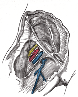
Right femoral sheath laid open to show its three compartments

Borders
The femoral triangle has three borders namely:[1]
- Superior (base of triangle): Inguinal ligament
- Lateral: Medial border of the sartorius muscle
- Medial: Medial border of the adductor longus
- Apex: Cross between medial and lateral border

Floor and roof
The floor of the femoral triangle is comprised of the adductor longus, pectineus (medially), psoas major and illiacus muscles (laterally).[1] The floor is gutter shaped since all the muscles forming the floor pass to the posterior aspect of the femur.[1]
The roof of the femoral triangle consists of skin, superficial and deep fascia (fascia lata, cribiform fascia) of the the thigh and subcutaneous tissue.[1]
Clinical Significance
The femoral triangle is a clinically significant anatomical region in the upper thigh for several reasons:
- It provides palpable access to the femoral artery, which is vital for assessing circulation.
- It's a common site for vascular access in medical procedures.
- Many important structures can be located here for various interventions.
- The femoral artery and vein in this area are used for catheterization in radiologic interventions such as: Coronary and peripheral angioplasty; Interventional embolization; Other endovascular procedures
- The large diameter of the vessels in the femoral triangle allows for the use of larger catheters.
- The femoral vein can be accessed for venipuncture when peripheral veins are not suitable.
- Ultrasound can easily visualize the femoral triangle, guiding procedures that require access to the femoral vessels.
- The area contains both deep and superficial inguinal lymph nodes, making it a target for lymphadenectomy in cancer staging involving inguinal lymphatic drainage.[3]
Conditions that can be found on ultrasound in the femoral triangle area include:
- Abscesses
- Cellulitis
- Hematoma
- Nodal enlargement
- Femoral artery aneurysm
- Thrombophlebitis of the femoral vein
These conditions can be identified through various sonographic features such as the appearance of masses, tissue swelling, or vascular abnormalities.[2]
References
- ↑ 1.0 1.1 1.2 1.3 1.4 1.5 Moore KL, Dalley AF, Agur AMR. Clinial oriented anatomy. Philadelphia: Wolters Kluwer, 2010.
- ↑ 2.0 2.1 Gitschlag KF, Sandler MA, Madrazo BL, Hricak H, Eyler WR. Disease in the femoral triangle: sonographic appearance. American Journal of Roentgenology 1982 Sep 1;139(3):515-9.
- ↑ Basinger H, Hogg JP. Anatomy, abdomen and pelvis, femoral triangle.Available:https://www.ncbi.nlm.nih.gov/books/NBK541140/ (accessed 9.5.2025)