Femoral Neck Hip Fracture: Difference between revisions
No edit summary |
No edit summary |
||
| Line 10: | Line 10: | ||
* Common injury sustained by older patients who are both more likely to have unsteadiness of [[gait]] and reduced [[bone]] mineral density, predisposing to fracture. | * Common injury sustained by older patients who are both more likely to have unsteadiness of [[gait]] and reduced [[bone]] mineral density, predisposing to fracture. | ||
* The biggest risk factors for a hip fracture are osteoporosis and [[Dementia|cognitive impairment.]]<ref name=":1">Radiopedia [https://radiopaedia.org/articles/femoral-neck-fracture NOF fractures] Available from:https://radiopaedia.org/articles/femoral-neck-fracture (last accessed 14.10.2020)</ref><ref name=":0" /> | * The biggest risk factors for a hip fracture are [[osteoporosis]] and [[Dementia|cognitive impairment.]]<ref name=":1">Radiopedia [https://radiopaedia.org/articles/femoral-neck-fracture NOF fractures] Available from:https://radiopaedia.org/articles/femoral-neck-fracture (last accessed 14.10.2020)</ref><ref name=":0" /> | ||
* About one-third of elderly people living independently fall every year, with 10% of these falls resulting in a hip fracture.<ref>Tinetti ME, Kumar C. The patient who falls: “It's always a trade- | * About one-third of elderly people living independently fall every year, with 10% of these falls resulting in a hip fracture.<ref>Tinetti ME, Kumar C. [https://jamanetwork.com/journals/jama/article-abstract/185213 The patient who falls:“It's always a trade-off”]. Jama. 2010 Jan 20;303(3):258-66.</ref> | ||
* A serious injury that occurs mostly in elderly people and complications can be life-threatening.<ref>Marks R. [https://www.researchgate.net/profile/Ray_Marks/publication/340793491_VITAMIN_C_AND_HIP_FRACTURES/links/5e9e058c299bf13079ad8850/VITAMIN-C-AND-HIP-FRACTURES.pdf Vitamin C and Hip Fractures-Review.] EC Orthopaedics. 2020;11:33-41.</ref><ref name=":0">Antapur | * A serious injury that occurs mostly in elderly people and complications can be life-threatening.<ref>Marks R. [https://www.researchgate.net/profile/Ray_Marks/publication/340793491_VITAMIN_C_AND_HIP_FRACTURES/links/5e9e058c299bf13079ad8850/VITAMIN-C-AND-HIP-FRACTURES.pdf Vitamin C and Hip Fractures-Review.] EC Orthopaedics. 2020;11:33-41.</ref><ref name=":0">Antapur P, Mahomed N, Gandhi R. [https://www.ncbi.nlm.nih.gov/pmc/articles/PMC3066247/ Fractures in the elderly: when is hip replacement a necessity?]. Clinical interventions in aging. 2011;6:1.</ref> | ||
* In younger patients femur fractures are the result of a high energy trauma, such as a motor accident, gunshot wound, or jump/fall from a height.<ref name=":0" /> | * In younger patients femur fractures are the result of a high energy trauma, such as a motor accident, gunshot wound, or jump/fall from a height.<ref name=":0" /> | ||
* High mortality, long-term disability and huge socio-economic burden are the main consequences of a hip fracture. | * High mortality, long-term disability and huge socio-economic burden are the main consequences of a hip fracture. | ||
| Line 22: | Line 22: | ||
* Very sturdy joint, due to the tight fitting of the bones and the strong surrounding [[Ligament|ligaments]] and [[Muscle|muscles]]. | * Very sturdy joint, due to the tight fitting of the bones and the strong surrounding [[Ligament|ligaments]] and [[Muscle|muscles]]. | ||
* The femur connects at the acetabulum of the pelvis and projects laterally before angling medially and inferiorly to form the [[knee]]. | * The femur connects at the acetabulum of the pelvis and projects laterally before angling medially and inferiorly to form the [[knee]]. | ||
A hip fracture occurs just below the head of femur (HOF) | A hip fracture occurs just below the head of femur (HOF), that is, the region of the femur called the femoral neck. A femoral neck fracture disconnects the HOF from the rest of the femur. <div> | ||
Click [[Hip Anatomy]] for more details | Click [[Hip Anatomy]] for more details | ||
| Line 40: | Line 40: | ||
== Classification of Hip Fractures == | == Classification of Hip Fractures == | ||
Hip fractures | Hip fractures is classified into intracapsular and extracapsular fractures<ref name=":7">Zuckerman JD. [https://www.nejm.org/doi/full/10.1056/NEJM199606063342307 Hip fracture]. New England journal of medicine. 1996 Jun 6;334(23):1519-25.</ref> | ||
'''Intracapsular fractures''' (femoral neck fractures): Occurs within the hip capsule; accounts for 45% of all acute hip fractures in the elderly<ref>Canale ST. ''Campbell's Operative Orthopaedics''. St. Louis, MO: Mosby;; 1998. pp. 2181–2223.</ref>; susceptible to malunion/[https://physio-pedia.com/Avascular_Necrosis avascular necrosis] of the HOF because of the limited blood supply to the area. [[Image:Fig5.png|right|frameless]]Intracapsular fractures are further classified as nondisplaced or displaced based on radiographic findings<ref>Garden RS. The structure and function of the proximal end of the femur. | '''Intracapsular fractures''' (femoral neck fractures): Occurs within the hip capsule; accounts for 45% of all acute hip fractures in the elderly<ref>Canale ST. ''Campbell's Operative Orthopaedics''. St. Louis, MO: Mosby;; 1998. pp. 2181–2223.</ref>; susceptible to malunion/[https://physio-pedia.com/Avascular_Necrosis avascular necrosis] of the HOF because of the limited blood supply to the area. [[Image:Fig5.png|right|frameless]]Intracapsular fractures are further classified as nondisplaced or displaced based on radiographic findings<ref>Garden RS. [https://online.boneandjoint.org.uk/doi/abs/10.1302/0301-620x.43b3.576 The structure and function of the proximal end of the femur]. The Journal of Bone and Joint Surgery. British volume. 1961 Aug;43(3):576-89.</ref> | ||
*Type 1: | *Type 1: non-displaced and incomplete fracture | ||
*Type 2: | *Type 2: non-displaced complete fracture | ||
*Type 3: complete fracture but incompletely displaced | *Type 3: complete fracture but incompletely displaced | ||
*Type 4: complete fracture and completely displaced | *Type 4: complete fracture and completely displaced | ||
'''Extracapsular fractures''': intertrochanteric fracture or subtronchanteric fracture. | '''Extracapsular fractures''': intertrochanteric fracture or subtronchanteric fracture. | ||
* Intertrochanteric fracture: occurs between the greater and the lesser trochanter<ref name=":7" />; intertrochanteric region has a good blood supply, [https://physio-pedia.com/Avascular_Necrosis avascular necrosis] or nonunion is rare. | |||
* Subtronchanteric fracture: occurs below the lesser trochanter, approximately 2.5 inches below. | |||
== Etiology == | == Etiology == | ||
Most commonly: | Most commonly: | ||
* Falls in the elderly: in elderly patients, the mechanism of injury various from falls directly onto the hip to a twisting mechanism in which the patient’s foot is planted and the body rotates. There is generally deficient elastic resistance in the fractured bone<ref name=":1" />. | * Falls in the elderly: in elderly patients, the mechanism of injury various from falls directly onto the hip to a twisting mechanism in which the patient’s foot is planted and the body rotates. There is generally deficient elastic resistance in the fractured bone<ref name=":1" />. | ||
* More than 50% of hip fracture patients have osteoporosis, and nearly all are osteopenia<ref name=":8">Rao, | * More than 50% of hip fracture patients have [[osteoporosis]], and nearly all are [[osteopenia]]<ref name=":8">Rao SS, Cherukuri M. [https://www.aafp.org/afp/2006/0615/p2195.html Management of hip fracture: the family physician's role]. American family physician. 2006 Jun 15;73(12):2195-200. </ref> | ||
* The majority of fragility hip fractures occurred inside the home<ref>Dhibar DP, Gogate Y, Aggarwal S, Garg S, Bhansali A, Bhadada SK. [https://www.ncbi.nlm.nih.gov/pmc/articles/PMC6683687/ Predictors and outcome of fragility hip fracture: | * The majority of fragility hip fractures occurred inside the home<ref>Dhibar DP, Gogate Y, Aggarwal S, Garg S, Bhansali A, Bhadada SK. [https://www.ncbi.nlm.nih.gov/pmc/articles/PMC6683687/ Predictors and outcome of fragility hip fracture: a prospective study from North India]. Indian Journal of Endocrinology and Metabolism. 2019 May;23(3):282.</ref> | ||
* Significant trauma (e.g. motor vehicle collisions) in younger patients | * Significant trauma (e.g. motor vehicle collisions) in younger patients | ||
* About 3% of hip fractures are related to localized bone weakness at the fracture site, secondary to tumor, followed by bone cysts, or Paget’s disease. | * About 3% of hip fractures are related to localized bone weakness at the fracture site, secondary to tumor, followed by bone cysts, or Paget’s disease. | ||
== Risk factors == | == Risk factors == | ||
Risk factors for hip fracture include<ref>Grisso, | Risk factors for hip fracture include<ref>Grisso JA, Kelsey JL, Strom BL, Ghiu GY, Maislin G, O'Brien LA, Hoffman S, Kaplan F. [https://www.nejm.org/doi/full/10.1056/NEJM199105093241905 Risk factors for falls as a cause of hip fracture in women]. New England journal of medicine. 1991 May 9;324(19):1326-31. </ref><ref>http://www.mayoclinic.com/health/hip-fracture/DS00185/DSECTION=risk-factors (visited on april 2016)</ref>: | ||
*Gender: prevalent in women; post[[Menopause|menopausal]] twice as likely as premenopausal to have hip fracture<ref>Banks E, Reeves GK, Beral V, Balkwill A, Liu B Roddam A. Million Women Study Collaborators. | *Gender: prevalent in women; post[[Menopause|menopausal]] twice as likely as premenopausal to have hip fracture<ref>Banks E, Reeves GK, Beral V, Balkwill A, Liu B, Roddam A, [https://journals.plos.org/plosmedicine/article?id=10.1371/journal.pmed.1000181 Million Women Study Collaborators. Hip fracture incidence in relation to age, menopausal status, and age at menopause: prospective analysis]. PLoS medicine. 2009 Nov 10;6(11):e1000181.</ref> | ||
*Reduced Bone density<ref>Angthong C, Suntharapa T, Harnroongroj T. [Major risk factors for the second contralateral hip fracture in the elderly] | *Reduced Bone density<ref>Angthong C, Suntharapa T, Harnroongroj T. [https://dergipark.org.tr/en/pub/aott/issue/18213/191450 Major risk factors for the second contralateral hip fracture in the elderly.] Acta orthopaedica et traumatologica turcica. 2009 May 1;43(3):193-8.</ref> | ||
*Falls<ref>Yang Y, Komisar V, Shishov N, Lo B, Korall AM, Feldman F, Robinovitch SN. [https://asbmr.onlinelibrary.wiley.com/doi/abs/10.1002/jbmr.4048 The Effect Of Fall Biomechanics On Risk For Hip Fracture In Older Adults: A Cohort Study Of Video‐Captured Falls In Long‐Term Care.] Journal of bone and mineral research. 2020 May 13.</ref> | *Falls<ref>Yang Y, Komisar V, Shishov N, Lo B, Korall AM, Feldman F, Robinovitch SN. [https://asbmr.onlinelibrary.wiley.com/doi/abs/10.1002/jbmr.4048 The Effect Of Fall Biomechanics On Risk For Hip Fracture In Older Adults: A Cohort Study Of Video‐Captured Falls In Long‐Term Care.] Journal of bone and mineral research. 2020 May 13.</ref> | ||
*[[Medication and Falls|Medications]]: Some medications can cause a decrease in bone density like cortisone. | *[[Medication and Falls|Medications]]: Some medications can cause a decrease in bone density like cortisone. | ||
| Line 82: | Line 83: | ||
== Diagnostic Procedures == | == Diagnostic Procedures == | ||
The diagnosis of a hip fracture is established based on patient history, physical examination, and radiography.<ref name=":9">Dinçel, V. | The diagnosis of a hip fracture is established based on patient history, physical examination, and radiography.<ref name=":9">Dinçel VE, Şengelen M, Sepici V, Çavuşoğlu T, Sepici B. [https://onlinelibrary.wiley.com/doi/abs/10.1002/ca.20680 The association of proximal femur geometry with hip fracture risk]. Clinical Anatomy: The Official Journal of the American Association of Clinical Anatomists and the British Association of Clinical Anatomists. 2008 Sep;21(6):575-80. | ||
</ref> | </ref> | ||
* Plain [[X-Rays|radiograph]]<nowiki/>s (sensitivity 93-98%) is the first-line investigation for suspected NOF fractures. | * Plain [[X-Rays|radiograph]]<nowiki/>s (sensitivity 93-98%) is the first-line investigation for suspected Neck of Femur(NOF) fractures. | ||
* In patients with a suspected occult NOF fracture, [[MRI Scans|MRI]] (sensitivity 99-100%) is recommended by many institutions as the second-line test if available within 24 hours, with CT or nuclear medicine bone scan third-line<ref name=":1" /> | * In patients with a suspected occult NOF fracture, [[MRI Scans|MRI]] (sensitivity 99-100%) is recommended by many institutions as the second-line test if available within 24 hours, with CT or nuclear medicine bone scan third-line<ref name=":1" /> | ||
| Line 97: | Line 98: | ||
[[Image:Figure4.jpg|right|frameless|400x400px]]The management of hip fracture is usually a combination of surgery and rehabilitation. | [[Image:Figure4.jpg|right|frameless|400x400px]]The management of hip fracture is usually a combination of surgery and rehabilitation. | ||
* Also depends on the location of the fracture and whether it is displaced. | * Also depends on the location of the fracture and whether it is displaced. | ||
* In comparison to conservative management (bed rest and traction), operative management results in a reduced length of hospital stay and improved rehabilitation.<ref>Handoll HH, Parker MJ. Conservative versus operative treatment for hip fractures in adults. Cochrane | * In comparison to conservative management (bed rest and traction), operative management results in a reduced length of hospital stay and improved rehabilitation.<ref>Handoll HH, Parker MJ. [https://www.cochranelibrary.com/cdsr/doi/10.1002/14651858.CD000337/abstract Conservative versus operative treatment for hip fractures in adults]. Cochrane database of systematic reviews. 2008(3).</ref> | ||
* Significant complications such as avascular necrosis and non-union are very common without surgical intervention. | * Significant complications such as avascular necrosis and non-union are very common without surgical intervention. | ||
* The treatment options include non-operative management, internal fixation or prosthetic replacement. | * The treatment options include non-operative management, internal fixation or prosthetic replacement. | ||
* Internal fixation can be performed with multiple pins, intramedullary hip screw (IHMS), crossed screw-nails or compression with a dynamic screw and plate. | * Internal fixation can be performed with multiple pins, intramedullary hip screw (IHMS), crossed screw-nails or compression with a dynamic screw and plate. | ||
* Replacing the femoral head is achieved with either [[Partial Hip Replacement|hemiarthroplasty]] and [[Total Hip Replacement|total hip arthroplasty.]] | * Replacing the femoral head is achieved with either [[Partial Hip Replacement|hemiarthroplasty]] and [[Total Hip Replacement|total hip arthroplasty.]] | ||
* A national pragmatic Randomised Control Trial: the '''SENSE''' trial will compare arthroplasties with internal fixation for undisplaced fracture neck femur in patients aged over 65<ref>Viberg B, Kold S, Brink O, Larsen MS, Hare KB, Palm H. Is arthroplaSty bEtter than interNal fixation for undiSplaced femoral nEck fracture? A national pragmatic RCT: the SENSE trial. BMJ open. 2020 Oct 1;10(10):e038442.</ref> | * A national pragmatic Randomised Control Trial: the '''SENSE''' trial will compare arthroplasties with internal fixation for undisplaced fracture neck femur in patients aged over 65<ref>Viberg B, Kold S, Brink O, Larsen MS, Hare KB, Palm H. [https://bmjopen.bmj.com/content/10/10/e038442.abstract Is arthroplaSty bEtter than interNal fixation for undiSplaced femoral nEck fracture?] A national pragmatic RCT: the SENSE trial. BMJ open. 2020 Oct 1;10(10):e038442.</ref> | ||
The high morbidity and mortality associated with hip and pelvic fractures after trauma has been well documented. | The high morbidity and mortality associated with hip and pelvic fractures after trauma has been well documented. | ||
* Prolonged bed rest can increase the risk of pressure sores, atelectasis, pneumonia, deconditioning, and thromboembolic complications. Weight-bearing immediately after hip fracture surgery is safe in most patients.<ref name=":6">Scheerlinck, | * Prolonged bed rest can increase the risk of pressure sores, atelectasis, pneumonia, deconditioning, and thromboembolic complications. Weight-bearing immediately after hip fracture surgery is safe in most patients.<ref name=":6">Scheerlinck T, Opdeweegh L, Vaes P, Opdecam P. [http://actaorthopaedica.be/assets/124/69_2-scheerlinck.pdf Hip fracture treatment: outcome and socio-economic aspects: a one-year survey in a Belgian university hospital]. Acta orthopaedica belgica. 2003 Apr 1;69(2):145-56.</ref> Complications following hip surgery involve blood clots, pneumonia, wound infections, and more, all of which can be reduced with activity<ref>Daniel Pendick. ‘’After hip fracture, exercise at home boosts day-to-day function’’ Harvard health publication (2014) </ref> | ||
* Prognosis is varied but is complicated by advanced age, as hip fractures increase the risk of death and major morbidity in the elderly. | * Prognosis is varied but is complicated by advanced age, as hip fractures increase the risk of death and major morbidity in the elderly. | ||
* Surgery <48 hours after admission may be associated with lower morbidity and may decrease hospital stay.<ref>Mak JC, Cameron ID, March LM; National Health and Medical Research Council (Australia). Evidence-based guidelines for the management of hip fractures in older persons: an update. Med J Aust. 2010;192:37-41.</ref><ref>Khan SK, Kalra S, Khanna A, | * Surgery <48 hours after admission may be associated with lower morbidity and may decrease hospital stay.<ref>Mak JC, Cameron ID, March LM; National Health and Medical Research Council (Australia). Evidence-based guidelines for the management of hip fractures in older persons: an update. Med J Aust. 2010;192:37-41.</ref><ref>Khan SK, Kalra S, Khanna A, Thiruvengada MM, Parker MJ. [https://www.sciencedirect.com/science/article/pii/S0020138309000114 Timing of surgery for hip fractures: a systematic review of 52 published studies involving 291,413 patients]. Injury. 2009 Jul 1;40(7):692-7.</ref>.<ref>Simunovic N, Devereaux PJ, Sprague S, Guyatt GH, Schemitsch E, DeBeer J, Bhandari M. [https://www.cmaj.ca/content/182/15/1609.short Effect of early surgery after hip fracture on mortality and complications: systematic review and meta-analysis]. Cmaj. 2010 Oct 19;182(15):1609-16.</ref><ref>Moja L, Piatti A, Pecoraro V, Ricci C, Virgili G, Salanti G, Germagnoli L, Liberati A, Banfi G. [https://journals.plos.org/plosone/article?id=10.1371/journal.pone.0046175 Timing matters in hip fracture surgery: patients operated within 48 hours have better outcomes]. A meta-analysis and meta-regression of over 190,000 patients.</ref> | ||
* The risk | * The risk of [[Avascular necrosis of the femoral head|avascular necrosis]] (AVN) depends on the type of fracture: | ||
# Transphyseal: ~90% risk of AVN | |||
# Subcapital: ~50% risk of AVN | |||
# Basicervical/transcervical: ~25% risk of AVN | |||
# Intertrochanteric: ~10% risk of AVN<ref name=":1" /> | |||
== Physical Examination == | == Physical Examination == | ||
| Line 119: | Line 125: | ||
* Tenderness to palpation around the inguinal area, over the femoral neck. This area may also be swollen. | * Tenderness to palpation around the inguinal area, over the femoral neck. This area may also be swollen. | ||
* Increased pain on the extremes of hip rotation, an abduction lurch, and an inability to stand on the involved leg | * Increased pain on the extremes of hip rotation, an abduction lurch, and an inability to stand on the involved leg | ||
For more on Hip examination, click [ | For more on Hip examination, click [[Hip Examination]] | ||
== Physical Therapy Management == | == Physical Therapy Management == | ||
[[File:Hip exercise 6.png|right|frameless]] | [[File:Hip exercise 6.png|right|frameless]] | ||
Rehabilitation begins promptly | Rehabilitation begins promptly. | ||
Two to three days postoperative | '''Two to three days postoperative''' | ||
* Instruct patient in deep breathing and cough. '''Goal:''' Prevent postoperative pneumonia and atelectasis. | * Instruct patient in deep breathing and cough. '''Goal:''' Prevent postoperative pneumonia and atelectasis. | ||
* Initiate isometrics and ankle pumps with involved extremity. '''Goal:''' Prepare patient for active exercise program. | * Initiate isometrics and ankle pumps with involved extremity. '''Goal:''' Prepare patient for active exercise program. | ||
* Initiate bedside sitting once physician has cleared patient for this activity. '''Goal:''' Prepare patient to begin transfer and progressive gait training processes.<ref>Luciani, | * Initiate bedside sitting once physician has cleared patient for this activity. '''Goal:''' Prepare patient to begin transfer and progressive gait training processes.<ref>Luciani D, Cadossi M, Mazzotti A, Chiarello E, Giannini S. [https://link.springer.com/article/10.1007/s40520-013-0079-9 The importance of rehabilitation after lower limb fractures in elderly osteoporotic patients.] Aging clinical and experimental research. 2013 Oct;25(1):113-5. | ||
</ref> | </ref> | ||
Three to five days postoperative | '''Three to five days postoperative''' | ||
* Gait train patient, observing weight-bearing precautions. Progress to walker or crutches. '''Goal:''' Establish independent gait with assistive device, using proper gait pattern on all surfaces and stairs. | * Gait train patient, observing weight-bearing precautions. Progress to walker or crutches. '''Goal:''' Establish independent gait with assistive device, using proper gait pattern on all surfaces and stairs. | ||
* Initiate training in activities of daily living, including bed mobility and transfers to and from bed and toilet. '''Goal:''' Achieve independence with all transfers. | * Initiate training in activities of daily living, including bed mobility and transfers to and from bed and toilet. '''Goal:''' Achieve independence with all transfers. | ||
| Line 138: | Line 144: | ||
* When internal fixation is performed, partial weight-bearing is recommended for a period of 8–10 weeks (according to the radiological evaluation of fracture healing), and after 3 months full weight-bearing should be allowed. | * When internal fixation is performed, partial weight-bearing is recommended for a period of 8–10 weeks (according to the radiological evaluation of fracture healing), and after 3 months full weight-bearing should be allowed. | ||
* The patient can also begin strengthening exercises based on the surgeon's orders (typically six weeks post-op). | * The patient can also begin strengthening exercises based on the surgeon's orders (typically six weeks post-op). | ||
Patients should also undergo [[Balance Training|balance]] and [[Proprioception|proprioceptive]] rehab and these abilities are quickly lost with inactivity. Rehabilitation classes for balance and [[Falls and Exercise|falls prevention]] are recommended | Patients should also undergo [[Balance Training|balance]] and [[Proprioception|proprioceptive]] rehab and these abilities are quickly lost with inactivity. Rehabilitation classes for balance and [[Falls and Exercise|falls prevention]] are recommended. | ||
Weight-bearing exercises are very important for mobility, balance, activities of daily living and quality of life<ref> | Weight-bearing exercises are very important for mobility, balance, activities of daily living and quality of life<ref>LeBlanc KE, Muncie Jr HL, LeBlanc LL. [https://www.aafp.org/afp/2014/0615/p945 Hip fracture: diagnosis, treatment, and secondary prevention]. American family physician. 2014 Jun 15;89(12):945-51. | ||
</ref>, examples: stepping in different directions, standing up and sitting down, tapping the foot and stepping onto and off a block. | </ref>, examples: stepping in different directions, standing up and sitting down, tapping the foot and stepping onto and off a block. | ||
[[File:Hip Abduction with IR.JPG|right|frameless]] | [[File:Hip Abduction with IR.JPG|right|frameless]] | ||
For patients who underwent a prosthetic replacement have to avoid for approximately 12 weeks: | For patients who underwent a prosthetic replacement have to avoid the following for approximately 12 weeks: | ||
*Hip flexion greater than 70–90° | *Hip flexion greater than 70–90° | ||
*External rotation of the leg | *External rotation of the leg | ||
*Adduction of the leg past midline | *Adduction of the leg past midline | ||
*Should not bend forward from the waist more than | *Should not bend forward from the waist more than 90° | ||
Rehabilitation program components<ref>Latham, | '''Rehabilitation program components'''<ref>Latham NK, Harris BA, Bean JF, Heeren T, Goodyear C, Zawacki S, Heislein DM, Mustafa J, Pardasaney P, Giorgetti M, Holt N. [https://jamanetwork.com/journals/jama/article-abstract/1829991 Effect of a home-based exercise program on functional recovery following rehabilitation after hip fracture: a randomized clinical trial]. Jama. 2014 Feb 19;311(7):700-8. | ||
</ref>: | </ref>: | ||
*Hip extension (theraband and manual exercise) | *Hip extension (theraband and manual exercise) | ||
| Line 160: | Line 166: | ||
*Stepping up and down step (vest, manual exercise and plyometric step) | *Stepping up and down step (vest, manual exercise and plyometric step) | ||
*Calf raises - both legs and one leg (manual exercise) | *Calf raises - both legs and one leg (manual exercise) | ||
Importance or rehabilitation/home exercise program: | '''Importance or rehabilitation/home exercise program''': | ||
* Moderate to large improvements in physical performance and quality of life was found in patients who had a 10- week home-based progressive resistance exercise program<ref>Mangione KK, Craik RL, Palombaro KM, Tomlinson SS, Hofmann MT. | * Moderate to large improvements in physical performance and quality of life was found in patients who had a 10- week home-based progressive resistance exercise program<ref>Mangione KK, Craik RL, Palombaro KM, Tomlinson SS, Hofmann MT. [https://agsjournals.onlinelibrary.wiley.com/doi/abs/10.1111/j.1532-5415.2010.03076.x Home‐based leg‐strengthening exercise improves function 1 year after hip fracture: a randomized controlled study]. Journal of the American Geriatrics Society. 2010 Oct;58(10):1911-7.</ref> | ||
* A meta-analyses, showed that balance training within 6 months improves person with hip fracture physical functioning, gait, lower limb strength, performance task, and activity of daily living.<ref>Wu JQ, Mao LB, Wu J. | * A meta-analyses, showed that balance training within 6 months improves person with hip fracture physical functioning, gait, lower limb strength, performance task, and activity of daily living.<ref>Wu JQ, Mao LB, Wu J. [https://josr-online.biomedcentral.com/articles/10.1186/s13018-019-1125-x Efficacy of balance training for hip fracture patients: a meta-analysis of randomized controlled trials]. Journal of orthopaedic surgery and research. 2019 Dec;14(1):1-1.</ref> | ||
* Among patients who had completed standard rehabilitation after hip fracture, the use of a home-based functionally oriented exercise program resulted in modest improvement in physical function at 6 months after randomization.<ref>Latham, | * Among patients who had completed standard rehabilitation after hip fracture, the use of a home-based functionally oriented exercise program resulted in modest improvement in physical function at 6 months after randomization.<ref>Latham NK, Harris BA, Bean JF, Heeren T, Goodyear C, Zawacki S, Heislein DM, Mustafa J, Pardasaney P, Giorgetti M, Holt N. [https://jamanetwork.com/journals/jama/article-abstract/1829991 Effect of a home-based exercise program on functional recovery following rehabilitation after hip fracture: a randomized clinical trial]. Jama. 2014 Feb 19;311(7):700-8. | ||
</ref> | </ref> | ||
Education and prevention are also important issues to address. | Education and prevention are also important issues to address. | ||
== Clinical Bottom Line == | == Clinical Bottom Line == | ||
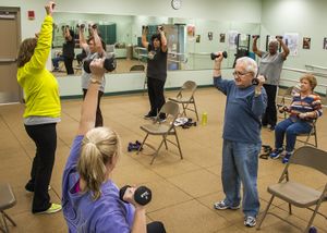
* [[File:Strengthing exercise for old people .jpg|right|frameless]]The number of hip fractures worldwide will increase up to 7-21 million incidences each year in 2050.<ref name=":4">Kannus, | * [[File:Strengthing exercise for old people .jpg|right|frameless]]The number of hip fractures worldwide will increase up to 7-21 million incidences each year in 2050.<ref name=":4">Kannus P, Parkkari J, Sievänen H, Heinonen A, Vuori I, Järvinen M. [https://www.sciencedirect.com/science/article/pii/8756328295003819 Epidemiology of hip fractures]. Bone. 1996 Jan 1;18(1):S57-63. </ref> | ||
* Mortality associated with a hip fracture is about 5-10% after one month. One year after fracture, about a third of patients will have died, compared with an expected annual mortality of about 10% in this age group.<ref name=":4" /> | * Mortality associated with a hip fracture is about 5-10% after one month. One year after fracture, about a third of patients will have died, compared with an expected annual mortality of about 10% in this age group.<ref name=":4" /> | ||
* Only a third of the deaths are directly attributable to the hip fracture itself. | * Only a third of the deaths are directly attributable to the hip fracture itself. | ||
Revision as of 23:20, 18 October 2022
Introduction[edit | edit source]

Hip Fracture:
Vernacular term for fracture of the femoral neck, typically resulting from a fall in an old person with osteoporosis
- More common in women; requires surgical repair with internal fixation and can lead to prolonged or permanent loss of mobility and shortened life span[1].
- Common injury sustained by older patients who are both more likely to have unsteadiness of gait and reduced bone mineral density, predisposing to fracture.
- The biggest risk factors for a hip fracture are osteoporosis and cognitive impairment.[2][3]
- About one-third of elderly people living independently fall every year, with 10% of these falls resulting in a hip fracture.[4]
- A serious injury that occurs mostly in elderly people and complications can be life-threatening.[5][3]
- In younger patients femur fractures are the result of a high energy trauma, such as a motor accident, gunshot wound, or jump/fall from a height.[3]
- High mortality, long-term disability and huge socio-economic burden are the main consequences of a hip fracture.
Clinically Relevant Anatomy[edit | edit source]

The hip joint is a
- Ball and socket joint, formed by the head of the femur and the acetabulum of the pelvis.
- Very sturdy joint, due to the tight fitting of the bones and the strong surrounding ligaments and muscles.
- The femur connects at the acetabulum of the pelvis and projects laterally before angling medially and inferiorly to form the knee.
A hip fracture occurs just below the head of femur (HOF), that is, the region of the femur called the femoral neck. A femoral neck fracture disconnects the HOF from the rest of the femur.
Click Hip Anatomy for more details
Epidemiology[edit | edit source]
Approximately 1.6 million hip fractures occur worldwide each year, by 2050 this number could reach between 4.5 million and 6.3 million.
- Between 1990 and 2000, there was nearly a 25% increase in hip fractures worldwide. The peak number of hip fractures occurred at 75-79 years of age for both sexes.
- Nearly 75% of all hip fractures occur in women.
- Men account for 25% of hip fractures occurring in the over 50 population.
- Hip fractures are invariably associated with chronic pain, reduced mobility, disability, and an increasing degree of dependence.
- After sustaining a hip fracture 10-20% of formerly community dwelling patients require long term nursing care, with the rate of nursing home admission rising with age.
- In white women, the lifetime risk of hip fracture is 1 in 6.
- A 50 year old woman has a 2.8% risk of death related to hip fracture during her remaining lifetime.
- 5-10% of patients experience a recurrent hip fracture, with the mean interval between the first and second fracture being 3.3 years .
- Up to 20% of patients die in the first year following hip fractures, mostly due to pre-existing medical conditions. Less than half those who survive the hip fracture regain their previous level of function[6]
Classification of Hip Fractures[edit | edit source]
Hip fractures is classified into intracapsular and extracapsular fractures[7]
Intracapsular fractures (femoral neck fractures): Occurs within the hip capsule; accounts for 45% of all acute hip fractures in the elderly[8]; susceptible to malunion/avascular necrosis of the HOF because of the limited blood supply to the area.

Intracapsular fractures are further classified as nondisplaced or displaced based on radiographic findings[9]
- Type 1: non-displaced and incomplete fracture
- Type 2: non-displaced complete fracture
- Type 3: complete fracture but incompletely displaced
- Type 4: complete fracture and completely displaced
Extracapsular fractures: intertrochanteric fracture or subtronchanteric fracture.
- Intertrochanteric fracture: occurs between the greater and the lesser trochanter[7]; intertrochanteric region has a good blood supply, avascular necrosis or nonunion is rare.
- Subtronchanteric fracture: occurs below the lesser trochanter, approximately 2.5 inches below.
Etiology[edit | edit source]
Most commonly:
- Falls in the elderly: in elderly patients, the mechanism of injury various from falls directly onto the hip to a twisting mechanism in which the patient’s foot is planted and the body rotates. There is generally deficient elastic resistance in the fractured bone[2].
- More than 50% of hip fracture patients have osteoporosis, and nearly all are osteopenia[10]
- The majority of fragility hip fractures occurred inside the home[11]
- Significant trauma (e.g. motor vehicle collisions) in younger patients
- About 3% of hip fractures are related to localized bone weakness at the fracture site, secondary to tumor, followed by bone cysts, or Paget’s disease.
Risk factors[edit | edit source]
Risk factors for hip fracture include[12][13]:
- Gender: prevalent in women; postmenopausal twice as likely as premenopausal to have hip fracture[14]
- Reduced Bone density[15]
- Falls[16]
- Medications: Some medications can cause a decrease in bone density like cortisone.
- Nutrition: It is well known that calcium and vitamin D increase bone mass, so a lack of it can cause several fractures, including hip fractures.
- Age: the older you get, the higher the risk is for hip fractures. 90% of these fractures occur in persons over 70 years old.
- Alcohol and tobacco: These products can reduce the bone mass, causing a higher risk to have a hip fracture
- Medical problems: Endocrine disorders can cause fragility of the bones
- Physical inactivity: Physical activity is very important for the muscle mass and the bone mass
- Stroke increases risk factor for falls which can cause a hip fracture.
- Parkinson’s disease increases risk factor for falls which can cause a hip fracture.
Characteristics/Clinical Presentation[edit | edit source]
- Dull ache in the groin and/or hip region[10]
- Inability to put weight on the injured leg causing immobility right after the fall[17]
- Shorter leg on the side of the injured hip
- External rotation of the injured leg[17]
- Stiffness, bruising and swelling in and around the hip
Diagnostic Procedures[edit | edit source]
The diagnosis of a hip fracture is established based on patient history, physical examination, and radiography.[17]
- Plain radiographs (sensitivity 93-98%) is the first-line investigation for suspected Neck of Femur(NOF) fractures.
- In patients with a suspected occult NOF fracture, MRI (sensitivity 99-100%) is recommended by many institutions as the second-line test if available within 24 hours, with CT or nuclear medicine bone scan third-line[2]
Outcome Measures[edit | edit source]
- Functional Independence Measure
- Berg Balance Scale
- Timed Up and Go Test (TUG)
- Patient Specific Functional Scale
- Falls Risk Assessment Tool
Treatment and Prognosis[edit | edit source]

The management of hip fracture is usually a combination of surgery and rehabilitation.
- Also depends on the location of the fracture and whether it is displaced.
- In comparison to conservative management (bed rest and traction), operative management results in a reduced length of hospital stay and improved rehabilitation.[18]
- Significant complications such as avascular necrosis and non-union are very common without surgical intervention.
- The treatment options include non-operative management, internal fixation or prosthetic replacement.
- Internal fixation can be performed with multiple pins, intramedullary hip screw (IHMS), crossed screw-nails or compression with a dynamic screw and plate.
- Replacing the femoral head is achieved with either hemiarthroplasty and total hip arthroplasty.
- A national pragmatic Randomised Control Trial: the SENSE trial will compare arthroplasties with internal fixation for undisplaced fracture neck femur in patients aged over 65[19]
The high morbidity and mortality associated with hip and pelvic fractures after trauma has been well documented.
- Prolonged bed rest can increase the risk of pressure sores, atelectasis, pneumonia, deconditioning, and thromboembolic complications. Weight-bearing immediately after hip fracture surgery is safe in most patients.[20] Complications following hip surgery involve blood clots, pneumonia, wound infections, and more, all of which can be reduced with activity[21]
- Prognosis is varied but is complicated by advanced age, as hip fractures increase the risk of death and major morbidity in the elderly.
- Surgery <48 hours after admission may be associated with lower morbidity and may decrease hospital stay.[22][23].[24][25]
- The risk of avascular necrosis (AVN) depends on the type of fracture:
- Transphyseal: ~90% risk of AVN
- Subcapital: ~50% risk of AVN
- Basicervical/transcervical: ~25% risk of AVN
- Intertrochanteric: ~10% risk of AVN[2]
Physical Examination[edit | edit source]
On physical examination, findings on the patient with a hip fracture may include the following:
- limited and painful hip range of motion, especially in internal rotation.
- the injured leg is shortened, externally rotated, and abducted in the supine position
- Pain is noted upon attempted passive hip motion.
- Ecchymosis may or may not be present.
- An antalgic gait pattern may be present.
- Tenderness to palpation around the inguinal area, over the femoral neck. This area may also be swollen.
- Increased pain on the extremes of hip rotation, an abduction lurch, and an inability to stand on the involved leg
For more on Hip examination, click Hip Examination
Physical Therapy Management[edit | edit source]

Rehabilitation begins promptly.
Two to three days postoperative
- Instruct patient in deep breathing and cough. Goal: Prevent postoperative pneumonia and atelectasis.
- Initiate isometrics and ankle pumps with involved extremity. Goal: Prepare patient for active exercise program.
- Initiate bedside sitting once physician has cleared patient for this activity. Goal: Prepare patient to begin transfer and progressive gait training processes.[26]
Three to five days postoperative
- Gait train patient, observing weight-bearing precautions. Progress to walker or crutches. Goal: Establish independent gait with assistive device, using proper gait pattern on all surfaces and stairs.
- Initiate training in activities of daily living, including bed mobility and transfers to and from bed and toilet. Goal: Achieve independence with all transfers.
- Initiate active range of motion/strengthening program. Individualize exercise program according to each patient's needs, but generally include the following. Goals: Increase strength of involved extremity; increase independence with exercise program.
- Supine: hip abduction and adduction, gluteal sets, quadriceps sets, straight leg raise, hip and knee flexion, short arc quadriceps, internal and external rotation.
- Sitting: Long arc quadriceps, hip flexion, ankle pumps[27].
- When internal fixation is performed, partial weight-bearing is recommended for a period of 8–10 weeks (according to the radiological evaluation of fracture healing), and after 3 months full weight-bearing should be allowed.
- The patient can also begin strengthening exercises based on the surgeon's orders (typically six weeks post-op).
Patients should also undergo balance and proprioceptive rehab and these abilities are quickly lost with inactivity. Rehabilitation classes for balance and falls prevention are recommended.
Weight-bearing exercises are very important for mobility, balance, activities of daily living and quality of life[28], examples: stepping in different directions, standing up and sitting down, tapping the foot and stepping onto and off a block.

For patients who underwent a prosthetic replacement have to avoid the following for approximately 12 weeks:
- Hip flexion greater than 70–90°
- External rotation of the leg
- Adduction of the leg past midline
- Should not bend forward from the waist more than 90°
Rehabilitation program components[29]:
- Hip extension (theraband and manual exercise)
- Heel raises onto toes (theraband and manual exercise)
- Resisted rowing (double arm lifting) (theraband and manual exercise)
- Standing diagonal reach (theraband and manual exercise)
- Modified get up and go (theraband and manual exercise)
- Overhead arm extensions (theraband and manual exercise)
- Repeated chair stands (vest and manual exercise)
- Lunges - forward and back (vest and manual exercise)
- Stepping up and down step (vest, manual exercise and plyometric step)
- Calf raises - both legs and one leg (manual exercise)
Importance or rehabilitation/home exercise program:
- Moderate to large improvements in physical performance and quality of life was found in patients who had a 10- week home-based progressive resistance exercise program[30]
- A meta-analyses, showed that balance training within 6 months improves person with hip fracture physical functioning, gait, lower limb strength, performance task, and activity of daily living.[31]
- Among patients who had completed standard rehabilitation after hip fracture, the use of a home-based functionally oriented exercise program resulted in modest improvement in physical function at 6 months after randomization.[32]
Education and prevention are also important issues to address.
Clinical Bottom Line[edit | edit source]
- The number of hip fractures worldwide will increase up to 7-21 million incidences each year in 2050.[33]
- Mortality associated with a hip fracture is about 5-10% after one month. One year after fracture, about a third of patients will have died, compared with an expected annual mortality of about 10% in this age group.[33]
- Only a third of the deaths are directly attributable to the hip fracture itself.
- More than 10% of survivors will be unable to return to their previous residence. Most of the remainder will have some residual pain or disability. Most people who sustain the injury require surgery followed by a period of rehabilitation.
- Treatment is generally surgical to replace or repair the broken bone.
- Some loss of function is to be expected in most patients.
- Rehabilitation plays a key role in treatment.

References[edit | edit source]
- ↑ Free dictionary Hip fracture: Available from:https://medical-dictionary.thefreedictionary.com/hip+fracture (last accessed 14.10.2020)
- ↑ 2.0 2.1 2.2 2.3 Radiopedia NOF fractures Available from:https://radiopaedia.org/articles/femoral-neck-fracture (last accessed 14.10.2020)
- ↑ 3.0 3.1 3.2 Antapur P, Mahomed N, Gandhi R. Fractures in the elderly: when is hip replacement a necessity?. Clinical interventions in aging. 2011;6:1.
- ↑ Tinetti ME, Kumar C. The patient who falls:“It's always a trade-off”. Jama. 2010 Jan 20;303(3):258-66.
- ↑ Marks R. Vitamin C and Hip Fractures-Review. EC Orthopaedics. 2020;11:33-41.
- ↑ IOF Facts and stats Available from:https://www.iofbonehealth.org/facts-statistics#category-16 (last accessed 14.10.2020)
- ↑ 7.0 7.1 Zuckerman JD. Hip fracture. New England journal of medicine. 1996 Jun 6;334(23):1519-25.
- ↑ Canale ST. Campbell's Operative Orthopaedics. St. Louis, MO: Mosby;; 1998. pp. 2181–2223.
- ↑ Garden RS. The structure and function of the proximal end of the femur. The Journal of Bone and Joint Surgery. British volume. 1961 Aug;43(3):576-89.
- ↑ 10.0 10.1 Rao SS, Cherukuri M. Management of hip fracture: the family physician's role. American family physician. 2006 Jun 15;73(12):2195-200.
- ↑ Dhibar DP, Gogate Y, Aggarwal S, Garg S, Bhansali A, Bhadada SK. Predictors and outcome of fragility hip fracture: a prospective study from North India. Indian Journal of Endocrinology and Metabolism. 2019 May;23(3):282.
- ↑ Grisso JA, Kelsey JL, Strom BL, Ghiu GY, Maislin G, O'Brien LA, Hoffman S, Kaplan F. Risk factors for falls as a cause of hip fracture in women. New England journal of medicine. 1991 May 9;324(19):1326-31.
- ↑ http://www.mayoclinic.com/health/hip-fracture/DS00185/DSECTION=risk-factors (visited on april 2016)
- ↑ Banks E, Reeves GK, Beral V, Balkwill A, Liu B, Roddam A, Million Women Study Collaborators. Hip fracture incidence in relation to age, menopausal status, and age at menopause: prospective analysis. PLoS medicine. 2009 Nov 10;6(11):e1000181.
- ↑ Angthong C, Suntharapa T, Harnroongroj T. Major risk factors for the second contralateral hip fracture in the elderly. Acta orthopaedica et traumatologica turcica. 2009 May 1;43(3):193-8.
- ↑ Yang Y, Komisar V, Shishov N, Lo B, Korall AM, Feldman F, Robinovitch SN. The Effect Of Fall Biomechanics On Risk For Hip Fracture In Older Adults: A Cohort Study Of Video‐Captured Falls In Long‐Term Care. Journal of bone and mineral research. 2020 May 13.
- ↑ 17.0 17.1 17.2 Dinçel VE, Şengelen M, Sepici V, Çavuşoğlu T, Sepici B. The association of proximal femur geometry with hip fracture risk. Clinical Anatomy: The Official Journal of the American Association of Clinical Anatomists and the British Association of Clinical Anatomists. 2008 Sep;21(6):575-80.
- ↑ Handoll HH, Parker MJ. Conservative versus operative treatment for hip fractures in adults. Cochrane database of systematic reviews. 2008(3).
- ↑ Viberg B, Kold S, Brink O, Larsen MS, Hare KB, Palm H. Is arthroplaSty bEtter than interNal fixation for undiSplaced femoral nEck fracture? A national pragmatic RCT: the SENSE trial. BMJ open. 2020 Oct 1;10(10):e038442.
- ↑ Scheerlinck T, Opdeweegh L, Vaes P, Opdecam P. Hip fracture treatment: outcome and socio-economic aspects: a one-year survey in a Belgian university hospital. Acta orthopaedica belgica. 2003 Apr 1;69(2):145-56.
- ↑ Daniel Pendick. ‘’After hip fracture, exercise at home boosts day-to-day function’’ Harvard health publication (2014)
- ↑ Mak JC, Cameron ID, March LM; National Health and Medical Research Council (Australia). Evidence-based guidelines for the management of hip fractures in older persons: an update. Med J Aust. 2010;192:37-41.
- ↑ Khan SK, Kalra S, Khanna A, Thiruvengada MM, Parker MJ. Timing of surgery for hip fractures: a systematic review of 52 published studies involving 291,413 patients. Injury. 2009 Jul 1;40(7):692-7.
- ↑ Simunovic N, Devereaux PJ, Sprague S, Guyatt GH, Schemitsch E, DeBeer J, Bhandari M. Effect of early surgery after hip fracture on mortality and complications: systematic review and meta-analysis. Cmaj. 2010 Oct 19;182(15):1609-16.
- ↑ Moja L, Piatti A, Pecoraro V, Ricci C, Virgili G, Salanti G, Germagnoli L, Liberati A, Banfi G. Timing matters in hip fracture surgery: patients operated within 48 hours have better outcomes. A meta-analysis and meta-regression of over 190,000 patients.
- ↑ Luciani D, Cadossi M, Mazzotti A, Chiarello E, Giannini S. The importance of rehabilitation after lower limb fractures in elderly osteoporotic patients. Aging clinical and experimental research. 2013 Oct;25(1):113-5.
- ↑ Physio treatment Hip Protocol Available from:https://www.physiotherapy-treatment.com/Femoral-Neck-Fracture-Physiotherapy.html (last accessed 15.10.2020)
- ↑ LeBlanc KE, Muncie Jr HL, LeBlanc LL. Hip fracture: diagnosis, treatment, and secondary prevention. American family physician. 2014 Jun 15;89(12):945-51.
- ↑ Latham NK, Harris BA, Bean JF, Heeren T, Goodyear C, Zawacki S, Heislein DM, Mustafa J, Pardasaney P, Giorgetti M, Holt N. Effect of a home-based exercise program on functional recovery following rehabilitation after hip fracture: a randomized clinical trial. Jama. 2014 Feb 19;311(7):700-8.
- ↑ Mangione KK, Craik RL, Palombaro KM, Tomlinson SS, Hofmann MT. Home‐based leg‐strengthening exercise improves function 1 year after hip fracture: a randomized controlled study. Journal of the American Geriatrics Society. 2010 Oct;58(10):1911-7.
- ↑ Wu JQ, Mao LB, Wu J. Efficacy of balance training for hip fracture patients: a meta-analysis of randomized controlled trials. Journal of orthopaedic surgery and research. 2019 Dec;14(1):1-1.
- ↑ Latham NK, Harris BA, Bean JF, Heeren T, Goodyear C, Zawacki S, Heislein DM, Mustafa J, Pardasaney P, Giorgetti M, Holt N. Effect of a home-based exercise program on functional recovery following rehabilitation after hip fracture: a randomized clinical trial. Jama. 2014 Feb 19;311(7):700-8.
- ↑ 33.0 33.1 Kannus P, Parkkari J, Sievänen H, Heinonen A, Vuori I, Järvinen M. Epidemiology of hip fractures. Bone. 1996 Jan 1;18(1):S57-63.

