Ligament
Original Editor -
Top Contributors - Lucinda hampton and Kim Jackson
Description[edit | edit source]
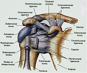
Ligaments are short bands of tough, flexible tissue, made up of lots of individual fibres, which connect the bones of the body together, being a dense type of connective tissue. Ligaments can be found connecting most of the bones in the body. The function of a ligament is to provide a passive limit to amount of movement between your bones. The human body has approximately 900 ligaments.[1] Once thought to be inert, they are in fact responsive to many local and systemic factors that influence their function.
Injury to a ligament results in a drastic change in its structure and physiology. The ligament is restored by the formation of scar tissue which is biologically and biomechanically inferior to the tissue it replaces.[2]
Structure[edit | edit source]
The basic building blocks of a ligament are collagen fibers. These fibers are very strong, flexible, and resistant to damage from pulling or compressing stresses. Collagen fibers are usually arranged in parallel bundles, which help multiply the strength of the individual fibers. The bundles of collagen are attached to the outer covering that surrounds all bones, the periosteum.[3] Skeletal ligaments vary in size, shape, orientation and location.
Their unique and complex bony attachments often involve unusual shapes on the bone that are likely critical to how the fibres within the ligament are recruited as the joint moves. While the ligament appears as a single structure, with joint movement, some fibres appear to tighten or loosen depending on the bone positions and the forces that are applied confirming that these structures are more complex than originally thought..
Ligaments are composed of cells called fibroblasts which are surrounded by matrix. The cells are responsible for matrix synthesis and they are relatively few in number and represent a small percentage of the total ligament volume. Although these cells may appear physically and functionally isolated, recent studies have indicated that normal ligament cells may communicate by means of prominent cytoplasmic extensions that extend for long distances and connect to cytoplasmic extensions from adjacent cells, thus forming an elaborate 3-dimensional architecture. Ligament microstructure can be visualized using polarized light that reveals collagen bundles aligned along the long axis of the ligament and displaying an underlying "waviness" or crimp along the length. Crimp is thought to play a biomechanical role relating to the ligaments loading state with increased loading resulting in some areas of the ligament uncrimping, allowing the ligament to elongate without sustaining damage.
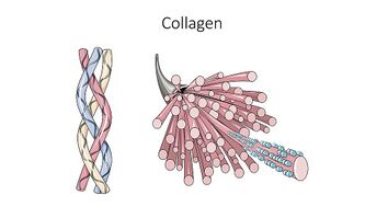
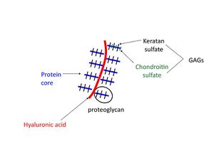
Biochemically, ligaments are approximately two-thirds water and one-third solid with the water contributing to cellular function and viscoelastic behaviour. The solid components of ligaments are principally type 1 collagen (image is of the triple helix, collagen) which accounts for approximately 75% of the dry weight with the balance being made up by proteoglycans (<1%), elastin and other proteins and glycoproteins.
Ultrastructural studies have revealed that collagen fibres are actually composed of smaller fibrils. Crosslink formation of the collagen fibres is the critical step that gives collagen fibres such incredible strength. During growth and development, crosslinks are relatively immature and soluble but with age they mature and become insoluble and increase in strength.
Epi-ligament: Ligaments often have a more vascular overlying layer termed the "epiligament" covering their surface and this layer is often indistinguishable from the actual ligament and merges into the periosteum of the bone around the attachment sites of the ligament. The epiligament is more vascular than the ligament and more cellular with more sensory and proprioceptive nerves. These nerves travel in close proximity to the blood vessels with more nerves closer to the bony ligament insertions.[2]
Ligament Injury[edit | edit source]
Ligaments are most often torn in traumatic joint injuries that can result in either partial or complete ligament discontinuities. The description below is of a complete tear.
Ligaments heal by a process which includes three phases:
- Hemorrhage with inflammation - involves retraction of the disrupted ligament ends, formation of a blood clot, which is subsequently resorbed, and replaced with a heavy cellular infiltrate. Subsequently, a considerable hypertrophic vascular response takes place in the gap between the disrupted ends and results in an increase in both vascularity and blood flow, both of which decrease with time.
- Matrix and cellular proliferation - defined as the production of "scar tissue" (dense, cellular, collagenous connective tissue matrix) by hypertrophic fibroblastic cells. This scar tissue is initially quite disorganized with more defects. After a few weeks of healing, the collagen becomes quite well aligned with the long axis of the ligament despite the fact that the types of collagen are abnormal and the collagen fibrils have smaller diameters in the proliferating tissue.
- Remodeling and maturation (matrix remodeling) - defects in the scar become filled in but although the matrix becomes more ligament-like with time, some major differences in composition, architecture and function persist. Differences which persist include altered proteoglycan and collagen types, failure of collagen crosslinks to mature, persistence of small collagen fibril diameters, altered cell connections, increased vascularity, abnormal innervation, increased cellularity and the incomplete resolution of matrix "flaws"[2]
The 3 Grades of Ligament Injury are:
Grade I sprains usually heal within a few weeks. Maximal ligament strength will occur after six weeks when the collagen fibres have matured. Resting from painful activity, icing the injury, and some anti-inflammatory medications are useful. Physiotherapy will help to hasten the healing process via electrical modalities, massage and exercise.
Grade II sprains are more significant and disabling. These injuries require load protection during the early healing phase. Depending on the ligament injury this may include the use of a weight-bearing brace or some supportive taping is common in early treatment (helps to ease the pain and avoid stretching of the healing ligament).
This may commonly take 6 to 12 weeks depending the injury and what sport or activity clients is to resume.
Grade III injury is a very significant injury often requiring an opinion from an Orthopaedic Surgeon to determine whether early surgical repair is required. If surgery is required, your rehabilitation will be guided by your surgeon and physiotherapist.
In non-surgical ligament injuries protection of the injury from weight-bearing stresses is important. The aim is to allow for ligament healing in a short/non-stressful position. Rehabilitation is slowly progressed as the ligament repairs and a gradual return to normal activities can occur.
Depending upon the ligament injury full level of activity can take 3 to 4 months or even up to 12 months. Very severe ligament injuries can even take longer. [4]
Common Ligament Injuries[edit | edit source]
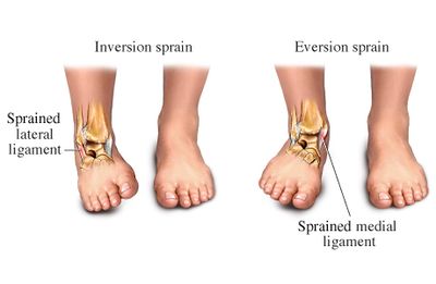
The most common torn ligaments are knee ligaments and ankle ligaments. This is because the joints are weight-bearing and under high stress with any change of direction sports or full contact sports. The shoulder, being inherently unstable, also has a high incidence of ligament injuries.
See also
Resources[edit | edit source]
References[edit | edit source]
- ↑ Southern hills medical centre Framing within our body Available from:https://southernhillshospital.com/about/newsroom/framing-within-our-bodies (last accessed 14.2.2020)
- ↑ 2.0 2.1 2.2 Frank CB. Ligament structure, physiology and function. Journal of Musculoskeletal and Neuronal Interactions. 2004 Jun;4(2):199. Available from:https://www.ismni.org/jmni/pdf/16/21FRANK.pdf?q=ligament (last accessed 13.2.2020)
- ↑ Study.com What are ligaments. Available from:https://study.com/academy/lesson/what-are-ligaments-definition-types-quiz.html (last accessed 13.2.2020)
- ↑ Physioworks Ligaments Available from:https://physioworks.com.au/Injuries-Conditions/Regions/ligament-injury (last accessed 13.2.2020)
