When refering to evidence in academic writing, you should always try to reference the primary (original) source. That is usually the journal article where the information was first stated. In most cases Physiopedia articles are a secondary source and so should not be used as references. Physiopedia articles are best used to find the original sources of information (see the references list at the bottom of the article).
If you believe that this Physiopedia article is the primary source for the information you are refering to, you can use the button below to access a related citation statement.
The periosteum is a membranous tissue that covers the surfaces of bones. It is an intricate structure composed of an outer fibrous layer that gives structural integrity and an inner cambium layer that possesses osteogenic potential. During growth and development it aides in bone elongation and modelling, and when a bone is fractured, participates in its recovery. It is not present in sesamoid bones and the intra-articular ends of bones.[2]
The periosteum separates the bone from surrounding structures. It contains small blood vessels that provide nourishment to the outer cortex of bones.
The periosteum has a plentiful supply of sensory nerves and is exceedingly sensitive to pain. This network of nerve fibers transmits messages throughout the body. It is the main structure responsible for the acute pain experienced during fractures.[3]
Outer Fibrous Layer
The outer layer of the periosteum is mostly made of elastic fibrous material, such as collagen. It also contains blood vessels and nerves.
The blood vessels of the periosteum contribute to the blood supply of the body’s bones. They can pass into the dense and compact layer of bone tissue below, called the bone cortex.
The nerves of the periosteum register pain when the tissue is injured or damaged. Some of the nerves of the periosteum travel alongside the blood vessels into the bone, although many remain in the outer layer of the periosteum.[4]
Inner Cambrium Layer
The inner cellular layer of the periosteum (described as the inner cambium layer by some authors) is made up of osteoprogenitor cells (fibroblast-like cells); it is also known as the osteogenetic layer. This layer contains osteoblasts in young developing bones and in adult bones, osteoblasts may be absent, but appear whenever required (e.g., fracture healing). The rate of regeneration for healing will be slower than in a child. This layer begins thinning in childhood and continues through to do so in adulthood. Often the inner layer becomes so thin that it’s indistinguishable from the outer layer of the periosteum.[5]
Periosteal Conditions
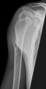
Periosteal ChondromaPeriostitis is an inflammation of the periosteum, and is caused by overuse or repetitive stress to muscles and connective tissue. It is often present with shin splints, a painful condition that tends to affect runners and dancers.[4]
Periosteal Chondromas are rare, benign, chondrogenic tumors. Occur on the surface of long bones, most frequently the proximal humerus. Patients are usually between the ages of 10 and 20 years of age with regional pain secondary to irritation of overlying tendons. Diagnosis is made on Xray. Treatment is usually observation. Marginal surgical excision of the lesion, including the underlying cortex, is indicated in patients with severe and progressive pain.[6]
↑Nahian A, Chauhan PR. Histology, Periosteum and Endosteum. InStatPearls [Internet] 2022 May 8. StatPearls Publishing.Available:https://www.ncbi.nlm.nih.gov/books/NBK557584/ (accessed 9.9.2022)