When refering to evidence in academic writing, you should always try to reference the primary (original) source. That is usually the journal article where the information was first stated. In most cases Physiopedia articles are a secondary source and so should not be used as references. Physiopedia articles are best used to find the original sources of information (see the references list at the bottom of the article).
If you believe that this Physiopedia article is the primary source for the information you are refering to, you can use the button below to access a related citation statement.
Ankle and foot injuries are common musculoskeletal injuries with a high prevalence among professional athletes, but they also occur in recreational sports and as a result of routine daily activities.[1] Typical injuries include sprains, fractures, tears, and inflammation. A good understanding of foot and ankle anatomy is necessary for the proper diagnosis and treatment of injuries.
Structure
The ankle is formed by three bones: the talus, tibia and fibula. The anatomical structure of the foot consists of the hindfoot, midfoot and forefoot. Each part of the foot is composed of several bones.
Ankle Bones
The lower leg and foot constitute the ankle. The following bony elements of the ankle joint are part of this structure:
The talus and calcaneus form the posterior aspect of the foot, which is called the hindfoot. The midfoot (located between the hindfoot and forefoot) is made up of five tarsal bones: the navicular, cuboid, and medial, intermediate, and lateral cuneiforms. The most anterior aspect of the foot including the metatarsals, phalanges, and sesamoid bones is called the forefoot. Each digit, except for the great toe, consists of a metatarsal and three phalanges. The great toe has only two phalanges.
Function
The foot and ankle provide various important functions which include bodyweight support, balance maintenance, shock absorption, response to ground reaction forces, or substitution for hand function in individuals with upper extremity amputation.[2] The foot and ankle have a crucial role in gait and posture and malalignment can cause other problems such as back pain or mobility limitations.[3][4]
Ankle and Foot Articulations
Basic Ankle and Foot Motion
The talocrural joint (TC or sometimes called a tibiotalar joint) is referred to as the ankle joint. The joint is framed laterally and medially by the lateral and the medial malleolus and from the top by the tibia and the talus.[5] Sagittal plane dorsiflexion and plantarflexion are the primary movements in this joint.
The hindfoot has a talus and calcaneus articulation called the subtalar joint (ST, also known as the talocalcaneal joint). Three facets of the talus and of the calcaneus are part of this joint. The main motions are inversion and eversion of the ankle and hindfoot.
The talonavicular and calcaneocuboid joints are known as Chopart's joint (MT, midtarsal or transverse tarsal joint), which is located between the hindfoot and midfoot. This joint allows forefoot rotation. The navicular articulates with all three cuneiform bones distally.[5] In addition to the navicular and cuneiforms bones, the cuboid bone has a distal articulation with the base of the fourth and fifth metatarsal bones.
The tarsometatarsal joint (TMT or Lisfranc's joint) connects the midfoot with the forefoot and originates from the lateral, intermediate and medial cuneiforms articulating with the bases of the three metatarsal bone (1st, 2nd, and 3rd). The small movement that occurs in the joint is described as dorsal and plantarflexion.[6] The bases of the remaining metatarsal bones (4th and 5th) connect with the cuboid bone.[5]
The five rays, metatarsal and corresponding phalanges create the forefoot medial and lateral columns where rays 1,2, and 3 belong to the middle column, rays 4 and 5 to the lateral column. The metatarsophalangeal joints (MTP joints) are the main components of the forefoot.[5] Each toe, except for the great toe, has proximal and distal interphalangeal joints (IP joints). The latter has only one IP joint.
Table 1 includes a summary of the ankle and foot articulations:
Joint
Type of Joint
Plane of Movement
Motion
TC joint
Hinge
Sagittal
Dorsiflexion and Plantarflexion
ST joint
Condyloid
Mainly transverse
Some sagittal
Inversion and Eversion
Dorsiflexion and Plantarflexion
MT joint
TN joint - Ball and socket
CC joint - Modified saddle
Largely in transverse
Some sagittal
Inversion and Eversion
Flexion and Extension
Forefoot rotation
TMT joint
Planar
MTP joint
Condyloid
Sagittal
Some Transverse
Flexion and Extension
Abduction and Adduction
IP joint
Hinge
Sagittal
Flexion and Extension
Ligaments
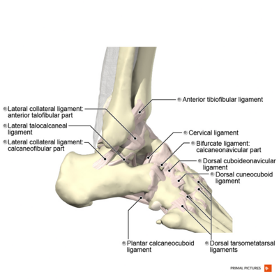
Acute and chronic ankle pain are often associated with either ligament injury or ligament laxity.[7] The ligaments of the ankle consist of the medial ligaments (the deltoid ligament), the lateral ligaments, and the ligaments connecting the distal epiphyses of the tibia and fibula called the tibiofibular syndesmosis.[7]
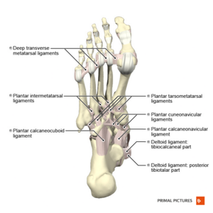
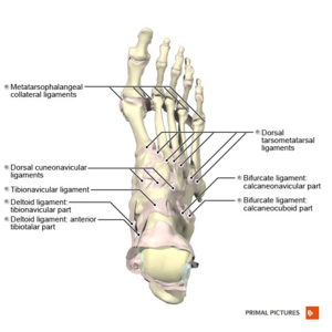
There are 4 main ligaments of the foot: the plantar fascia, the plantar calcaneonavicular ligament (known as a spring ligament), the calcaneocuboid ligament, and the Lisfranc ligaments.
Medial Collateral Ligament (Deltoid Ligament)
The deltoid ligament plays a crucial role in maintaining medial ankle stability by preventing valgus tilt and axial rotation of the talus. [8]
Main components of the medial collateral ligament: 1. Tibionavicular ligament; 2. tibiospring ligament; 3. tibiocalcaneal ligament; 4. deep posterior tibiotalar ligament; 5. spring ligament complex (plantar and superomedial calcaneonavicular ligaments)[9]
There are various descriptions of the medial collateral ligament (MCL), but the definition provided by Milner and Soames[10] is the most commonly accepted. The authors divide the MCL into two layers: superficial and deep. The ligaments consist of bands that are either always present or sometimes present (bands that are always present are shown in italics):[10]
Superficial: tibiospring ligament, tibionavicular ligament, superficial posterior tibiotalar ligament, tibiocalcaneal ligament. It stabilises the medial tibiotalar, subtalar, and talonavicular joints[8]
Deep: deep posterior tibiotalar ligament, deep anterior tibiotalar ligament. It crosses strictly the ankle joint [8]
It is important to mention that the dissection revealed the continuous nature of these ligaments and individual bands were impossible to distinguish.[7]
The MCL has the following characteristics:
multifascicular
originates from the medial malleolus and inserts in the talus
tendon sheath of the posterior tibial muscle covers the posterior and middle part of the deltoid ligament
tendons cover most of the MCL as it inserts at the foot.[7]
Lateral Collateral Ligament
The lateral collateral ligament complex (LCL) is composed of three ligaments: the anterior talofibular, calcaneofibular, and posterior talofibular ligaments.
Anterior talofibular ligament (ATFL):
is made up of two bandsLateral ankle ligament
originates at the anterior margin of the lateral malleolus and inserts on the talus
lays horizontally to the ankle
limits anterior displacement of the talus and ankle plantarflexion
the ligament is relaxed in a neutral ankle position and during plantarflexion, and it is taut in dorsiflexion.
Interconnections between the ATFL, CFL, and PTFL via medial connecting fibres lead to the formation of an anatomical and functional lateral fibulotalocalcaneal ligament complex.[11][12]
Tibiofibular Syndesmosis
The tibiofibular syndesmosis articulation allows medial rotation of the fibula during maximum ankle dorsiflexion and inversion of the fibula during ankle plantarflexion. In addition, it provides stability between the tibia and the fibula at their distal ends.[13] It includes:
Anteroinferior tibiofibular ligament
originates in the anterior tubercle of the tibia and inserts in the anterior margin of the lateral malleolus
made up of several fascicles
when damaged can cause ankle instability.
Posteroinferior tibiofibular ligament
has two independent parts: superficial and deep
superficial component (a true posteroinferior tibiofibular ligament) originates at the posterior edge of the lateral malleolus and is inserted in the posterior tibial tubercle
deep component (transverse ligament) originates in the proximal malleolar fossa and inserts in the posterior edge of the tibia
provides talocrural joint stability and prevents posterior talar translation.
Interosseous tibiofibular ligament
short fibres running between the tibia and fibula
distal continuation of the interosseous membrane
responsible for maintaining ankle stability.[7][14]
A fatty synovial fringe located distal to the ligament insertion is also part of this structure. Its upward and downward movement occurs during ankle movement and alters its placement between the tibia and the fibula. This adipose tissue is considered a cause of a syndesmotic impingement following an ankle sprain.
Ligaments of the Foot
The main function of the short and long plantar ligaments and the calcaneonavicular ligament (spring ligament) is to assist in the passive maintenance of the arch.
originates in the medial calcaneal tubercle and ends at the five metatarsal bones
considered the primary passive plantar tissue that supports the medial longitudinal arch
extremely strong ligament
absorbs the impact of the ground reaction force in running or jumping
maintains and stores energy in the foot
disperses stress throughout the metatarsus
dysfunction of plantar fascia affects the height and shape of the medial longitudinal arch[15]
any morphological or mechanical changes in the plantar fascia will produce lesions.[16]
Plantar calcaneonavicular ligament (SL, spring ligament)Ligaments of the foot-dorsal surface
a passive stabiliser of the plantar arch in the talocalcaneal joint
supports compression forces
the strongest medial ligament
assists with preventing talus medial rotation and plantar flexion
limits navicular dorsiflexion, eversion, and abduction
pes planus may be produced by isolated SL injuries.[17]
Calcaneocuboid ligament
consists of the medial calcaneocuboid ligament (a component of the bifurcate ligament), the dorsolateral calcaneocuboid ligament, the plantar calcaneocuboid ligament (SPL or short plantar ligament), and the long plantar ligament (LPL)
LPL originates on the inferior surface of the calcaneus and inserts into the bases of the second to fourth metatarsals (MTs) and not the distal cuboid.
SPL originates at the anterior tubercle of the calcaneus and attaches to the plantar surface of the cuboid
Supports the medial and lateral longitudinal arches
Can be damaged during inversion injury of the foot.[18]
Ankle fractures mainly affect two groups: young active males and older females. In the first group, fractures occur as a consequence of sporting activities, whereas in the latter group it is an osteoporotic injury.[21]
Injuries to the ankle are painful and cause significant mobility limitations.[22][23]
When compared with healthy individuals and copers, individuals with chronic ankle instability demonstrate impairments in visual memory, inhibitory control, and attention.[24]
Patient-reported outcome measures (PROM) are used in clinical trials to measure the effect of treatment after lower limb fracture.[25]
One third of all ankle fractures can result in tibiotalar joint dislocations.[26]
Chronic ankle ligament injury is difficult to diagnose, but using ultrasound showed high diagnostic accuracy.[27]
Chronic ankle instability may be a result of poorly managed acute ankle ligament injury.[28]
After 1 to 5 years post-acute ligament sprain, the individual can still suffer from pain, functional instability, mechanical instability or recurrent sprain.[29]
Resources
Anatomy of the Ankle Ligaments: A Pictorial Essay[9] - In this pictorial essay, the ligaments around the ankle are grouped, depending on their anatomic orientation, and each of the ankle ligaments is discussed in detail.
References
↑Vuurberg G, Hoorntje A, Wink LM, Van Der Doelen BF, Van Den Bekerom MP, Dekker R, Van Dijk CN, Krips R, Loogman MC, Ridderikhof ML, Smithuis FF. Diagnosis, treatment and prevention of ankle sprains: update of an evidence-based clinical guideline. British journal of sports medicine. 2018 Aug 1;52(15):956-.
↑Houglum PA, Bertotti DB. Brunnstrom's clinical kinesiology. FA Davis; 2012
↑Dalmau-Pastor M, Malagelada F, Calder J, Manzanares MC, Vega J. The lateral ankle ligaments are interconnected: the medial connecting fibres between the anterior talofibular, calcaneofibular and posterior talofibular ligaments. Knee Surg Sports Traumatol Arthrosc. 2020 Jan;28(1):34-39.
↑Yammine K, Jalloul M, Assi C.Distal tibiofibular syndesmosis: A meta-analysis of cadaveric studies. Morphologie, 2021.
↑Hoefnagels EM, Waites MD, Wing ID, Belkoff SM, Swierstra BA. Biomechanical comparison of the interosseous tibiofibular ligament and the anterior tibiofibular ligament. Foot Ankle Int. 2007 May;28(5):602-4.
↑Lavrentiieva O, Filiptsova K. RESTRICTION OF MOBILITY OF THE ANKLE JOINT IN MATURE MEN: RELEVANCE OF THE PROBLEM AND WAYS OF EFFECTIVE REHABILITATION. Collection of scientific papers «SCIENTIA». 2025 Oct 24(October 24, 2025; Florence, Italia):204-9.
↑Al Shdefat S, Arshad Z, Khan R, Haq II, Bhatia M. Patient reported outcome measures in the foot and ankle literature: a systematic review. The Foot. 2025 Sep 29:102209.