Calcaneus
Introduction[edit | edit source]

The calcaneus is one of the 7 articulating bones that make up the tarsus. The calcaneus is located in the hindfoot with the talus and is the largest bone of the foot[1].
- It is commonly referred to as the heel.
- Numerous ligaments and muscles attach to the calcaneus and help with its role in human bipedal biomechanics[2]
- It articulates with the talus superiorly and the cuboid anteriorly and shares a joint space with the talocalcaneonavicular joint.
- The calcaneus transfers most of the body weight from the lower limb to the ground.[3]
Structure[edit | edit source]

The calcaneus is an irregular, roughly box-shaped bone sitting below the talus. Its long axis is orientated along the mid-line of the foot, however, deviates lateral to the mid-line anteriorly. It projects posteriorly to form the core of the heel[1].
The posterior part of the calcaneus is circular, with three facets (superior, middle and inferior):
- The superior facet is separated from the calcaneal tendon by the retrocalcaneal bursa.
- The middle facet provides the attachment site for the calcaneal tendon (Achilles tendon).
- The inferior facet curves anteriorly and is continuous with calcaneal tuberosity on the plantar surface.
The plantar surface of the calcaneal tuberosity projects forward on the plantar surface as a medial (larger) and lateral (smaller) process and at its most anterior projection is the calcaneal tubercle, where the short plantar ligament attaches[1].
On the lateral aspect of the calcaneus is the peroneal tubercle, anterior to the middle of the surface, where the tendons of the fibularis brevis and longus muscles pass above and below respectively.
Protruding anteromedially from upper margin of the medial surface is the sustentaculum tali which supports the more posterior part of the head of the talus.
At its inferior aspect is a groove accommodating the flexor hallucis longus tendon.
Superiorly is the middle talar articular facet for the corresponding middle facet of the head of talus as part of the subtalar joint.
The anterior and posterior facets of the talocalcaneal joint are on the superior surface of the calcaneus. The anterior facet is small and the posterior facet is large. Between these two facets runs a fairly deep sulcus, the calcaneal sulcus, which together with the opposing talar sulcus forms the tarsal sinus.
The anterior surface has a convex articular facet for the cuboid bone[3].

Articulations[edit | edit source]
- Superiorly, the calcaneus articulates with the talus at the subtalar joint, making contact at anterior, middle and posterior facets[1].
- The anterior part of the talocalcaneal joint and the talonavicular joint are collectively known as the talocalcaneonavicular joint.
- Anteriorly, the calcaneus articulates with the cuboid, forming the calcaneocuboid joint.[3]
Attachments[edit | edit source]
Muscular[edit | edit source]

- Triceps surae: gastrocnemius and soleus inserts onto the middle facet of posterior surface of calcaneus through calcaneal/Achilles tendon
- Abductor hallucis: originates from the medial process of calcaneal tuberosity.
- Flexor digitorum brevis: originates from the medial process of calcaneal tuberosity and plantar aponeurosis.
- Quadratus plantae: originates the plantar surface of calcaneus
- Abductor digiti minimi: originates from the medial and lateral process of calcaneal tuberosity
- Extensor digitorum brevis: origin - dorsolateral surface
- Extensor hallucis brevis: origin - dorsal surface, tarsal sinus
Ligamentous[edit | edit source]
- Lateral: Lateral collateral/calcaneofibular ligament of the ankle
- Inferior: Short plantar ligament (at calcaneal tubercle), long plantar ligament (in front of calcaneal tuberosity), plantar aponeurosis (medial process of calcaneal tuberosity proximal to flexor digitorum brevis)
- Superior: Tarsal sinus ligaments, including:
- Cervical ligament
- Talocalcaneal interosseous ligament,
- Lateral, intermediate, and medial roots of the inferior extensor retinaculum
- Bifurcate ligament
- Anterior: plantar calcaneonavicular ligament (anterior margin of the sustentaculum tali of the calcaneus)[3]
Blood Supply[edit | edit source]
- Medial side of the calcaneus is supplied by perforating branches from the posterior tibial artery.
- Lateral side of the calcaneus is supplied by the lateral calcaneal artery (can be a branch of the posterior tibial artery or the peroneal artery).
- Within the bone, there is a watershed area where the medial and lateral intra-osseous arteries anastomose in the midline.[4]
Palpation[edit | edit source]
The posterior aspect of the calcaneus can be palpated directly inferior to the Achilles tendon.
Palpation is also possible around the inferior aspect of the calcaneus particularly the calcaneal tuberosity where the plantarfascia arises.
On the medial aspect the sustentaculum tali is possible to be palpated by palpating the medial malleolus and travelling directly inferior approximately 1-2cm.
Related Pathology[edit | edit source]
- Calcaneal fracture
- Pseudo-tumour of the calcaneus
- Talocalcaneal coalition
- Calcaneonavicular coalition
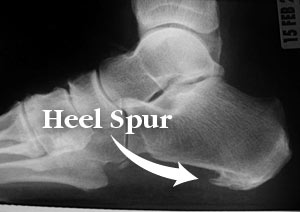
- Calcaneal/heel spur[3]
- Plantarfascia pain: a condition that involves a non-inflammatory structural breakdown of the connective tissue that makes up the plantar aponeurosis or fascia of the foot. Often found in association with heel spurs.[2]
- A Haglund deformity is a bony exostosis that extends from the posterior superior calcaneus where the Achilles attaches. The etiology of the condition is unknown but hypothesized causes are Achilles tendinopathy, high arched feet and other hereditary factors.[2]

References[edit | edit source]
- ↑ 1.0 1.1 1.2 1.3 Palastanga N, Field D, Soames R. Anatomy and human movement. 4th edition. Oxford. Butterworth-Heinemann; 2002. p242.
- ↑ 2.0 2.1 2.2 Gupton M, Terreberry RR. Anatomy, bony pelvis and lower limb, calacaneous. InStatPearls [Internet] 2018 Dec 6. StatPearls Publishing. Available from:https://www.statpearls.com/kb/viewarticle/18764 (last accessed 12.3.2020)
- ↑ 3.0 3.1 3.2 3.3 3.4 Radiopedia. Calcaneus. Available from:https://radiopaedia.org/articles/calcaneus?lang=gb (accessed 12/03/2020).
- ↑ Razik A, Harris M, Trompeter A. Calcaneal fractures: Where are we now?. Strategies in Trauma and Limb Reconstruction. 2018 Apr 1;13(1):1-1.

