Navicular
Original Editor - Alex Benham
Top Contributors - Alex Benham, Lucinda hampton, Fasuba Ayobami and Kim Jackson
Introduction[edit | edit source]
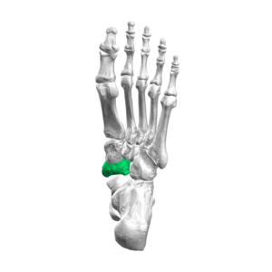
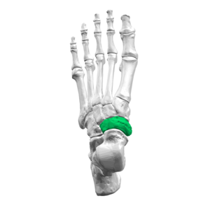
The navicular bone is one of the seven bones which make up the tarsus of the Ankle and Foot. It is located on the medial aspect of the foot, next to the cuboid bone, anterior to the head of the talus and posterior to the cuneiform bones. It is one of the five bones of the midfoot[1]. It is the last bone of the foot to ossify fully .[2]
Structure[edit | edit source]
The navicular is a small irregular bone with its shape being described as pyriform[3]. Its posterior surface is concave and there are two faint ridges anteriorly to correspond with the articulation with the three cuneiform bones. There is a large protuberance on the inferomedial aspect of the navicular bone called the navicular tuberosity [1] which is often simple to palpate in the area directly anteroinferior to the medial malleolus.
Function[edit | edit source]
The navicular is sometimes referred to as the keystone of the medial longitudinal arch of the foot, corresponding to its location at the peak of the arch and its role in maintaining the arch of the foot.
Articulations[edit | edit source]
The acetabulum pedis is the common name for the talocalcaneonavicular joint and forms the subtalar articular complex along with the posterior talocalcaneal joint. It has some of the morphological characteristics of a ball and socket joint which led to its name[4]. The skeletal structures included are the posterior articular surface of the navicular bone, and the anterior and middle articular surface of the calcaneus that articulate with the head and the anteromedial surface of the talus. These bones are stabilized by the plantar calcaneonavicular ligament (spring ligament) on the medial aspect and the lateral calcaneonavicular ligament (a component of the bifurcate ligament) on the lateral aspect.[4]
Muscle and Ligamentous Attachments[edit | edit source]
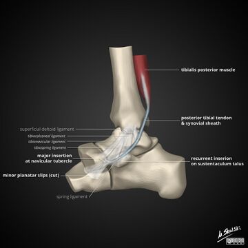
The only muscle attachment on the navicular is the tendon of the tibialis posterior. This a muscle located within the deep compartment of the posterior aspect of the tibia and the tendon extends inferiorly around the medial malleolus to insert on the navicular tuberosity. [3]
The dorsal cuneonavicular ligament and plantar cuneonavicular ligament are two ligaments that join each cuneiform bone to the navicular. The first/medial cuneiform bone also is joined to the navicular by the medial cuneonavicular ligament.[2]
Arterial Supply[edit | edit source]
A branch of dorsalis pedis artery gives off three to five smaller branches which supply the navicular from the medial side. On the lateral aspect of the bone, small branches arise from the posterior tibial artery. Branches of the medial plantar artery also supply the plantar surface of the bone.
In the central navicular there is a region of watershed blood supply which predisposes the bone to stress fractures.
Clinical relevance[edit | edit source]
There are several clinical conditions which involve the navicular directly.
- The navicular is often subject to developing stress fractures and can lead to Navicular stress syndrome.
- Müller Weiss syndrome is a spontaneous adult onset osteonecrosis of the navicular bone which may be picked up radiographically. In children, there is also a similar condition, although distinct from Müllers Weiss syndrome called Koehlers syndrome[5].
One anatomical variant is the Accessory Navicular Bone.
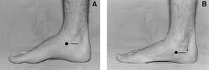
The navicular is also used clinically within the Navicular Drop Test which may be used as a measure of foot pronation.
References[edit | edit source]
- ↑ 1.0 1.1 Soames RW. Anatomy and Human Movement E-Book: Structure and function. Elsevier Health Sciences; 2018 Aug 22.
- ↑ 2.0 2.1 Radiopedia Navicular Available:https://radiopaedia.org/articles/navicular (accessed 8.6.2022)
- ↑ 3.0 3.1 Golano P, Fariñas O, Sáenz I. The anatomy of the navicular and periarticular structures. Foot and ankle clinics. 2004 Mar 1;9(1):1-23.
- ↑ 4.0 4.1 Epeldegui T, Delgado E. Acetabulum pedis. Part I: Talocalcaneonavicular joint socket in normal foot. Journal of pediatric orthopedics. Part B. 1995;4(1):1-0.
- ↑ Samim M, Moukaddam HA, Smitaman E. Imaging of Mueller-Weiss syndrome: a review of clinical presentations and imaging spectrum. American Journal of Roentgenology. 2016 Aug;207(2):W8-18.
- ↑ Viren Kariya. Navicular Bone. Available from: https://www.youtube.com/watch?v=xf3ZdsyGaiI [Last accessed 21/09/2020]


