When refering to evidence in academic writing, you should always try to reference the primary (original) source. That is usually the journal article where the information was first stated. In most cases Physiopedia articles are a secondary source and so should not be used as references. Physiopedia articles are best used to find the original sources of information (see the references list at the bottom of the article).
If you believe that this Physiopedia article is the primary source for the information you are refering to, you can use the button below to access a related citation statement.
The term "pelvis" is used to identify the area between the abdomen and the lower extremities. It can be divided into the greater pelvis and the lesser pelvis.[1] The pelvis consists of the sacrum, the coccyx, the ischium, the ilium, and the pubis.[2][3] The structure of the pelvis supports the contents of the abdomen while also helping to transfer the weight from the spine to the lower limbs.[4] During gait, the joints within the pelvis work together to decrease the amount of force transferred from the ground and lower extremities to the spine and upper extremities.[4]
The inlet to the pelvic canal is at the level of the sacral promontory and superior aspect of the pubic bones.
The outlet is formed by the pubic arch, ischial spines, sacrotuberous ligaments, and the coccyx.
The enclosed space between the inlet and outlet is called the true pelvis, with the plane of the inlet being at right angles to the plane of the outlet.
The female true pelvis differs from the male in being shallower, having straighter sides, a wider angle between the pubic rami at the symphysis, and a proportionately larger pelvic outlet.
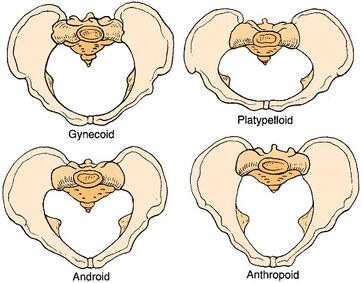
The shape of the female bony pelvis can be classified into four broad categories: gynecoid, anthropoid, android, and platypelloid[3].
Gynecoid/genuine pelvis
the brim is round, wider, and both ischial spines are less prominent this allows easy baby delivery.
Android pelvis
like a heart-shaped brim with a narrow pelvic cavity than presented in gynecoid one, get narrower at the supra-pubic arch, and both ischial spines are prominent. During delivery, she needs to take an active role.
Anthropoid pelvis
oval brim, slightly narrower pelvic cavity than above with larger outlet diameter.
Platypelloid pelvis
shallow pelvic cavity, and decreased antero-posterior diameter, broad outlet, during labour there will be difficult for the baby to enter the pelvis at the beginning.
In women, the pelvis houses the uterus, tubes, ovaries and vagina.
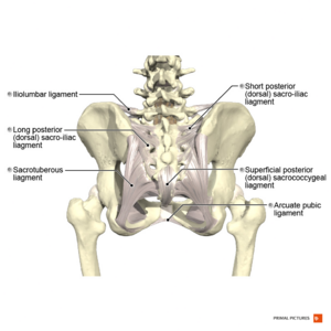
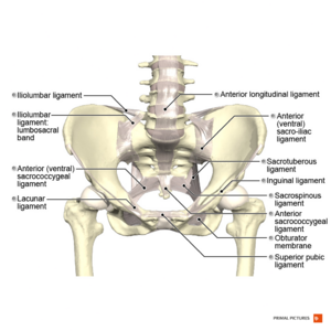
Superior pubic ligament - runs between pubic tubercles[17]
Inferior pubic ligament (aka arcuate pubic ligament) - runs between inferior pubic rami and blends with the fibrocartilaginous disc of the pubic symphysis[17]
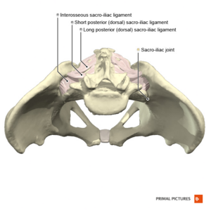
The upper portion (short posterior sacroiliac ligament) - from 1st and 2nd transverse tubercles of the sacrum to the tuberosity of ilium
The lower portion (long dorsal posterior sacroiliac ligament) - from 3rd transverse tubercle of the sacrum to the posterior superior iliac spine (PSIS)
Interosseous sacroiliac ligament- lies deep to posterior SI ligament and runs between the tuberosities of the sacrum and ilium[21]
Iliolumbar Ligament
from the tip of the transverse process of L5 to posterior aspect of the inner lip of iliac crest[7]
2 main bands attach to the pelvis
Lower band - attaches to the base of sacrum, contiguous with anterior sacroiliac ligament
Upper band - attaches to iliac crest anterior to the sacro-iliac articulation, contiguous with lumbodorsal fascia
Deep portion - from the inside sacral canal at the 5th sacral segment to the dorsal surface of the coccyx; continuation of the posterior longitudinal ligament of the spine
Superficial portion - from free margin of sacral hiatus to dorsal surface of the coccyx; corresponds with the ligamentum flavum of the spine
Lateral sacrococcygeal ligament
from the inferior lateral angle of the sacrum to the transverse process of the 1st coccygeal vertebra
Muscles of the Pelvis
There are 36 muscles that attach to the sacrum or innominates. The purpose of these muscles is primarily to provide stability to the joint not to produce movement.[22]
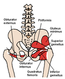
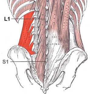
Muscles that attach to the sacrum or innominates are:
The urogenital diaphragm, also called the triangular ligament, is a strong, muscular membrane that occupies the area between the symphysis pubis and ischial tuberosities and stretches across the triangular anterior portion of the pelvic outlet.[32] The urogenital diaphragm is external and inferior to the pelvic diaphragm.
The pelvic diaphragm is a wide but thin muscular layer of tissue that forms the inferior border of the abdominopelvic cavity. Composed of a broad, funnel-shaped sling of fascia and muscle, it extends from the symphysis pubis to the coccyx and from one lateral sidewall to the other.
Levator Ani Muscle (pubococcygeus aka pubovisceral, pubovaginalis, puboanalis, puborectalis, iliococcygeus)
Perineal Body[31] - a fibromuscular structure located between the vagina/testicles and the anus, attaching to the sides of the ischiopubis rami by the deep transverse perineal muscle. It is known as the central tendon of the pelvis because many pelvic floor structures intersect with the perineum at this structure.
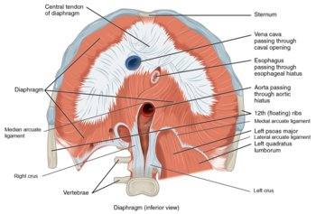
Diaphragm - Inferior view.Contributes to the health of the pelvic girdle by "pistoning" motion in produces in abdominal "canister"
Peripherally
Attaches to 11th & 12th ribs and the costal cartilages 7 to 10, xiphoid process and lumbar vertebrae
Centrally via R and L crura of the central tendon
L1-3 intervertebral discs
Physiological sphincter of the oesophagus
The inferior surface of the pericardium
References
↑ 1.01.1Madsen C, Gordon DA. Anatomy, Neuroanatomy, and Biomechanics of the Pelvis. InPelvic Floor Dysfunction and Pelvic Surgery in the Elderly 2017 (pp. 3-26). Springer, New York, NY.
↑ 10.010.110.2Kiapour A, Joukar A, Elgafy H, Erbulut DU, Agarwal AK, Goel VK. Biomechanics of the sacroiliac joint: anatomy, function, biomechanics, sexual dimorphism, and causes of pain. International journal of spine surgery. 2020 Feb 1;14(s1):S3-13.
↑Wong M, Kiel J. Anatomy, Abdomen and Pelvis, Sacroiliac Joint. InStatPearls [Internet] 2019 Feb 6. StatPearls Publishing.
↑Waldman LE, Maluli I, Moon CN, Skalski M, Matcuk GR. Sacroiliac joint dysfunction: anatomy, pathophysiology, differential diagnosis, and treatment approaches. Skeletal Radiology. 2025 Jun;54(6):1195-213.
↑ 21.021.121.2Poilliot AJ, Zwirner J, Doyle T, Hammer N. A Systematic Review of the Normal Sacroiliac Joint Anatomy and Adjacent Tissues for Pain Physicians. Pain physician. 2019 Jul;22(4):E247-74.
↑ 23.023.1Buckthorpe M, Stride M, Della Villa F. Assessing and treating gluteus maximus weakness–a clinical commentary. International journal of sports physical therapy. 2019 Jul;14(4):655.
↑Klahsen O. Sacroiliac joint pain: Where are we now?. SURG Journal. 2019 Jun 20;11.
↑ 25.025.125.225.325.425.5Lifshitz L, Sela SB, Gal N, Martin R, Klar MF. Iliopsoas the Hidden Muscle: Anatomy, Diagnosis, and Treatment. Current Sports Medicine Reports. 2020 Jun 1;19(6):235-43.
↑KenHub Learn Human Anatomy. Iliopsoas Muscle: Action / Function, Anatomy & Innervation - Human Anatomy | Kenhub. Available from https://www.youtube.com/watch?v=cHWjpQ06-cE (last accessed 14 November 2o20)
↑Poor AE, Roedl JB, Zoga AC, Meyers WC. Core muscle injuries in athletes. Current sports medicine reports. 2018 Feb 1;17(2):54-8.
↑Riff AJ, Movassaghi K, Beck EC, Neal WH, Inoue N, Coleman SH, Nho SJ. Surface mapping of the musculotendinous attachments at the pubic symphysis in cadaveric specimens: implications for the treatment of core muscle injury. Arthroscopy: The Journal of Arthroscopic & Related Surgery. 2019 Aug 1;35(8):2358-64.












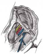

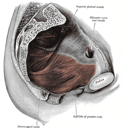 Pelvic Floor
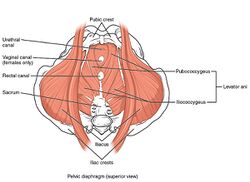 Pelvic Floor Muscles
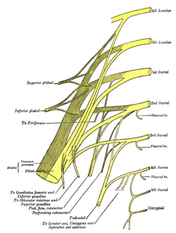 Sacral and Coccygeal Plexus