When refering to evidence in academic writing, you should always try to reference the primary (original) source. That is usually the journal article where the information was first stated. In most cases Physiopedia articles are a secondary source and so should not be used as references. Physiopedia articles are best used to find the original sources of information (see the references list at the bottom of the article).
If you believe that this Physiopedia article is the primary source for the information you are refering to, you can use the button below to access a related citation statement.
The pelvic floor is a dome-shaped muscular sheet separating the pelvic cavity above from the perineal region below. This cavity encloses the pelvic viscera - bladder, intestines, and uterus(in females).[1]
The main function of the pelvic floor muscles are:
To support the abdominal and pelvic viscera
To maintain the continence of urine and faeces
Allows voiding, defaecation, sexual activity, and childbirth[2]
Osteology, Ligaments and Fascia
Osteology
The Pelvis Consists Of
Two innominate bones and the sacrum to which coccyx is attached.
The inlet to the pelvic canal is at the level of the sacral promontory and superior aspect of the pubic bones.
The outlet is formed by the pubic arch, ischial spines, sacrotuberous ligaments, and the coccyx.
The enclosed space between the inlet and outlet is called the true pelvis, with the plane of the inlet being at right angles to the plane of the outlet.
The female true pelvis differs from the male in being shallower, having straighter sides, a wider angle between the pubic rami at the symphysis, and a proportionately larger pelvic outlet.
The shape of the female bony pelvis can be classified into four broad categories: gynecoid, anthropoid, android, and platypelloid[2].
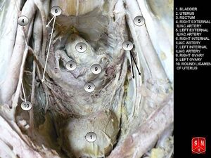
In women, the pelvis houses the uterus, tubes, ovaries and vagina.
Gynecoid/ genuine pelvis, the brim is round, more wider, and both ischial spines are less prominent this allows easy baby delivery.
Android pelvis, like a heart-shaped brim with narrow pelvic cavity than presented in gynecoid one, get narrower at the supra-pubic arch, and both ischial spines are prominent. During delivery, she needs to take an active role.
Anthropoid pelvis, oval brim, slightly narrower pelvic cavity than above with larger outlet diameter.
Platypelloid, shallow pelvic cavity, and decreased antero-posterior diameter, broad outlet, during labor there will be difficult for the baby to enter the pelvis at the beginning.
Female Pelvic CavityLigaments and Pelvic Fascia[3][4]
Ligaments of the Pelvis
Iliolumbar ligament - from the tip of the transverse process of L5 to posterior aspect of the inner lip of iliac crest; strengthens the lumbo-sacral joint.
Sacrospinous ligament - from the ischial spine to lateral margins of the sacrum
Sacroiliac Ligaments
Ventral/Anterior sacroiliac ligament- from antero-lateral aspect of the sacrum to auricular surface of the ilium
Dorsal/Posterior sacroiliac ligament
The upper portion (short posterior sacroiliac ligament) - from 1st and 2nd transverse tubercles of the sacrum to the tuberosity of ilium
The lower portion (long posterior sacroiliac ligament) - from 3rd transverse tubercle of the sacrum to the posterior superior iliac spine (PSIS)
Interosseous sacroiliac ligament- lies deep to posterior SI ligament and runs between the tuberosities of the sacrum and ilium
Sacrococcygeal Ligaments
Ventral/Anterior sacrococcygeal ligament -from the anterior surface of sacrum to the front of the coccyx; continuation of the anterior longitudinal ligament of the spine
Dorsal sacrococcygeal ligament
Deep portion - from the inside sacral canal at the 5th sacral segment to the dorsal surface of the coccyx; continuation of the posterior longitudinal ligament of the spine
Superficial portion - from free margin of sacral hiatus to dorsal surface of the coccyx; corresponds with the ligamentum flavum of the spine
Lateral sacrococcygeal ligament - from the inferior lateral angle of the sacrum to the transverse process of the 1st coccygeal vertebra
Pubic Symphysis Ligaments
Superior pubic ligament - runs between pubic tubercles
Inferior pubic ligament (aka arcuate public ligament) - runs between inferior pubic rami and blends with the fibrocartilaginous disc of the pubic symphysis
Anterior pubic ligament
Posterior pubic ligament - a membranous structure which blends with periosteum
Endopelvic Fascia - Ligaments
Pubovesical ligaments - attach bladder to pubic symphysis
Sacrogenital - these are thickenings of the fascia, not specific ligaments
Uterosacral ligaments - attach upper vagina, upper portion of cervix and uterus to the 3rd sacral vertebra
Cardinal ligaments (aka Mackenrodt's) - attach upper vagina, cervix and uterus to the sidewalls of the pelvis
Broad ligaments - attach the uterus to the medial aspect of the ilium
Urachus ligament - attaches bladder to the umbilicus (formed from the remnants of the umbilical vein)
Penile Suspensory Ligaments
Penile Suspensory ligament - attaches to the anterior aspect of the interpubic disc and divides in two to sling around the penis
Fundiform ligament - extends from inferior linea alba and divides to wrap around the penis
Parietal pelvic fascia - lines the internal surface of the muscles of the pelvic floor and walls
Visceral pelvic fascia - invests each pelvic organ
The parietal and visceral fascia is continuous where organs penetrate the pelvic floor. They thicken to form the arcus tendinous, arches of fascia running adjacent to the viscera from the pubis to the sacrum.
Endopelvic fascia- a meshwork of smooth muscle, ligaments, blood vessels, and connective tissue lying between the parietal and visceral fascia, sometimes condensing to form fibrous fascial septa which separate and suspend the organs.
Anatomists use the name subserous fascia whereas surgeons refer to this layer of retroperitoneal fascia as endopelvic fascia.
Hypogastric sheath - separates retropubic space from presacral space; a conduit for vessels and nerves
Transverse cervical (cardinal) ligaments - part of hypogastric sheath; runs from lateral pelvic wall to uterine cervix and vagina; transmits uterine artery and provides passive support for the uterus
The urogenital diaphragm, also called the triangular ligament, is a strong, muscular membrane that occupies the area between the symphysis pubis and ischial tuberosities and stretches across the triangular anterior portion of the pelvic outlet. The urogenital diaphragm is external and inferior to the pelvic diaphragm.
Urethral sphincter (sphincter urethrae)
Compressor urethrae
Sphincter urethral vaginalis
Deep transverse perineal
Perineal membrane
Layer Three - Pelvic Diaphragm
The pelvic diaphragm is a wide but thin muscular layer of tissue that forms the inferior border of the abdominopelvic cavity. Composed of a broad, funnel-shaped sling of fascia and muscle, it extends from the symphysis pubis to the coccyx and from one lateral sidewall to the other.
Levator Ani Muscle (pubococcygeus aka pubovisceral, pubovaginalis, puboanalis, puborectalis, iliococcygeus)
Perineal Body - a fibromuscular structure located between the vagina/testicles and the anus, attaching to the sides of the ischiopubis rami by the deep transverse perineal muscle. It is known as the central tendon of the pelvis because many pelvic floor structures intersect with the perineum at this structure.
↑Anatomy Zone. Pelvic Floor Part 2 - Perineal Membrane and Deep Perineal Pouch - 3D Anatomy Tutorial. Available from: http://www.youtube.com/watch?v=q0Ax3rLFc6M[last accessed 11/04/14]