Pyramidalis Muscle
Original Editor - Khloud Shreif
Top Contributors - Khloud Shreif, Lucinda hampton, Kim Jackson, Oyemi Sillo and Leana Louw
Description[edit | edit source]
The paired pyramidalis muscles are small triangular-shaped muscles that lie between the anterior surface of the rectus abdominus and the posterior surface of the rectus sheath. The defined function of pyramidalis muscle is vague, but it is thought to tense the linea alba.[1]
The muscles are not always present (absent in ~20% of cases ), or are often unilateral, and vary greatly in size.[2] [3]
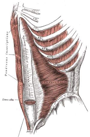
Image 1: Muscles at the front of the abdomen, showing the pyramidalis at the bottom centre.
Gross Anatomy[edit | edit source]
The pyramidalis muscle has its origin from the bony pelvis, where it is attached to the pubic symphysis and pubic crest. The fibres run superiorly and medially to insert into the linea alba at a point midway between umbilicus and pubis.
It lies within the rectus sheath, anterior to the rectus abdominis muscle.[3]
Nerve[edit | edit source]
The nerve supply to the muscle is from ventral part of nerve T12 although this has been reported as being quite variable.[3]
Artery[edit | edit source]
The main arterial supply from the inferior epigastric supply and the deep circumflex iliac artery to a lesser extent.
Function[edit | edit source]
When they contract tense the linea alba, contract with other abdominal muscle to increase positive abdominal pressure.[4]
Many authors have related the pyramidalis muscle with erection of the penis or assumption of upright posture in humans.[1]
Clinical relevance[edit | edit source]
- It is used as a surgical landmark as in cesarean section to define the midline of the linea alba[5].
- After long-term cryopreservation, pyramidalis muscle specimens have been used as a source of striated muscle stem cells for the treatment of post-prostatectomy stress urinary incontinence.[1]
- can be used for microsurgical transfer to treat small recalcitrant wounds in the foot/ankle region because of reduced donor site morbidity as compared to traditional free flaps.[1]
- There is a strong direct connection between the pyramidalis muscle and adductor longus tendon via the anterior pubic ligament, which introduces the new anatomical concept of the pyramidalis–anterior pubic ligament–adductor longus complex (PLAC). [6]
Image 2: In muscular individuals the linea alba can be seen on the skin, forming the depression between the left and right halves of a “six pack”
Vestigal Muscle[edit | edit source]
The pyramidalis muscle is a vestigal muscle. Vestigial muscles in human beings are those muscles which are tendinous in larger part or reduced in size compared to the homologous muscles in other species or is frequently absent within or between populations.
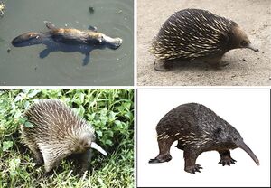
Phylogenetically, pyramidalis is linked to the pouch inside monotremes such as hedgehog and the platypus and marsupials such as the koala or kangaroo.[1]
Image 3: Four of the five extant monotreme species: platypus (top-left), short-beaked echidna (top-right), western long-beaked echidna (bottom-left), and eastern long-beaked echidna (bottom-right)
Resources[edit | edit source]
References[edit | edit source]
- ↑ 1.0 1.1 1.2 1.3 1.4 Das SS, Saluja S, Vasudeva N. Biometrics of pyramidalis muscle and its clinical importance. Journal of clinical and diagnostic research: JCDR. 2017 Feb;11(2):AC05.Available: https://www.ncbi.nlm.nih.gov/pmc/articles/PMC5376864/ (accessed 20.12.2021)
- ↑ Lovering RM, Anderson LD. Architecture and fiber type of the pyramidalis muscle. Anatomical science international. 2008 Dec;83(4):294-7. Available: https://www.ncbi.nlm.nih.gov/pmc/articles/PMC3531545/(accessed 20.12.20210
- ↑ 3.0 3.1 3.2 Radiopedia Pyramidalis Available:https://radiopaedia.org/articles/pyramidalis-muscle (accessed 20.12.2021)
- ↑ https://www.kenhub.com/en/library/anatomy/pyramidalis-muscle
- ↑ Das SS, Saluja S, Vasudeva N. Biometrics of Pyramidalis Muscle and its Clinical Importance. Journal of clinical and diagnostic research: JCDR. 2017 Feb;11(2):AC05.
- ↑ Schilders E, Bharam S, Golan E, Dimitrakopoulou A, Mitchell A, Spaepen M, Beggs C, Cooke C, Holmich P. The pyramidalis–anterior pubic ligament–adductor longus complex (PLAC) and its role with adductor injuries: a new anatomical concept. Knee Surgery, Sports Traumatology, Arthroscopy. 2017 Dec 1;25(12):3969-77.
- ↑ Kenhub - Learn Human Anatomy. Pyramidalis Muscle Overview and Function- Human Anatomy | Kenhub. Available from: http://www.youtube.com/watch?v=NR037gEOsWg[last accessed 17/5/2020]


