Semimembranosus
Original Editor - George Prudden
Top Contributors - George Prudden, Riccardo Ugrin, Kim Jackson, Evan Thomas, WikiSysop, Vidya Acharya and Rucha Gadgil
Description[edit | edit source]
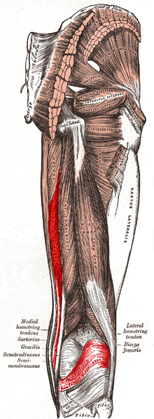
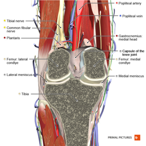
Semimembranosis is one of a group of muscles called the Hamstrings. It is located on the posteromedial side of the thigh deep to Semitendinosus and medial to the Biceps Femoris. Its origin is the ischial tuberosity on the inferior pelvis and it has a complex distal insertion connected to Popliteus Muscle, to the Medial Meniscus, to the Medial Collateral Ligament and to the Tibia . In the lower part of the thigh, semitendinosus and semimembranosus together form the upper medial boundary of the popliteal fossa.[1]
It's primary action is knee flexion,knee internal rotation and hip extension [2][1] . It also plays a main role providing knee stability. Infact the semimembranosus distal tendon is a major component of the posteromedial corner of the knee.
Origin[edit | edit source]
A strong membranous tendon attaches to the upper lateral facet on the rough part of the ischial tuberosity.[2]
Insertion[edit | edit source]
The anatomy of the distal insertion of the semimembranosus can be described as the distal semimembranosus complex: infact six different semimembranosus insertions can be identified. Anatomic dissection revealed six insertions of the distal semimembranosus tendon: direct arm, anterior arm, posterior oblique ligament extension, oblique popliteal ligament extension (capsular arm), distal tibial expansion (popliteus aponeurosis), and meniscal arm[3]:
- The direct arm attaches in a groove at the posteromedial aspect of the tibia;
- The anterior arm courses more anteriorly and attaches to the tibia deep to the medial collateral ligament.
- A broad component extends into the oblique popliteal ligament, which courses along the posterior capsule of the knee. Some authors suggest more precise nomenclature renaming this ligament as oblique popliteal tendon/expansion because its function[4][5].
- The insertion that is directed towards the components of the posterior oblique ligament has been described as the capsular arm. The entire posterior oblique ligament is inseparable from the capsule
- A short meniscal arm attaches to the meniscotibial ligament (coronary ligament) of the posterior horn of the medial meniscus.
- A distal arm also extends over the popliteus muscle and its aponeurosis. Is a fascia-like structure extending from the inferior aspect of the direct arm over the popliteus muscle.
- Additional connections to the posterior capsule and lateral meniscus have been reported. Infact a tendinous branch of the semimembranosus inserting into the posterior horn of the lateral meniscus was found in 43.2% of the knees.[6]
A bursae separate the muscle from the medial head of the tibia and the medial head of the Gastrocnemius[2]
Nerve[edit | edit source]
Tibial division of the Sciatic Nerve (root value L5, S1 and 2).[2]
Nerve supply for the skin covering the muscle is L2.[2]
Artery[edit | edit source]
Branches from the internal iliac, popliteal, and profunda femoris arteries.[1]
Function[edit | edit source]
The functions of distal semimembranosus complex can be summarized as following:
- Hamstrings generally play a role in hip extension and knee flexion. Otherwise the semimembranosus alone is an hip extensor and a knee flexor/intrarotator.
- In knee extension, the semimembranosus tendon prevents valgus, whereas in knee flexion it prevents external rotation.
- The distal semimembranosus complex stabilizes the posterior capsule through the oblique popliteal ligament. Infact on knee flexion during running, cutting and pivoting, the capsular arm of the semimembranosus actively tightens the posterior oblique ligament providing dynamic stability. Moreover the oblique popliteal ligament plays significant roles in preventing both excessive external knee rotation and hyperextension and in strengthening the stability of the knee[8].
- The semimembranosus acts synergistically with the popliteus muscle through the fibrous extension towards this muscle.
- The distal semimembranosus complex actively pulls the posterior horn of the medial meniscus, protecting it from being crushed between the medial femoral condyle and the posterior medial tibial plateau during knee flexion. Furthermore it provides an element of posterior traction over the posterior horn of the lateral meniscus, along with the structures forming the stabilizing mechanism of the posterolateral aspect of the knee[6].
- Hip extension
- Agonists: Gluteus Maximus, semitendinosus, Biceps Femoris (long head), and Adductor Magnus (posterior part)
- Antagonists: Psoas Major and Iliacus
- Knee flexion
- Agonists: biceps femoris (long head), biceps femoris (short head), and semitendinosus. However the dynamic synergy of the semimembranosus complex and the biceps femoris also provides to pulling the posterior horns of the medial and lateral menisci.[9]
- Antagonists: Vastus Lateralis, Vastus Medialis, Vastus Intermedius, and Rectus Femoris, Gracilis, Sartorius, Popliteus, gastrocnemius, and plantaris assist with flexion of the knee.
- Knee internal rotation of the knee when it is flexed
- Agonists: popliteus and semitendinosus
- Antagonists: biceps femoris (long head) and biceps femoris (short head).
- Sartorius and Gracilis assist with internal rotation of the knee when the knee is flexed.
Functional movements[edit | edit source]
- Stand from sitting
- Walking upstairs
- Standing jump forwards
- Standing jump upwards
Clinical relevance[edit | edit source]
Hamstring Syndrome[edit | edit source]
This pathology commonly affects athletes who present with localised pain near the ischial tuberosity. The pathophysiology is thought to be that of an insertional tendopathy at the ischium but there may also be involvement of sciatic nerve compression. The pain in hamstring syndrome radiates down the posterior thigh or popliteal region and is exacerbated when the hamstrings are on tension. This is often seen in sprinters or hurdlers. On examination there is exquisite tenderness over the ischial tuberosity and percussion in that region may reproduce the sciatic distribution of pain. Treatment involves rest, anti-inflammatory agents and steroid injections. [1]
For more information please see Proximal Hamstring Tendinopathy page
Baker's cyst[edit | edit source]
The bursae that separates the muscle from the medial head of the tibia and the medial head of the Gastrocnemius may at times become enlarged with distended fluid. This swelling is termed 'Baker's cyst' (described by Morrant Baker in the 19th century as a cystic mass in the popliteal fossae of children).[1]
For more information please see Baker's Cyst page.
Assessment[edit | edit source]
Palpation[edit | edit source]
Semimembranous lies deep to semitendinosus and is difficult to palpate, but can be felt easier when the knee is flexed.[1]
Strenght[edit | edit source]
The evaluation of strenght is made with patient lying prone, knee extended. Patient actively flexes the knee through range while the terapist applies resistance through the distal tibia and fibula in a direction opposite to flexion. To be more specific on the semimembranosus it can be useful combine the knee bend maintaning the tibia in internal rotation[10].
Knee flexion assessment. [11]
Please see Manual Muscle Testing: Knee Flexion for more information.
Length[edit | edit source]
Straight leg raise[edit | edit source]
The patient is positioned in supine with the hip and knee extended. One hand is placed over the anterior thigh to maintain full extension throughout the movement. The hip is flexed until firm muscular resistance to further motion is felt. To emphasise the stretch of the semimembranosus it is possible to add a little bit of external rotation of the hip and adduction. A goniometer can then be aligned as follows[12]:
- Stationary arm: Lateral midline of trunk
- Axis: Greater trochanter of the femur
- Moving arm: Lateral epicondyle of femur
Maximal hip flexion can then be documented.
Knee extension test[edit | edit source]
The patient is positioned in supine with the hip to 90o and the contralateral limb should be placed on a supporting surface with the knee extended. The knee is then extended through full range of motion whilst the hip is maintained in 90o flexio[12]n.
- Active test: the patient performs active knee extension of the knee until myoclonus is observed in the hamstring
- Passive test: the knee is passively extended until firm muscular resistance to further motion is felt
A goniometer can then be aligned as follows:
- Stationary arm: Greater trochanter of femur
- Axis: Lateral epicondyle of femur
- Moving arm: Lateral malleolus
Maximal knee extension can be documented.
Treatment[edit | edit source]
The hamstring strain is a common injury in athletes. The physiotherapic intervention must be comprehensive both with physical therapies and exercises of strenghtening/stretching.
For more information please see the Hamstring Strain page.
| Hamstring Strengthening Exercises & Stretches [13] | 4 Stages for Hamstring Exercises [14] |
Resources[edit | edit source]
- ↑ 1.0 1.1 1.2 1.3 1.4 1.5 Anatomy.tv | 3D Human Anatomy | Primal Pictures [Internet]. Anatomy.tv. 2018 [cited 30 April 2018]. Available from: http://www.anatomy.tv/
- ↑ 2.0 2.1 2.2 2.3 2.4 Palastanga N, Field D, Soames R. Anatomy and human movement: structure and function. Elsevier Health Sciences; 2012.
- ↑ De Maeseneer M, Shahabpour M, Lenchik L, Milants A, De Ridder F, De Mey J, Cattrysse E. Distal insertions of the semimembranosus tendon: MR imaging with anatomic correlation. Skeletal Radiol. 2014 Jun;43(6):781-91
- ↑ Benninger B, Delamarter T. Distal semimembranosus muscle-tendon-unit review: morphology, accurate terminology, and clinical relevance. Folia Morphol (Warsz). 2013 Feb;72(1):1-9
- ↑ Benninger B, Delamarter T. The "oblique popliteal ligament": a macro- and microanalysis to determine if it is a ligament or a tendon. Anat Res Int. (2012); 2012:151342.
- ↑ 6.0 6.1 Kim YC, Yoo WK, Chung IH, Seo JS, Tanaka S. Tendinous insertion of semimembranosus muscle into the lateral meniscus. Surg Radiol Anat. 1997;19(6):365-9.
- ↑ Anatomy Of The Semimembranosus Muscle - Everything You Need To Know - Dr. Nabil Ebraheim. Available from https://www.youtube.com/watch?v=eEguukkdAPo
- ↑ Wu XD, Yu JH, Zou T, Wang W, LaPrade RF, Huang W, Sun SQ. Anatomical Characteristics and Biomechanical Properties of the Oblique Popliteal Ligament. Sci Rep. 2017 Feb 16;7:42698.
- ↑ Beltran J, Matityahu A, Hwang K, Jbara M, Maimon R, Padron M, Mota J, Beltran L, Sundaram M. The distal semimembranosus complex: normal MR anatomy, variants, biomechanics and pathology. Skeletal Radiol. 2003 Aug;32(8):435-45
- ↑ Hislop H, Avers D, Brown M. Daniels and Worthingham's muscle Testing-E-Book: Techniques of manual examination and performance testing. Elsevier Health Sciences; 2013 Sep 2
- ↑ Knee Flexion MMT. Available from:https://www.youtube.com/watch?v=Cyej-jC-bCg
- ↑ 12.0 12.1 Reese NB, Bandy WD. Joint Range of Motion and Muscle Length Testing-E-Book. Elsevier Health Sciences; 2016 Mar 31.
- ↑ Hamstring Strengthening Exercises & Stretches - Ask Doctor Jo. Available from: https://www.youtube.com/watch?v=fPCTajPbux4
- ↑ Proximal Hamstring Tendinopathy Rehab (4 Stages). Available from:https://www.youtube.com/watch?v=uzbg4ZOWwoQ


