Erector Spinae: Difference between revisions
No edit summary |
Kim Jackson (talk | contribs) No edit summary |
||
| (45 intermediate revisions by 3 users not shown) | |||
| Line 4: | Line 4: | ||
'''Top Contributors''' - {{Special:Contributors/{{FULLPAGENAME}}}}
| '''Top Contributors''' - {{Special:Contributors/{{FULLPAGENAME}}}}
| ||
</div> | </div> | ||
== Description == | |||
[[File:Muscles of the back erector spinae group Primal.png|thumb|343x343px|alt=|Erector spinae group]][[File:Muscles of the cervical region intermediate muscles Primal.png|thumb|Spinalis capitis, spinalis cervicis, longissimus capitis|251x251px]]The erector spinae (ES) is one of the [[Core Muscles|core]] and [[Paraspinal Muscles|paraspinal]] muscles, is a large and superficial muscle that lies just deep to the [[Thoracolumbar Fascia|thoracolumbar fascia]] and arises from the erector spinae [[aponeurosis]] (ESA). | |||
The | The ESA is a common aponeurosis that blends with the thoracolumbar fascia, with a proximal attachment on the sacrum and the spinous processes of the lumbar vertebrae. | ||
# Covered by thoracolumbar fascia, [[serratus posterior]] inferior, [[rhomboids]], and [[Splenius Capitis|splenii]] muscle groups | The ES relationships: | ||
# Between the posterior and middle layers of the thoracolumbar fascia in the lumbar regions<ref>Radiopedia Erector Spinae Available: https://radiopaedia.org/articles/erector-spinae-group?lang=us<nowiki/>(accessed 5.3.2022)</ref> | # Covered by thoracolumbar fascia, [[Serratus Posterior|serratus posterior]] inferior, [[rhomboids]], and [[Splenius Capitis|splenii]] muscle groups | ||
# Between the posterior and middle layers of the thoracolumbar fascia in the lumbar regions <ref>Radiopedia Erector Spinae Available: https://radiopaedia.org/articles/erector-spinae-group?lang=us<nowiki/>(accessed 5.3.2022)</ref> | |||
The ES is formed of 3 muscles with its fibres running more or less vertically throughout the [[lumbar]], [[Thoracic Anatomy|thoracic]] and [[Cervical Anatomy|cervical]] regions. It lies in the groove to the side of the vertebral column. <ref name=":0">Whitmore I. [https://onlinelibrary.wiley.com/doi/full/10.1002/%28SICI%291097-0185%2819990415%29257%3A2%3C50%3A%3AAID-AR4%3E3.0.CO%3B2-W Terminologia anatomica: new terminology for the new anatomist]. The Anatomical Record: An Official Publication of the American Association of Anatomists. 1999 Apr 15;257(2):50-3.</ref> Its muscle mass is poorly differentiated but divides into three sections in the upper lumbar area named: | |||
[[ | # [[Iliocostalis]], most lateral | ||
# [[Longissimus]], the intermediate column | |||
# [[Spinalis]], most medial <ref>Musculoskeletal Key Lumbar Musculature Available: https://musculoskeletalkey.com/lumbar-musculature-anatomy-and-function/ (accessed 5.3.2022)</ref> | |||
=== Origin / Insertion === | |||
{| | {| | ||
|'''Muscles''' | |||
|'''Origin''' | |||
|'''Insertion''' | |||
|- | |- | ||
![[Spinalis Capitis|Spinalis capitis]] | |||
!Medial aspect between the superior and inferior nuchal lines of the occiput <ref name=":12">Radiopedia Erector spinae group Available: https://radiopaedia.org/articles/erector-spinae-group?lang=us<nowiki/>(accessed 4.2.2022)</ref> | |||
| | ![[Vertebra Prominens|C7]], T1 | ||
|- | |- | ||
|[[Spinalis Cervicis|Spinalis cervicis]] | |[[Spinalis Cervicis|Spinalis cervicis]] | ||
|Spinous process of C7 | |Spinous processes of the [[axis]] and sometimes the C3, C4<ref name="wh3">Spinalis Cervicis : Wheeless' Textbook of Orthopaedics [Internet]. Wheeless' Textbook of Orthopaedics. 2021 [cited 30 November 2021]. Available from: https://www.wheelessonline.com/bones/spine/spinalis-cervicis/</ref> <ref name=":14">Radiopedia Erector Spinae Available:https://radiopaedia.org/articles/erector-spinae-group?lang=us (accessed 4.2.2022)</ref> | ||
| | |Lower part of [[ligamentum nuchae]] (C4 to C6) and spinous process of [[Vertebra Prominens|C7]] to [[Thoracic Vertebrae|T2]] <ref name=":03">spinalis muscle | anatomy [Internet]. Encyclopedia Britannica. 2021 [cited 30 November 2021]. Available from: https://www.britannica.com/science/spinalis-muscle</ref><ref name="wh4">Spinalis Cervicis : Wheeless' Textbook of Orthopaedics [Internet]. Wheeless' Textbook of Orthopaedics. 2021 [cited 30 November 2021]. Available from: https://www.wheelessonline.com/bones/spine/spinalis-cervicis/</ref> | ||
|- | |- | ||
|[[Spinalis Thoracis|Spinalis thoracis]] | |[[Spinalis Thoracis|Spinalis thoracis]][[File:Spinalis.png|thumb|375x375px|Spinalis thoracis|left]] | ||
|Spinous | |Spinous processes of T11-[[Lumbar Vertebrae|L2]] <ref name=":02">Spinalis muscle [Internet]. Kenhub. 2021 [cited 30 November 2021]. Available from: https://www.kenhub.com/en/library/anatomy/spinalis-muscle</ref> <ref name=":13">Palastanga, N., & Soames, R. (2012). Anatomy and human movement (6th ed.). Edinburgh: Churchill Livingstone.</ref> | ||
|Spinous processes of [[Thoracic Vertebrae|T2-T8]] <ref name=":15">Palastanga, N., & Soames, R. (2012). Anatomy and human movement (6th ed.). Edinburgh: Churchill Livingstone.</ref> | |||
|- | |- | ||
|[[Longissimus Capitis|Longissimus capitis]] | |[[Longissimus Capitis|Longissimus capitis]] | ||
|C4 | |Posterior surface of transverse processes of T1 to T5 and the articular tubercle of C4 to C7<ref name="wh">http://www.wheelessonline.com/ortho/longissimus_capitis_1</ref> | ||
|Posterior | |Posterior margin of mastoid process and the temporal bone <ref name="wh2">http://www.wheelessonline.com/ortho/longissimus_capitis_1</ref><ref name="PP">http://www.primalonlinelearning.com/cedaandp/muscular_system/muscles_of_the_back.aspx#longissimuscapitis</ref> | ||
|- | |- | ||
|Longissimus cervicis | |Longissimus cervicis | ||
|T1- | |Transverse processes of vertebrae T1-T5 | ||
|C2 | |Transverse processes of vertebrae C2-C6 <ref name=":7">Gordana Sendić, Adrian Rad. Longissimus muscle [Internet]. Ken Hub; c2023 [updated 2022 Jul 19]. Available from:https://www.kenhub.com/en/library/anatomy/longissimus-muscle</ref> | ||
|- | |- | ||
|[[Longissimus Thoracis|Longissimus thoracis]] | |[[Longissimus Thoracis|Longissimus thoracis]]: | ||
| | |Lumbal part: Lumbar intermuscular aponeurosis, medial part of sacropelvic surface of ilium, posterior sacroiliac ligament | ||
|Transverse process of | Thoracic part: Spinous and transverse processes of vertebrae L1-L5, median sacral crest, posterior surface of sacrum and posterior iliac crest | ||
|Lumbal part: Accessory and transverse processes of vertebrae L1-L5 | |||
Thoracic part: Transverse process of vertebrae T1-T12, Angles of ribs 7-12 <ref name=":7" /> | |||
|- | |- | ||
|Iliocostalis cervicis | |[[Iliocostalis Cervicis|Iliocostalis cervicis]] | ||
|Angle of ribs 3-6 | |Angle of ribs 3-6 | ||
|Transverse process of C4-C6 | |Transverse process of C4-C6 | ||
|- | |- | ||
|Iliocostalis thoracis | |[[Iliocostalis Thoracis|Iliocostalis thoracis]] | ||
|Angle of lower six ribs | |Angle of lower six ribs | ||
|Angles of upper six ribs and transverse process of C7 | |Angles of upper six ribs and transverse process of C7 | ||
|- | |- | ||
|[[Iliocostalis Lumborum|Iliocostalis lumborum]] | |[[Iliocostalis Lumborum|Iliocostalis lumborum]] | ||
|Iliac crest | |Iliac crest | ||
|L1-L4 lumbar transverse processes, angle of 4-12 ribs and thoracolumbar fascia | |L1-L4 lumbar transverse processes, angle of 4-12 ribs and thoracolumbar fascia | ||
|} | |} | ||
=== | === Nerve === | ||
[[ | Dorsal rami of [[Thoracic Spinal Nerves|spinal nerves]].<ref name=":2">Henson B, Edens MA. [https://www.ncbi.nlm.nih.gov/books/NBK537074/ Anatomy, Back, Muscles.] InStatPearls [Internet] 2018 Dec 23. StatPearls Publishing.</ref> | ||
=== Artery === | |||
Branches of the [[Vertebral Artery|vertebral]], deep cervical, occipital, transverse cervical, posterior intercostal, subcostal, lumbar and lateral sacral arteries. <ref name=":1">Drake R, Vogl AW, Mitchell AW. Gray's Anatomy for Students E-Book. Elsevier Health Sciences; 2009 Apr 4.</ref> | |||
== Function == | |||
== | # [[File:Lower back extension.jpeg|thumb|Back Extension]]Bilateral contraction of the erector spinae muscles causes back and head extension.<ref name=":3">Palastanga, N., & Soames, R. (2012). Anatomy and human movement (6th ed.). Edinburgh: Churchill Livingstone.</ref> | ||
# It controls the forward flexion of the thorax, which can occur secondary to gravity. <ref name=":6">Henson B, Kadiyala B, Edens MA. Anatomy, Back, Muscles.[Updated 2020 Aug 10]. StatPearls [Internet]. Treasure Island (FL): StatPearls Publishing. 2021. Available:https://www.ncbi.nlm.nih.gov/books/NBK537074/ (accessed 5.3.2022)</ref> | |||
# The actions of the cervical and capitis groups are disputed. These muscles are small when compared to the larger cervical muscle groups and have little force capacity. | |||
# The unilateral contraction causes ipsilateral side flexion and rotation of the vertebral column and head. <ref name=":1" /><ref name=":3" /> | |||
# Spinalis connects the spinous process of the adjacent vertebrae. <ref name=":1" /> | |||
# Longissimus forms the middle part of the erector spinae muscles, lateral to the spinalis. The longissimus muscle forms the main meat of the erector group. It attaches along the transverse process of the vertebrae. <ref name=":1" /> | |||
# Iliocostalis is the most lateral part of the erector spinae muscles. It attaches to the ribs. <ref name=":1" /> Due to the lateral position, tightness in iliocostalis muscles can force the ipsilateral hip into a superior position, or bring the ribcage inferior toward the hip. | |||
== Clinical Relevance == | |||
= | |||
[[File:Back pain lady.jpeg|thumb|Back Pain]] | [[File:Back pain lady.jpeg|thumb|Back Pain]] | ||
The | The main pathology associated with the back muscles is [[Pain Behaviours|pain]]. | ||
* These muscles can develop spasms that can be debilitating. | * These muscles can develop spasms that can be debilitating. | ||
* The lower back muscles are a common cause of low back pain. This entity is often misdiagnosed and involves millions of people of all ages and gender. Patients often undergo exhaustive workups, including an [[MRI Scans|MRI]], often unwarranted.<ref name=":6" /> | * The lower back muscles are a common cause of [[Low Back Pain|low back pain (LBP)]]. This entity is often misdiagnosed and involves millions of people of all ages and gender. Patients often undergo exhaustive workups, including an [[MRI Scans|MRI]], often unwarranted. <ref name=":6" /> | ||
The erector spinae muscles play an important role in the [[Lumbar Instability|spinal stability]] and [[Low Back Pain|LBP]]. | |||
* In patients with [[Low Back Pain|LBP]], there is decreased activity and atrophy of the [[Lumbar Multifidus|multifidus]] muscle which compromises the [[Lumbar Instability|spinal stability]]. <ref name=":4">Ranger TA, Cicuttini FM, Jensen TS, Peiris WL, Hussain SM, Fairley J, Urquhart DM. [https://pubmed.ncbi.nlm.nih.gov/28756299/ Are the size and composition of the paraspinal muscles associated with low back pain? A systematic review.] The spine journal. 2017 Nov 1;17(11):1729-48.</ref> The spinal control is compensated for by the increased activity of the erector spinae muscle to stabilize the [[Lumbar Anatomy|lumbar spine]]. <ref name=":5" /> eg erector spinae contract to compensate for the delay in increasing the stiffness of the [[Lumbar Anatomy|lumbar spine]]. | |||
* This increased activity of erector spinae increases the compression load on the vertebral column, stimulating the [[Nociception|nociceptors]] of the spinal structures continuously which may increase the risk of injury. <ref name=":5">Mazis N. [https://www.omicsonline.org/open-access/does-a-history-of-non-specific-low-back-pain-influence-electromyographic-activity-of-the-erector-spinae-muscle-group-during-functional-movements-2165-7025-226.php?aid=32007 Does a history of non specific low back pain influence electromyographic activity of the erector spinae muscle group during functional movements]. J. Nov. Physiother. 2014;4:226.</ref> | |||
[[File:Exercise stretching erector spine.png|thumb|Stretching erector spine|326x326px]] | |||
Erector Spinae Flexion-Relaxation Phenomenon: The flexion-relaxation phenomenon is defined as the silencing of the erector spinae myoelectric activity during full trunk flexion. | |||
* In healthy individuals with no low back pain, the erector spinae muscles relax in a range from an upright position to full-lumbar flexion, due to the deep back muscles ([[Lumbar Multifidus|multifidus]]) acting to stabilize the lumbar spine. | |||
* In individuals with [[Low Back Pain|low back pain]], the erector spinae flexion-relaxation phenomenon is absent. As the erector spinae functions to stabilize the lumbar spine due to laxity of the passive structures and changes in the neuromuscular activation pattern. | |||
See also [[Lumbar Instability]]. | |||
== Assessment == | |||
[[Manual Muscle Testing: Trunk Extension]] | |||
Watch this brief video.{{#ev:youtube|pfbm_-fgylo}} <ref>Sheena Livingstone. Erector Spinae Muscle Test. Available from: http://www.youtube.com/watch?v=pfbm_-fgylo [last accessed 26/03/14]</ref> | |||
# | |||
[[Exercises for Lumbar Instability|Lumbar stabilization exercise]]<nowiki/>s can restore the erector spinae flexion-relaxation phenomenon by strengthening the multifidus muscle.<ref>Park SS, Choi BR. [https://www.ncbi.nlm.nih.gov/pmc/articles/PMC4932040/ Effects of lumbar stabilization exercises on the flexion-relaxation phenomenon of the erector spinae.] Journal of physical therapy science. 2016;28(6):1709-11.</ref> | == Treatment == | ||
[[Exercises for Lumbar Instability|Lumbar stabilization exercise]]<nowiki/>s can restore the erector spinae flexion-relaxation phenomenon by strengthening the multifidus muscle. <ref>Park SS, Choi BR. [https://www.ncbi.nlm.nih.gov/pmc/articles/PMC4932040/ Effects of lumbar stabilization exercises on the flexion-relaxation phenomenon of the erector spinae.] Journal of physical therapy science. 2016;28(6):1709-11.</ref> | |||
[[Myofascial Release|Myofascial release]] of the erector spinae muscles in patients with non specific chronic low back pain normalized the flexion-relaxation response and decreased low back pain.<ref>Arguisuelas MD, Lison JF, Domenech-Fernandez J, Martinez-Hurtado I, Coloma PS, Sanchez-Zuriaga D. [https://pubmed.ncbi.nlm.nih.gov/30784788/ Effects of myofascial release in erector spinae myoelectric activity and lumbar spine kinematics in non-specific chronic low back pain: randomized controlled trial.] Clinical Biomechanics. 2019 Mar 1;63:27-33.</ref> | [[Myofascial Release|Myofascial release]] of the erector spinae muscles in patients with non-specific chronic low back pain normalized the flexion-relaxation response and decreased low back pain. <ref>Arguisuelas MD, Lison JF, Domenech-Fernandez J, Martinez-Hurtado I, Coloma PS, Sanchez-Zuriaga D. [https://pubmed.ncbi.nlm.nih.gov/30784788/ Effects of myofascial release in erector spinae myoelectric activity and lumbar spine kinematics in non-specific chronic low back pain: randomized controlled trial.] Clinical Biomechanics. 2019 Mar 1;63:27-33.</ref> | ||
[[Back Exercises]] play an important role. | |||
== Resources == | |||
= | This is a 5-minute video on "Function and Training of the Erector Spinae Muscles": {{#ev:youtube|l9XwHX3ma5A}} <ref>3StrongVideos. Function and Training of the Erector Spinae Muscles - Coach. Available from: http://www.youtube.com/watch?v=l9XwHX3ma5A [last accessed 26/03/14]</ref> | ||
== References == | == References == | ||
Latest revision as of 13:29, 9 April 2024
Original Editor - Aarti Sareen
Top Contributors - Sehriban Ozmen, Kim Jackson, Lucinda hampton, Abbey Wright, Laura Ritchie, Aarti Sareen, Lilian Ashraf, Wendy Walker, Joanne Garvey, Admin, Scott Buxton, Naomi O'Reilly, Mariam Hashem and WikiSysop
Description[edit | edit source]
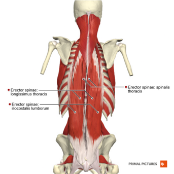
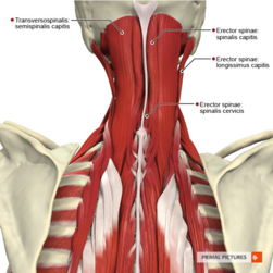
The erector spinae (ES) is one of the core and paraspinal muscles, is a large and superficial muscle that lies just deep to the thoracolumbar fascia and arises from the erector spinae aponeurosis (ESA).
The ESA is a common aponeurosis that blends with the thoracolumbar fascia, with a proximal attachment on the sacrum and the spinous processes of the lumbar vertebrae.
The ES relationships:
- Covered by thoracolumbar fascia, serratus posterior inferior, rhomboids, and splenii muscle groups
- Between the posterior and middle layers of the thoracolumbar fascia in the lumbar regions [1]
The ES is formed of 3 muscles with its fibres running more or less vertically throughout the lumbar, thoracic and cervical regions. It lies in the groove to the side of the vertebral column. [2] Its muscle mass is poorly differentiated but divides into three sections in the upper lumbar area named:
- Iliocostalis, most lateral
- Longissimus, the intermediate column
- Spinalis, most medial [3]
Origin / Insertion[edit | edit source]
| Muscles | Origin | Insertion |
| Spinalis capitis | Medial aspect between the superior and inferior nuchal lines of the occiput [4] | C7, T1 |
|---|---|---|
| Spinalis cervicis | Spinous processes of the axis and sometimes the C3, C4[5] [6] | Lower part of ligamentum nuchae (C4 to C6) and spinous process of C7 to T2 [7][8] |
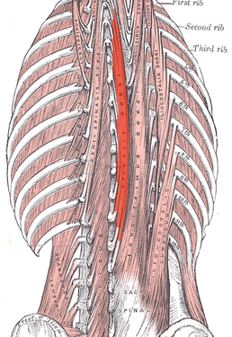
| Spinalis thoracis | Spinous processes of T11-L2 [9] [10] | Spinous processes of T2-T8 [11] |
| Longissimus capitis | Posterior surface of transverse processes of T1 to T5 and the articular tubercle of C4 to C7[12] | Posterior margin of mastoid process and the temporal bone [13][14] |
| Longissimus cervicis | Transverse processes of vertebrae T1-T5 | Transverse processes of vertebrae C2-C6 [15] |
| Longissimus thoracis: | Lumbal part: Lumbar intermuscular aponeurosis, medial part of sacropelvic surface of ilium, posterior sacroiliac ligament
Thoracic part: Spinous and transverse processes of vertebrae L1-L5, median sacral crest, posterior surface of sacrum and posterior iliac crest |
Lumbal part: Accessory and transverse processes of vertebrae L1-L5
Thoracic part: Transverse process of vertebrae T1-T12, Angles of ribs 7-12 [15] |
| Iliocostalis cervicis | Angle of ribs 3-6 | Transverse process of C4-C6 |
| Iliocostalis thoracis | Angle of lower six ribs | Angles of upper six ribs and transverse process of C7 |
| Iliocostalis lumborum | Iliac crest | L1-L4 lumbar transverse processes, angle of 4-12 ribs and thoracolumbar fascia |
Nerve[edit | edit source]
Dorsal rami of spinal nerves.[16]
Artery[edit | edit source]
Branches of the vertebral, deep cervical, occipital, transverse cervical, posterior intercostal, subcostal, lumbar and lateral sacral arteries. [17]
Function[edit | edit source]
- Bilateral contraction of the erector spinae muscles causes back and head extension.[18]
- It controls the forward flexion of the thorax, which can occur secondary to gravity. [19]
- The actions of the cervical and capitis groups are disputed. These muscles are small when compared to the larger cervical muscle groups and have little force capacity.
- The unilateral contraction causes ipsilateral side flexion and rotation of the vertebral column and head. [17][18]
- Spinalis connects the spinous process of the adjacent vertebrae. [17]
- Longissimus forms the middle part of the erector spinae muscles, lateral to the spinalis. The longissimus muscle forms the main meat of the erector group. It attaches along the transverse process of the vertebrae. [17]
- Iliocostalis is the most lateral part of the erector spinae muscles. It attaches to the ribs. [17] Due to the lateral position, tightness in iliocostalis muscles can force the ipsilateral hip into a superior position, or bring the ribcage inferior toward the hip.
Clinical Relevance[edit | edit source]
The main pathology associated with the back muscles is pain.
- These muscles can develop spasms that can be debilitating.
- The lower back muscles are a common cause of low back pain (LBP). This entity is often misdiagnosed and involves millions of people of all ages and gender. Patients often undergo exhaustive workups, including an MRI, often unwarranted. [19]
The erector spinae muscles play an important role in the spinal stability and LBP.
- In patients with LBP, there is decreased activity and atrophy of the multifidus muscle which compromises the spinal stability. [20] The spinal control is compensated for by the increased activity of the erector spinae muscle to stabilize the lumbar spine. [21] eg erector spinae contract to compensate for the delay in increasing the stiffness of the lumbar spine.
- This increased activity of erector spinae increases the compression load on the vertebral column, stimulating the nociceptors of the spinal structures continuously which may increase the risk of injury. [21]
Erector Spinae Flexion-Relaxation Phenomenon: The flexion-relaxation phenomenon is defined as the silencing of the erector spinae myoelectric activity during full trunk flexion.
- In healthy individuals with no low back pain, the erector spinae muscles relax in a range from an upright position to full-lumbar flexion, due to the deep back muscles (multifidus) acting to stabilize the lumbar spine.
- In individuals with low back pain, the erector spinae flexion-relaxation phenomenon is absent. As the erector spinae functions to stabilize the lumbar spine due to laxity of the passive structures and changes in the neuromuscular activation pattern.
See also Lumbar Instability.
Assessment[edit | edit source]
Manual Muscle Testing: Trunk Extension
Watch this brief video.
Treatment[edit | edit source]
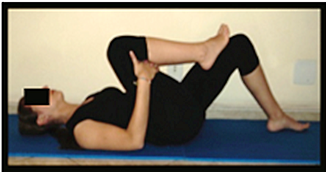
Lumbar stabilization exercises can restore the erector spinae flexion-relaxation phenomenon by strengthening the multifidus muscle. [23]
Myofascial release of the erector spinae muscles in patients with non-specific chronic low back pain normalized the flexion-relaxation response and decreased low back pain. [24]
Back Exercises play an important role.
Resources[edit | edit source]
This is a 5-minute video on "Function and Training of the Erector Spinae Muscles":
References[edit | edit source]
- ↑ Radiopedia Erector Spinae Available: https://radiopaedia.org/articles/erector-spinae-group?lang=us(accessed 5.3.2022)
- ↑ Whitmore I. Terminologia anatomica: new terminology for the new anatomist. The Anatomical Record: An Official Publication of the American Association of Anatomists. 1999 Apr 15;257(2):50-3.
- ↑ Musculoskeletal Key Lumbar Musculature Available: https://musculoskeletalkey.com/lumbar-musculature-anatomy-and-function/ (accessed 5.3.2022)
- ↑ Radiopedia Erector spinae group Available: https://radiopaedia.org/articles/erector-spinae-group?lang=us(accessed 4.2.2022)
- ↑ Spinalis Cervicis : Wheeless' Textbook of Orthopaedics [Internet]. Wheeless' Textbook of Orthopaedics. 2021 [cited 30 November 2021]. Available from: https://www.wheelessonline.com/bones/spine/spinalis-cervicis/
- ↑ Radiopedia Erector Spinae Available:https://radiopaedia.org/articles/erector-spinae-group?lang=us (accessed 4.2.2022)
- ↑ spinalis muscle | anatomy [Internet]. Encyclopedia Britannica. 2021 [cited 30 November 2021]. Available from: https://www.britannica.com/science/spinalis-muscle
- ↑ Spinalis Cervicis : Wheeless' Textbook of Orthopaedics [Internet]. Wheeless' Textbook of Orthopaedics. 2021 [cited 30 November 2021]. Available from: https://www.wheelessonline.com/bones/spine/spinalis-cervicis/
- ↑ Spinalis muscle [Internet]. Kenhub. 2021 [cited 30 November 2021]. Available from: https://www.kenhub.com/en/library/anatomy/spinalis-muscle
- ↑ Palastanga, N., & Soames, R. (2012). Anatomy and human movement (6th ed.). Edinburgh: Churchill Livingstone.
- ↑ Palastanga, N., & Soames, R. (2012). Anatomy and human movement (6th ed.). Edinburgh: Churchill Livingstone.
- ↑ http://www.wheelessonline.com/ortho/longissimus_capitis_1
- ↑ http://www.wheelessonline.com/ortho/longissimus_capitis_1
- ↑ http://www.primalonlinelearning.com/cedaandp/muscular_system/muscles_of_the_back.aspx#longissimuscapitis
- ↑ 15.0 15.1 Gordana Sendić, Adrian Rad. Longissimus muscle [Internet]. Ken Hub; c2023 [updated 2022 Jul 19]. Available from:https://www.kenhub.com/en/library/anatomy/longissimus-muscle
- ↑ Henson B, Edens MA. Anatomy, Back, Muscles. InStatPearls [Internet] 2018 Dec 23. StatPearls Publishing.
- ↑ 17.0 17.1 17.2 17.3 17.4 Drake R, Vogl AW, Mitchell AW. Gray's Anatomy for Students E-Book. Elsevier Health Sciences; 2009 Apr 4.
- ↑ 18.0 18.1 Palastanga, N., & Soames, R. (2012). Anatomy and human movement (6th ed.). Edinburgh: Churchill Livingstone.
- ↑ 19.0 19.1 Henson B, Kadiyala B, Edens MA. Anatomy, Back, Muscles.[Updated 2020 Aug 10]. StatPearls [Internet]. Treasure Island (FL): StatPearls Publishing. 2021. Available:https://www.ncbi.nlm.nih.gov/books/NBK537074/ (accessed 5.3.2022)
- ↑ Ranger TA, Cicuttini FM, Jensen TS, Peiris WL, Hussain SM, Fairley J, Urquhart DM. Are the size and composition of the paraspinal muscles associated with low back pain? A systematic review. The spine journal. 2017 Nov 1;17(11):1729-48.
- ↑ 21.0 21.1 Mazis N. Does a history of non specific low back pain influence electromyographic activity of the erector spinae muscle group during functional movements. J. Nov. Physiother. 2014;4:226.
- ↑ Sheena Livingstone. Erector Spinae Muscle Test. Available from: http://www.youtube.com/watch?v=pfbm_-fgylo [last accessed 26/03/14]
- ↑ Park SS, Choi BR. Effects of lumbar stabilization exercises on the flexion-relaxation phenomenon of the erector spinae. Journal of physical therapy science. 2016;28(6):1709-11.
- ↑ Arguisuelas MD, Lison JF, Domenech-Fernandez J, Martinez-Hurtado I, Coloma PS, Sanchez-Zuriaga D. Effects of myofascial release in erector spinae myoelectric activity and lumbar spine kinematics in non-specific chronic low back pain: randomized controlled trial. Clinical Biomechanics. 2019 Mar 1;63:27-33.
- ↑ 3StrongVideos. Function and Training of the Erector Spinae Muscles - Coach. Available from: http://www.youtube.com/watch?v=l9XwHX3ma5A [last accessed 26/03/14]


