When refering to evidence in academic writing, you should always try to reference the primary (original) source. That is usually the journal article where the information was first stated. In most cases Physiopedia articles are a secondary source and so should not be used as references. Physiopedia articles are best used to find the original sources of information (see the references list at the bottom of the article).
If you believe that this Physiopedia article is the primary source for the information you are refering to, you can use the button below to access a related citation statement.
Respiratory failure is a clinical condition where the lungs fail to effectively exchange gases, resulting in insufficient oxygen (O₂) intake and/or inadequate removal of carbon dioxide (CO₂) from the blood.
This is reflected in two key measurements:
PaO₂ (partial pressure of oxygen), the amount of oxygen in the blood. In respiratory failure, the PaO₂ drops below 60 mmHg, meaning there isn’t enough oxygen in the bloodstream to meet the body's needs.
PaCO₂ (partial pressure of carbon dioxide), the amount of carbon dioxide in the blood. In some types of respiratory failure, PaCO₂ rises above 50 mmHg, indicating that the lungs aren’t removing enough carbon dioxide from the blood, which can lead to dangerous levels of CO₂ in the body.
Respiratory failure is classified into two types according to blood gas abnormalities (levels of O₂ and CO₂ in the blood).
Type 1 - (hypoxemic) respiratory failure occurs when the PaO2 is less than 60 mmHg while the PaCO2 remains normal or lower than normal. In this type, the gas exchange is impaired at the level of alveolar-capillary membrane. Examples of type I respiratory failures include conditions like pulmonary oedema (cardiogenic or non-cardiogenic), ARDs (acute respiratory distress syndrome), COVID-19 and severe pneumonia.
Type 2 - (hypercapnic) respiratory failure where PaCO2 is higher than 50 mmHg. Hypoxemia (low oxygen) is often present, and it happens due to failure of the respiratory pump to work properly. This is often caused by an obstruction such as in conditions like COPD (chronic obstructive pulmonary disease), or by musculoskeletal problems such as chest wall deformities, neurological and respiratory muscle weakness.
Respiratory failure is a significant cause of illness and death with the risk increasing as people age or when other health conditions are present.
Pathophysiology
Respiratory failure arises due to several underlying mechanisms that impair gas exchange:
Hypoventilation: in which PaCO2 and PaO2 and alveolar-arterial PO2 gradient (difference between the calculated oxygen pressure available in the alveolus and the arterial oxygen tension, measures the efficiency of gas exchange) are normal. Depression of CNS (central nervous system) from drugs (eg opiod use disorder) is an example of this condition.
Ventilation/Perfusion (V/Q) mismatch: this is the most common cause of hypoxemia. It occurs when air (ventilation) and blood flow (perfusion) to the lungs are not properly matched. The V/Q ratio describes the balance between air reaching the alveoli and blood flow through the lung capillaries. When this balance is disrupted, gas exchange is impaired, leading to low oxygen levels [1]. Administering 100% O2 usually corrects hypoxemia caused by a V/Q mismatch.
Shunt: A shunt is a pathological mechanism in which the alveoli are perfused with blood but not ventilated, leading to impaired gas exchange. The deoxygenated blood bypasses the ventilated areas of the lung and remains unoxygenated even with 100% O2 inhalation. This results in persistent hypoxemia and occurs in conditions such as pulmonary oedema, pneumonia and atelectasis[2]
Types
Type I (Hypoxemic) respiratory failure is characterised by low levels of oxygen, and normal or low carbon dioxide levels. This occurs due to damage to lung tissue that impairs the lung's ability to oxygenate the blood. Conditions including pulmonary oedema, pneumonia, acute respiratory distress syndrome (ARDS), and Idiopathic pulmonary fibrosis (IPF) are common causes. In these cases, the damaged parts of the lung cannot transfer oxygen effectively, leading to hypoxaemia (low blood oxygen). However, the remaining healthy lung tissue can still remove carbon dioxide. This is possible because less functioning lung tissue is required for carbon dioxide excretion than is needed for oxygenation of the blood [3].
Type II (Hypercapnic) respiratory failure is characterised by low oxygen levels, with high carbon dioxide levels. It occurs when alveolar ventilation is insufficient to excrete the carbon dioxide being produced, resulting in CO₂ retention. The underlying issue is typically due to reduced alveolar ventilation either from respiratory muscle weakness (pump failure) common in conditions such as Guillain-Barre syndrome; chest wall deformities, or central nervous system depression (e.g. from heroin overdose). COPD is one of the most common causes. Inadequate ventilation is due to reduced ventilatory effort, or inability to overcome increased resistance to ventilation – it affects the lung as a whole, and thus carbon dioxide accumulates in the blood, leading to respiratory acidosis and if untreated, damage to vital organs due to a combination of hypoxaemia and hypercapnia. [4]
Epidemiology
The exact prevalence of respiratory failure is difficult to determine since it is a syndrome rather than a specific disease [2]. However, it is a major cause of morbidity and mortality, particularly in older adults and those with underlying chronic conditions such as chronic obstructive pulmonary disease (COPD) or heart disease. The incidence of respiratory failure increases with age, and hospital admissions for acute respiratory failure are common in critical care settings. Advances in treatments have improved outcomes, but it remains a significant public health concern. The prevalence of COPD is projected to increase by 23% from 2020 to 2050, with the largest growth expected among female and low and middle income regions, likely leading to an increase in cases of respiratory failure [5].
Etiology
Respiratory failure may be due to pulmonary or extra-pulmonary causes which include:
Central nervous system (CNS) depression can lead to respiratory failure by diminishing the neural drive to breathe. This may occur due to traumatic brain injury, stroke, narcotic or sedative overdose, severe infections (e.g., encephalitis), or neurodegenerative diseases such as Parkinson's disease or ALS [6]
Disorders of the peripheral nervous system: Respiratory muscle and chest wall weakness as in cases of Guillian-Barre syndrome and myasthenia gravis.
Renal function tests and liver function tests- may indicate the etiology of respiratory failure or identify complications associated with it.
Pulmonary Function Test- identifies obstruction, restriction, and gas diffusion abnormalities. Normal values for forced expiratory volume in 1 second(FEV1) and forced vital capacity(FVC) suggest a disturbance in respiratory control. Decrease in FEV1 to FVC ratio indicates airflow obstruction. A decrease in FEV1 and FVC and maintenance of FEV1 to FVC ratio suggest restrictive lung disease.
Gastrointestinal: haemorrhage, gastric distention, ileus, diarrhoea, pneumoperitoneum and duodenal ulceration- caused by stress is common in patients with acute respiratory failure
Infectious: noscomial- pneumonia, urinary tract infection and catheter-related sepsis. Usually occurs with use of mechanical devices.
Renal: acute renal failure, abnormalities of electrolytes and acid-base balance.
Nutritional: malnutrition and complications relating to parenteral or enteral nutrition and complications associated with NG tube- abdominal distention and diarrhoea
Management
This includes supportive measures and treatment of the underlying cause.
Supportive measures focus on maintaining airway management to ensure adequate ventilation and correct blood gas abnormalities
1. Correction of Hypoxemia
The goal is to maintain adequate tissue oxygenation, generally achieved with an arterial oxygen tension (PaO2) of 60 mm Hg or arterial oxygen saturation (SaO2), about 90%.
Un-controlled oxygen supplementation can result in oxygen toxicity and CO2 (carbon dioxide) narcosis. Inspired oxygen concentration should be adjusted at the lowest level, which is sufficient for tissue oxygenation.
Oxygen can be delivered by several routes depending on the clinical situations in which we may use a nasal cannula, simple face mask nonrebreathing mask, or high flow nasal cannula.
Extracorporeal membrane oxygenation may be needed in refractory cases
2. Correction of hypercapnia and respiratory acidosis
This may be achieved by treating the underlying cause or providing ventilatory support.
3. Ventilatory support for the patient with respiratory failure
The goals of ventilatory support in respiratory failure are:
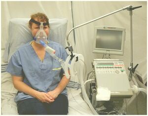
Non-invasive respiratory support: is ventilatory support without tracheal intubation/ via upper airway. Considered in patients with mild to moderate respiratory failure. Patients should be conscious, have an intact airway and airway protective reflexes. Noninvasive positive pressure ventilation(NIPPV) has been shown to reduce complications, duration of ICU stay and mortality(). It has been suggested that NIPPV is more effective in preventing endotracheal intubation in acute respiratory failure due to COPD than other causes. The etiology of respiratory failure is an important predictor of NIPPV failure.[8]
Invasive respiratory support: indicated in persistent hypoxemia despite receiving maximum oxygen therapy, hypercapnia with impairment of conscious level. Intubation is associated with complications such as aspiration of gastric content, trauma to the teeth, barotraumas, trauma to the trachea etc
Permissive hypercapnia[9] - A ventilatory strategy that allows arterial carbon dioxide(PaCO2) to rise by accepting a lower alveolar minute ventilation to avoid the risk of ventilator-associated lung injury in patients with ALI and minimize intrinsic positive end-expiratory pressure (auto PEEP) in patients with COPD thereby protecting the lungs from barotrauma. Permissive hypercapnia could increase survival in immunocompromised children with severe ARDS[10]
Physiotherapy Management
Physio-therapeutic interventions aim to maximize function in pump and ventilatory systems and improve quality of life.
Main indications for physiotherapy are excessive pulmonary secretions and atelectasis.
Timely physical therapy interventions may improve gas exchange and reverse pathological progression thereby avoiding ventilation.
Interventions include:
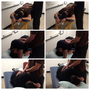
Positioning: the use of specific body position aimed at improving ventilation/perfusion(V/Q) matching, promoting mucociliary clearance, improving aeration via increased lung volumes and reducing the work of breathing.[11] These include:
Prone: helps to improve V/Q matching, redistribute oedema and increase functional residual capacity(FRC) in patients with acute respiratory distress syndrome. It has been shown to result in oxygenation for 52-92% of patients with severe acute respiratory failure.[12][13]
Side-lying: with affected lungs uppermost to improve aeration through increased lung volumes in patients with unilateral lung disease.
Semi-recumbent: 450 head-up position serves to prevent the risk of gastroesophageal reflux and aspiration.
Upright: helps to improve lung volumes and decrease work of breathing in patients that are being weaned from mechanical ventilator.
Suction: used for clearing secretions when the patient cannot do so independently (Guglielminotti et al recommended a saw-tooth pattern seen on ventilator’s flow-volume loop and/or respiratory sounds over the trachea as good indicators for tracheal suction in a ventilated patient).[14]
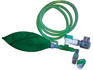
Manual Hyperinflation: aims to re-inflate atelectatic areas of the lung as such improving pulmonary compliance[15] and facilitate the clearance of pulmonary secretions when used with other techniques.
Limb exercises: passive, active-assisted, active exercises may optimize oxygen transport and reduce the effects of immobility.[11]
Inspiratory muscle training: aims to improve inspiratory muscle strength and it facilitates weaning from mechanical ventilation. It has be shown to improve whole body exercise performance, particularly in less fit subjects.[17] The study by Hoffman L et showed positive outcomes with inspiratory muscle training in patients with advanced lung disease.[18]
Early mobilisation: improves function, mobility and quality of life[19][20][21]
Respiratory failure is associated with poor prognosis but advances in mechanical ventilation and airway management have improved prognosis. It is dependent on the underlying cause of respiratory failure.
Mortality rates of approximately 40-45% which has remained constant over the years occur with acute respiratory distress syndrome(ARDS)[22][23] with younger patients(< 60 years) having better survival rates.
About two-thirds of the patients who survive an episode of ARDS show some impairment of pulmonary function one or more years of post-recovery.
For COPD and acute respiratory failure. overall mortality has reportedly declined from 26% to 10%.
Significant mortality occurs in patients with hypercapnic respiratory failure due to associated co-morbidities and poor nutritional status.[24]
↑Agarwal R, Gupta R, Aggarwal AN, Gupta D. Noninvasive positive pressure ventilation in acute respiratory failure due to COPD vs other causes: effectiveness and predictors of failure in a respiratory ICU in North India. Int J Chron Obstrut Pulmon Dis. 2008;3(4):737-43.
↑Mackenzie I, editor. Core Topics in Mechanical Ventilation. Cambridge: Cambridge University Press, 2008. p153.
↑Fuchs H, Rossmann N, Schmid MB, Hoenig M, Thome U, Mayer B, et al. Permissive hypercapnia for severe acute respiratory distress syndrome in immunocompromised children: A single center experience. PLoS One. 2017;12(6):e0179974.
↑ 11.011.1Dean E. Oxygen transport: a physiologically-based conceptual framework for the practice of cardiopulmonary Physiotherapy. Physiotherapy. 1994; 80(6): 347-354
↑Jolliet P, Bulpa P, Chevrolet JC. Effects of the prone position on gas exchange and hemodynamics in severe acute respiratory distress syndrome. Crit Care Med. 1998; 26(12):1977-1985
↑Mure M, Martling CR, Lindahl SG. Dramatic effect on oxygenation in patients with severe acute lung insufficiency treated in prone position.Crit Care Med. 1997; 25(9):1539-1544
↑Guglielminotti J, Alzieu M, Maury E, Guidet B, Offenstadt G. Bedside detection of retained tracheobronchial secretions in patients receiving mechanical ventilation: is it time for tracheal suctioning?Chest. 2000;118(4):1095-9.
↑Clarke RCN, Kelly BE. Ventilatory characteristics in mechanically ventilated patients during manual hyperinflation for chest physiotherapy. Anaesthesia. 1999; 54: 936-940
↑Inal-Ince D, Savci S, Topeli A, Arikan H. Active cycle of breathing techniques in non-invasive ventilation for acute hypercapnic respiratory failure. Aust J Physiother. 2004;50(2):67-73.
↑Illi SK, Held U,Frank I, Spengler CM. Effect of respiratory muscle training on exercise performance in healthy individuals: a systematic review and meta-analysis. Sport Med. 2012;42(8):707-24.
↑Hodgson CL, Bailey M, Bellomo R, Berney S, Buhr H, Denehy L, et al. A binational multicenter pilot feasibility randomized controlled trial of early goal-directed mobilization in the ICU. Crit Care Med. 2016; 44(6):1145-1152
↑Schaller SJ, Anstey M, Blobner M, Edrich T, Grabitz SD, Gradwohl-Matis I, et al. Early, goal-directed mobilisation in the surgical intensive care unit: a randomised controlled trial. Lancet. 2016;388(10052):1377-1388
↑Kayambu G, Boots R, Paratz J. Early physical rehabilitation in intensive care patients with sepsis syndromes: a pilot randomised controlled trial. Intensive Care Med. 2015; 41(15):865-874
↑Phua J, Badia JR, Adhikari NK. Has mortality from acute respiratory distress syndrome decreased over time? A systematic review. Am J Respir Crit Care Med. 2009; 179(3): 220-227.
↑Acute Respiratory Distress Syndrome Network, Brower RG, Matthay MA, Morris A, Schoenfeld D, Thompson BT, et al. Ventilation with lower tidal volumes as compared with traditional tidal volumes for acute lung injury and acute respiratory distress syndrome. N Engl J Med. 2000; 342(18):1301-8.
↑Noveanu M, Breidthardt T, Reichlin H, Gayat E, Potocki M, Pragger H, et al. Effect of oral beta-blocker on short term and long term mortality in patients with acute respiratory failure: results from the BASEL-II-ICU study. Crit Care. 2010; 14(6): R198.doi: 10.1186/cc9317.