Paget's Disease: Difference between revisions
No edit summary |
No edit summary |
||
| (3 intermediate revisions by the same user not shown) | |||
| Line 5: | Line 5: | ||
</div> | </div> | ||
== Introduction == | == Introduction == | ||
[[File:Pagets Disease.jpeg|right|frameless]] | |||
Paget's disease of the [[bone]] is a [[Metabolic and Endocrine Disorders|metabolic]] bone disease caused by increased bone resorption followed by excessive unrestricted bone formation, due to activated osteoclasts. The normal bone marrow is replaced by increased and unorganized [[collagen]] and fibrous tissue, which lacks the structural stability of normal bone. This increased bone mass formation leads to complications eg [[Fracture|fractures]], [[Osteoarthritis|arthritis]], deformities, [[Pain Behaviours|pain]], and to a patient's weakened condition. Paget's disease is the second most common metabolic bone disease to [[osteoporosis]] <ref name="Goodman and Fuller">Goodman C, Fuller K. Pathology: Implications for the Physical Therapist. 3rd ed. St. Louis, Missouri: Saunders Elsevier;2009.</ref><ref name="Josse">Josse RG, Hanley DA, Kendler D, Marie LG, Adachi JD, Brown J. [https://cimonline.ca/index.php/cim/article/view/2897 Diagnosis and treatment of Paget’s disease of bone.] Clinical and Investigative Medicine. 2007 Oct 1:E210-23.</ref><ref name="Seitz">Seitz S, Priemel M, von Domarus C, Beil FT, Barvencik F, Amling M, Rueger JM. [https://link.springer.com/content/pdf/10.1007/s00068-008-8208-4.pdf The second most common bone disease: a review on Paget’s disease of bone.] European Journal of Trauma and Emergency Surgery. 2008 Dec 1;34(6):549-53.</ref> | |||
The prognosis for patients who are treated is good, especially if the disease is in its early stages. There is no cure for Paget disease but the disorder can be controlled from progressing.<ref name=":0" /> | |||
== Etiology == | == Etiology == | ||
The exact cause of Paget's disease is unknown, though it is thought to be a slow, viral bone infection due to risk factors that include a gene-environment interaction. <ref name="Goodman and Fuller" /><ref name="Josse" /><ref name="Goodman and Snyder">Goodman C, Snyder T. Differential Diagnosis for Physical Therapists: Screening for Referral. 6th ed. St. Louis, Missouri: Saunders Elsevier, 2007.</ref> The prevalence and causes of Paget's disease has been associated with genetic and geographical factors. A positive family history is reported in as many as 40% of patients with Paget's disease <ref name="Goodman and Fuller" /><ref name="Josse" /><ref name="Goodman and Snyder" /><ref name="Schneider">Schneider D, Hofmann MT,Peterson JA. Diagnosis and Treatment of Paget’s Disease of Bone. American Family Physician. 2002; 65(10):2069-72</ref>. Paget's disease is mostly seen in an autosomal dominant distribution, and there have been three identifiable chromosomal regions associated with Paget's disease. <ref name="Goodman and Fuller" /> Geographically, populations in European, British, and Australian origin, and a migratory influence also play an important role especially in countries which of the early population migrated from Britain (United States, Australia, New Zealand, Canada).<ref name="Goodman and Fuller" /> | The exact cause of Paget's disease is unknown, though it is thought to be a slow, [[Viral Infections|viral]] bone infection (the family of paramyxoviruses) due to risk factors that include a gene-environment interaction. <ref name="Goodman and Fuller" /><ref name="Josse" /><ref name="Goodman and Snyder">Goodman C, Snyder T. Differential Diagnosis for Physical Therapists: Screening for Referral. 6th ed. St. Louis, Missouri: Saunders Elsevier, 2007.</ref> Some research suggests that the osteoclastic abnormalities are due to a cytokine is known as IL-6, found solely in the bone marrow of Paget's disease populations.<ref name=":0" /> The prevalence and causes of Paget's disease has been associated with genetic and geographical factors. A positive family history is reported in as many as 40% of patients with Paget's disease <ref name="Goodman and Fuller" /><ref name="Josse" /><ref name="Goodman and Snyder" /><ref name="Schneider">Schneider D, Hofmann MT,Peterson JA. Diagnosis and Treatment of Paget’s Disease of Bone. American Family Physician. 2002; 65(10):2069-72</ref>. Paget's disease is mostly seen in an autosomal dominant distribution, and there have been three identifiable chromosomal regions associated with Paget's disease. <ref name="Goodman and Fuller" /> Geographically, populations in European, British, and Australian origin, and a migratory influence also play an important role especially in countries which of the early population migrated from Britain (United States, Australia, New Zealand, Canada).<ref name="Goodman and Fuller" /> | ||
== Epidemiology == | == Epidemiology == | ||
| Line 14: | Line 17: | ||
== Characteristics/Clinical Presentation == | == Characteristics/Clinical Presentation == | ||
[[File:Paget's disease of Right Hip Bone.jpg|alt=This file is licensed under the Creative Commons Attribution-Share Alike 4.0 International license.|thumb|Paget's R Hip Bone]]A patient with Paget's disease will often present as asymptomatic. However, the clinical presentation of a symptomatic patient varies greatly, due to the different levels of severity of this condition. The spine and pelvis are commonly affected and among the long bones, the [[femur]] is often involved. Symptomatic patients can present with the following: | |||
* [[Pain Assessment|Pain]] involving the bones and joints | |||
* Diffuse joint stiffness | |||
* | * Abnormally enlarged [[skull]] | ||
* | * Musculoskeletal deformities | ||
* | * Loss of hearing (due to the involvement of the petrous temporal bone) | ||
* | * [[Migraine Headache|Migraines]] | ||
* | * [[Fracture|Fractures]] | ||
* | * [[Heart Failure|Heart failure]] | ||
* | * [[Cranial Nerves|Cranial nerve]] [[neuropathies]] | ||
* | * [[Headaches and Dizziness|Headaches]] | ||
* Enlarged skull | |||
* Skull and jaw deformity | |||
The [[Lumbar Spine Fracture|lumbar spine]], [[sacrum]], and skull are involved in most cases. Pain is a common feature and is worse with [[Weight bearing|weight-bearing]]. Incomplete fractures are common in Paget disease and seen in the tibia and femur. Even mild injuries can result in [[Insufficiency Fracture|fractures.]] [[Femoral Neck Fractures|Femur fractures]] often involve the subtrochanteric region.<ref name=":0" /> | |||
== | == Diagnosis == | ||
Tests to assist in the diagnosis of Paget disease include: | Tests to assist in the diagnosis of Paget disease include: | ||
| Line 63: | Line 53: | ||
Plain x-rays may reveal arthritis or fractures of gross bony lesions. | Plain x-rays may reveal arthritis or fractures of gross bony lesions. | ||
Bone scans can help document the extent of disease and should be used to follow treatment. In addition, a bone scan can pick up early changes in bone even before the patient develops symptoms. | Bone scans can help document the extent of disease and should be used to follow treatment. In addition, a bone scan can pick up early changes in bone even before the patient develops symptoms.<ref name="Goodman and Fuller" /> | ||
| |||
== Management == | == Management == | ||
| Line 125: | Line 64: | ||
* Calcium and vitamin D – are both important for bone health. You can get calcium through the diet and vitamin D through safe exposure to sunlight. | * Calcium and vitamin D – are both important for bone health. You can get calcium through the diet and vitamin D through safe exposure to sunlight. | ||
Surgery is only offered as an option to patients diagnosed with Paget disease when there is a progression into osteosarcoma. <ref name=":0" /> | Surgery is only offered as an option to patients diagnosed with Paget disease when there is a progression into osteosarcoma. <ref name=":0" /> | ||
There is no specific diet for patients with Paget disease, however those who are prescribed bisphosphonates should ensure adequate intake of calcium and vitamin D.<ref name="Schneider" /> | |||
== Physical Therapy Management == | == Physical Therapy Management == | ||
[[File:Walking dog.jpg|thumb|Walking, a safe exercise.]] | |||
Treatment Options Include: | |||
A physical therapist can also assist a patient with Paget's disease in home modifications to make the patient safer with mobility around the home<ref name="Chow" /><br> | * Encourage Client to Stay active – exercise helps to maintain bone health and joint mobility, as well as strengthen muscles. <ref name="Goodman and Fuller" />Aggressive physical activity is not recommended, as the risk of fracture is high. Certain forms of exercise are not suitable for people with Paget’s disease. eg avoid activities such as jogging, running, jumping, and aggressive forward bending and twisting exercises, if the spine is affected by Paget's disease <ref name="Goodman and Fuller" /><ref name="Schneider" /> | ||
* Provide a tailored exercises plan, and also provide techniques and/or devices that can help to improve movement, reduce pain and make everyday activities easier. eg a walking stick to reduce the weight placed through affected bones, braces to correct position, foot orthotics to support and correct abnormal foot position or motion. | |||
* Educate on a healthy well-balanced diet – this can help client reach and maintain a healthy weight and reduce your risk of other health problems. Make sure they include calcium-rich foods. | |||
* Teach new ways to manage pain. eg heat packs can help ease muscle pain, cold packs can help with inflammation, gentle exercise can help relieve muscle tension, transcutaneous electrical nerve stimulation (TENS), and massage . Try different techniques until client finds the things that work best for them<ref name="Chow">Chow, David. Emedicine. Med Web:Paget Disease. http://emedicine.medscape.com/article/311688-overview. Updated December 18,2008. Accessed April 2, 2010.</ref>. | |||
* Encourage client to stay at work, it’s good for health and wellbeing. Discuss ways to help client get back to or stay at work<ref>MSK Pagets Disease Available:https://msk.org.au/pagets-disease/ (accessed 13.5.2022)</ref> | |||
* A physical therapist can also assist a patient with Paget's disease in home modifications to make the patient safer with mobility around the home<ref name="Chow" /><br> | |||
== References == | == References == | ||
Latest revision as of 06:04, 26 March 2023
Original Editors - Kevin Schoenfeld from Bellarmine University's Pathophysiology of Complex Patient Problems project.
Top Contributors - Kevin Schoenfeld, Admin, Lucinda hampton, Elaine Lonnemann, Dave Pariser, Priya Gulla, Nikhil Benhur Abburi, Kim Jackson, WikiSysop, Uchechukwu Chukwuemeka, Aminat Abolade, Rachael Lowe, Wendy Walker, Mariam Hashem and Claire Knott
Introduction[edit | edit source]
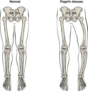
Paget's disease of the bone is a metabolic bone disease caused by increased bone resorption followed by excessive unrestricted bone formation, due to activated osteoclasts. The normal bone marrow is replaced by increased and unorganized collagen and fibrous tissue, which lacks the structural stability of normal bone. This increased bone mass formation leads to complications eg fractures, arthritis, deformities, pain, and to a patient's weakened condition. Paget's disease is the second most common metabolic bone disease to osteoporosis [1][2][3]
The prognosis for patients who are treated is good, especially if the disease is in its early stages. There is no cure for Paget disease but the disorder can be controlled from progressing.[4]
Etiology[edit | edit source]
The exact cause of Paget's disease is unknown, though it is thought to be a slow, viral bone infection (the family of paramyxoviruses) due to risk factors that include a gene-environment interaction. [1][2][5] Some research suggests that the osteoclastic abnormalities are due to a cytokine is known as IL-6, found solely in the bone marrow of Paget's disease populations.[4] The prevalence and causes of Paget's disease has been associated with genetic and geographical factors. A positive family history is reported in as many as 40% of patients with Paget's disease [1][2][5][6]. Paget's disease is mostly seen in an autosomal dominant distribution, and there have been three identifiable chromosomal regions associated with Paget's disease. [1] Geographically, populations in European, British, and Australian origin, and a migratory influence also play an important role especially in countries which of the early population migrated from Britain (United States, Australia, New Zealand, Canada).[1]
Epidemiology[edit | edit source]
Paget disease is usually seen in individuals older than 50 years. It is common in Caucasians of northern European descent. Paget disease is equally common in males and females. In the US, it affects 1-3 million people with most being asymptomatic. The disorder is slightly more common in white males. The disorder usually presents in the 4-5 decade of life but the diagnosis is often made a decade later[4].
Characteristics/Clinical Presentation[edit | edit source]
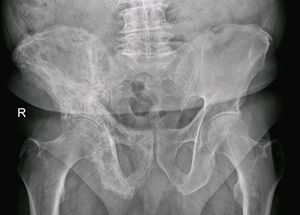
A patient with Paget's disease will often present as asymptomatic. However, the clinical presentation of a symptomatic patient varies greatly, due to the different levels of severity of this condition. The spine and pelvis are commonly affected and among the long bones, the femur is often involved. Symptomatic patients can present with the following:
- Pain involving the bones and joints
- Diffuse joint stiffness
- Abnormally enlarged skull
- Musculoskeletal deformities
- Loss of hearing (due to the involvement of the petrous temporal bone)
- Migraines
- Fractures
- Heart failure
- Cranial nerve neuropathies
- Headaches
- Enlarged skull
- Skull and jaw deformity
The lumbar spine, sacrum, and skull are involved in most cases. Pain is a common feature and is worse with weight-bearing. Incomplete fractures are common in Paget disease and seen in the tibia and femur. Even mild injuries can result in fractures. Femur fractures often involve the subtrochanteric region.[4]
Diagnosis[edit | edit source]
Tests to assist in the diagnosis of Paget disease include:
- Bone scan
- Bone x-ray
- Elevated markers of bone breakdown like N-telopeptide
This disease also may also present with the following findings:
- Elevated ALP (alkaline phosphatase)
- Normal Serum calcium and Phosphate
Hyperuricemia is common and is due to a high turnover of bone.
Secondary hyperparathyroidism occurs in about 10% of patients due to inadequate calcium in the face of increased demand.
Plain x-rays may reveal arthritis or fractures of gross bony lesions.
Bone scans can help document the extent of disease and should be used to follow treatment. In addition, a bone scan can pick up early changes in bone even before the patient develops symptoms.[1]
Management[edit | edit source]
Although there’s no cure for Paget’s disease of bone, there are treatments available to help people live well and manage the symptoms.
Medications
- Bisphosphonates are used to slow the progression of Paget’s disease. They help the body control the bone-building process to stimulate more normal bone growth.
- Pain relievers (analgesics) and non-steroidal anti-inflammatory drugs (NSAIDs) – are used to provide temporary pain relief.
- Calcium and vitamin D – are both important for bone health. You can get calcium through the diet and vitamin D through safe exposure to sunlight.
Surgery is only offered as an option to patients diagnosed with Paget disease when there is a progression into osteosarcoma. [4]
There is no specific diet for patients with Paget disease, however those who are prescribed bisphosphonates should ensure adequate intake of calcium and vitamin D.[6]
Physical Therapy Management[edit | edit source]
Treatment Options Include:
- Encourage Client to Stay active – exercise helps to maintain bone health and joint mobility, as well as strengthen muscles. [1]Aggressive physical activity is not recommended, as the risk of fracture is high. Certain forms of exercise are not suitable for people with Paget’s disease. eg avoid activities such as jogging, running, jumping, and aggressive forward bending and twisting exercises, if the spine is affected by Paget's disease [1][6]
- Provide a tailored exercises plan, and also provide techniques and/or devices that can help to improve movement, reduce pain and make everyday activities easier. eg a walking stick to reduce the weight placed through affected bones, braces to correct position, foot orthotics to support and correct abnormal foot position or motion.
- Educate on a healthy well-balanced diet – this can help client reach and maintain a healthy weight and reduce your risk of other health problems. Make sure they include calcium-rich foods.
- Teach new ways to manage pain. eg heat packs can help ease muscle pain, cold packs can help with inflammation, gentle exercise can help relieve muscle tension, transcutaneous electrical nerve stimulation (TENS), and massage . Try different techniques until client finds the things that work best for them[7].
- Encourage client to stay at work, it’s good for health and wellbeing. Discuss ways to help client get back to or stay at work[8]
- A physical therapist can also assist a patient with Paget's disease in home modifications to make the patient safer with mobility around the home[7]
References[edit | edit source]
- ↑ 1.0 1.1 1.2 1.3 1.4 1.5 1.6 1.7 Goodman C, Fuller K. Pathology: Implications for the Physical Therapist. 3rd ed. St. Louis, Missouri: Saunders Elsevier;2009.
- ↑ 2.0 2.1 2.2 Josse RG, Hanley DA, Kendler D, Marie LG, Adachi JD, Brown J. Diagnosis and treatment of Paget’s disease of bone. Clinical and Investigative Medicine. 2007 Oct 1:E210-23.
- ↑ Seitz S, Priemel M, von Domarus C, Beil FT, Barvencik F, Amling M, Rueger JM. The second most common bone disease: a review on Paget’s disease of bone. European Journal of Trauma and Emergency Surgery. 2008 Dec 1;34(6):549-53.
- ↑ 4.0 4.1 4.2 4.3 4.4 Bouchette P, Boktor SW. Paget disease. InStatPearls [Internet] 2021 Jul 13. StatPearls Publishing. Available:https://www.ncbi.nlm.nih.gov/books/NBK430805/(accessed 13.5.2022)
- ↑ 5.0 5.1 Goodman C, Snyder T. Differential Diagnosis for Physical Therapists: Screening for Referral. 6th ed. St. Louis, Missouri: Saunders Elsevier, 2007.
- ↑ 6.0 6.1 6.2 Schneider D, Hofmann MT,Peterson JA. Diagnosis and Treatment of Paget’s Disease of Bone. American Family Physician. 2002; 65(10):2069-72
- ↑ 7.0 7.1 Chow, David. Emedicine. Med Web:Paget Disease. http://emedicine.medscape.com/article/311688-overview. Updated December 18,2008. Accessed April 2, 2010.
- ↑ MSK Pagets Disease Available:https://msk.org.au/pagets-disease/ (accessed 13.5.2022)


