Midbrain
Original Editor - Lucinda hampton
Top Contributors - Lucinda hampton, Uchechukwu Chukwuemeka and Kim Jackson
Introduction[edit | edit source]
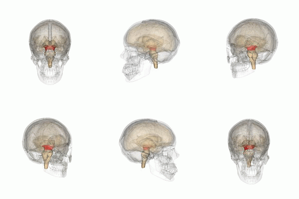
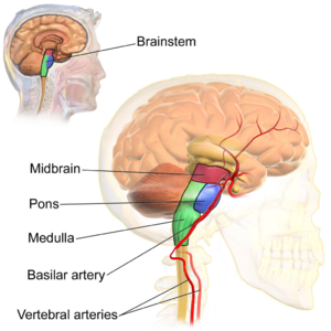
The midbrain is a part of the central nervous system, located below the cerebral cortex and at the topmost part of the brainstem. This small but important structure plays a crucial role in processing visual and auditory signals.
- Midbrain functions involves movement of body and head, as it provides passage for downward pathways for the cerebral cortex.
- It is a channel for the spinal cord transmitting stimuli (sensory) from the head and body to the direct brain.
- Midbrain function psychology invokes survival instincts by identifying potential hazardous objects and behaviour[1].
Divisions[edit | edit source]
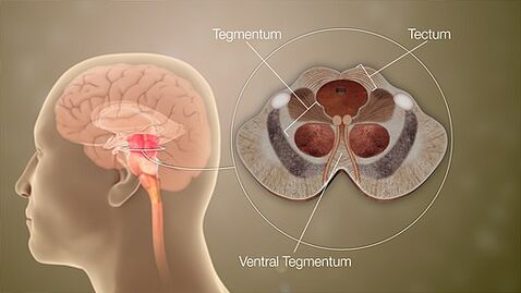
When viewed in cross-section, the midbrain can be divided into three portions:
- Tectum (posterior)
- Tegmentum
- Cerebral peduncles (anterior)[2]
Tectum[edit | edit source]
Sitting posteriorly, the tectum (Latin for "roof" or "covering") is composed of the tectal plate and superior and inferior colliculi. The tectum is unique to the midbrain and does not have a counterpart in the rest of the brainstem.
- Nerve cells within the superior colliculi process vision signals from the retina of the eye before channeling them on to the occipital lobe located at the back of the head.
- The inferior colliculi is responsible for processing auditory (hearing) signals before they are channeled through the thalamus and eventually to the primary auditory cortex in the temporal lobe
Tegmentum[edit | edit source]
The tegmentum is the phylogenetically-old part of the brainstem and runs through the pons and medulla oblongata. Its contents include:
- A variety of ascending and descending tracts pass through the midbrain (eg the medial lemniscus and the anterolateral tracts)
- Reticular formation: This highly diverse and integrative area contains a network of nuclei responsible for many vital functions including arousal, consciousness, sleep-wake cycles, coordination of certain movements, and cardiovascular control.
- Spinothalamic tract: This major nerve pathway carries information about pain and temperature sensation from the body to the thalamus of the brain.
- Corticospinal tract: This major nerve pathway carries movement-related information from the brain to the spinal cord.
- The red nucleus is one of the brainstem nuclei, and part of the extrapyramidal system, and is a portion of the motor system specifically dedicated to the modulation and regulation of movement.
- Nuclei for two cranial nerves: cranial nerve III (oculomotor nerve) and cranial nerve IV (trochlear nerve).
- Part of the raphe nuclei (clusters of serotonin-producing neurons found in the brainstem that send serotonin throughout the central nervous system);
- Substantia nigra: crescent-shaped mass of nerve cells in the midbrain with a dynamic role. Although it is the smallest portion of the midbrain, it is involved in the regulation of gathering auditory and visual sensory information, motor control (primarily informed by the production and distribution of dopamine), reward-based learning patterns, and the Circadian rhythm.
- Ventral tegmental area (VTA): This structure contains dopamine-producing cell bodies and plays a key role in the reward system.
Cerebral Peduncles[edit | edit source]
Cerebral peduncles: Anterior to the tegmentum are the cerebral peduncles which are composed of the large ascending and descending tracts that run to and from the cerebrum[3]. It includes
- The cerebral peduncles contain the substantia nigrae, which (like the ventral tegmental area) contain large collections of dopamine-producing neurons.
- Periaqueductal gray (PAG) matter: This area plays a primary role in processing pain signals, autonomic function, and behavioral responses to fear and anxiety. Recently, this structure has been linked to controlling the defensive reactions associated with post-traumatic stress disorder (PTSD)[3].
Blood Supply[edit | edit source]
The midbrain is supplied by the vertebrobasilar circulation, from small penetrating branches of the:
- basilar artery
- superior cerebellar artery
- posterior cerebral artery[2]
Associated Conditions[edit | edit source]
The midbrain may be affected by a number of different pathological processes including stroke, tumor, a demyelinating process, infection, or a neurodegenerative disease.
Examples of specific conditions include the following:
- Multiple sclerosis (MS): If the brainstem is affected, a patient may experience symptoms like: Vision changes, including diplopia; Problems swallowing (dysphagia); Problems speaking (dysarthria); Altered sensation or weakness of the face; Hearing difficulties; Ataxia; Headache that resembles a migraine; Rarely, problems that affect vital functions (e.g., breathing or heart rate).
- Parkinson’s disease is a progressive neurological disease, caused by the death of dopamine-producing nerve cells in the substantia nigra. As a result of this dopamine depletion, various symptoms may develop, including: Resting tremor; Bradykinesia; Stiffness and shuffling gait; micrographia; Sleep troubles.
- Oculomotor (Third) Nerve Palsy
- Trochlear (Fourth) Nerve Palsy[4]
Resources[edit | edit source]
- bulleted list
- x
or
- numbered list
- x
References[edit | edit source]
- ↑ Vedantu Midbrain Available:https://www.vedantu.com/biology/midbrain (accessed 23.4.20220
- ↑ 2.0 2.1 Radiopedia Midbrain Available: https://radiopaedia.org/articles/midbrain(accessed 23.4.2022)
- ↑ 3.0 3.1 Neuroscientifically challenged Know your midbrain Available:https://neuroscientificallychallenged.com/posts/know-your-brain-midbrain (accessed 23.4.2022)
- ↑ Very Well health Midbrain Available:https://www.verywellhealth.com/midbrain-anatomy-5093684 (accessed 23.4.2022)


