Apoptosis
Original Editor - Lucinda hampton
Top Contributors - Lucinda hampton and Kim Jackson
Introduction[edit | edit source]
Apoptosis is an evolutionarily conserved cell death pathway that is responsible for the proper culling of cells during normal development and the maintenance of homeostasis.
- In vertebrates, apoptosis is important for proper development, maintenance of tissue homeostasis. To maintain normal physiology and tissue function, cells that are damaged, dysfunctional or no longer necessary are constantly cleared via regulated cell death and ideally replaced by new, healthy cells. When these normal processes of cell death go awry, the consequences can be disastrous.
- Many of the diseases that constitute the leading causes of death and disability worldwide eg neurodegenerative, cardiovascular, autoimmuneiandnfectious diseases and cancer,in volve either excessive or insufficient cell removal.
- Despite the undeniable importance of maintaining the survival of our healthy cells or eliminating those that are damaged/potentially dangerous, our understanding of cell death processes and their regulation is still nascent[1].

Mechanism[edit | edit source]
Apoptosis is a complex process. During apoptosis, a cell triggers a process from within that will allow it to commit suicide.
Apoptosis involves the following pathways. All the pathways involve activation of caspases (an enzyme that cuts or cleaves proteins) as the final tool.
- Intrinsic Pathway (Mitochondrial Pathway)
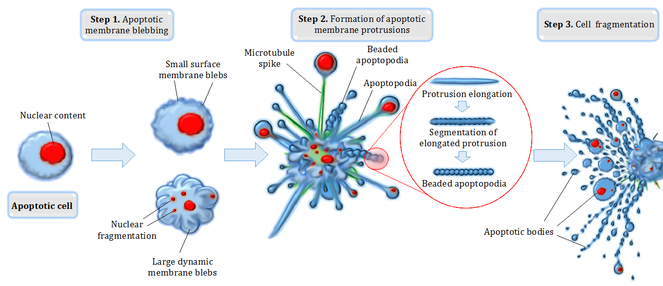

- It is activated when the cell is stressed from the inside due to an array of factors like DNA damage from x-ray exposure or UV light exposure; chemotherapeutic agents; hypoxia; accumulation of misfolded proteins inside the cell as eg.Alzheimer disease, Parkinson disease, or Huntington disease; and many others. When the cell is stressed signals are released which cause mitochondria to release apoptosis-inducing proteins. As a result, the cell undergoes a reduction in size as its cellular components and organelles break down and condense.
- Bubble-shaped balls called blebs appear on the surface of the cell membrane. Once the cell shrinks, it breaks down into smaller fragments called apoptotic bodies and sends out distress signals to the body. These fragments are enclosed in membranes so as not to harm nearby cells. The distress signal is answered by vacuum cleaners known as macrophages. The macrophages clean away the shrunken cells, leaving no trace, so these cells have no chance to cause cellular damage or an inflammatory reaction.
2. Extrinsic Pathway
- This pathway is triggered when the cell receives death signals from the other cell(s). The extrinsic pathway can be triggered externally by chemical substances that bind to specific receptors on the cell surface. This is how white blood cells combat infection and activate apoptosis in infected cells[1].[2][3]
Why do cells undergo apoptosis?[edit | edit source]
Many cells in the human body have the built-in ability to undergo apoptosis (in the same way that they have the built-in ability to copy their DNA or break down fuels). Basically, apoptosis is a general and convenient way to remove cells that should no longer be part of the organism.
- Some cells need to be “deleted” during development: eg to whittle an intricate structure like a hand out of a larger block of tissue; during brain development, the body creates millions of more cells than it needs; the ones that do not form synaptic connections can undergo apoptosis so that the remaining cells can function well.
- Some cells are abnormal and could hurt the rest of the organism if they survive eg. Cells may recognize viruses and gene mutations/DNA damage and can induce death to prevent the damage from spreading.
- Cells in an adult organism may be eliminated to maintain balance making way for new cells or remove cells needed only for temporary tasks[4].
Health Implications[edit | edit source]

Too little or too much apoptosis can have serious clinical implications such as the following:
Cancer:
- A decrease in apoptosis leads to increased cell-survival rates, leading to the development of cancers.
- Tumor viruses change cells by integrating their genetic material with the host cell's DNA. Cancer cells are usually a permanent insertion in the genetic material. These viruses can sometimes initiate the production of proteins that stop apoptosis from occurring. eg papilloma viruses, which have been linked with cervical cancer. Cancer cells that do not develop from viral infection can also produce substances that inhibit apoptosis and promote uncontrolled growth.
- Targeting apoptosis is a new standard in cancer drug development.[5]
- A decrease in apoptosis of self-reactive immune cells can lead to the development of autoimmune diseases like rheumatoid arthritis, systemic lupus erythematosus (SLE), autoimmune lymphoproliferative syndrome, and others.
- Cell death has also been implicated in many neurodegenerative disorders. Both necrosis and apoptosis occur in an acute neurologic disease like acute ischemic syndrome. In chronic neurodegenerative disorders neuronal cell death mainly by apoptosis has been implicated in examples like Parkinson disease, Alzheimer disease, and Huntington disease.
- Necrosis was long considered the sole cause of myocardial infarction, but recent studies have shown that apoptosis also occurs mainly during the reperfusion phase after the acute infarction, leading to further myocardial damage[3].
Conclusion[edit | edit source]
Apoptosis is a form of programmed cell death, or “cellular suicide" (different from necrosis, in which cells die due to injury). Apoptosis is not the only form of programmed cell death, but it is the form we understand best.
Apoptosis is an orderly process in which the cell’s contents break down and are packaged into small packets of membrane for “garbage collection” by immune cells (contrasting with necrosis in which the dying cell’s contents spill out and cause inflammation).
Apoptosis removes cells during development. It also eliminates pre-cancerous and virus-infected cells. Apoptosis maintains the balance of cells in the human body and is particularly important in the immune system[4].
References[edit | edit source]
- ↑ 1.0 1.1 Singh R, Letai A, Sarosiek K. Regulation of apoptosis in health and disease: the balancing act of BCL-2 family proteins. Nature Reviews Molecular Cell Biology. 2019 Mar;20(3):175-93.Available from: https://www.ncbi.nlm.nih.gov/pmc/articles/PMC7325303/(accessed 11.1.2021)
- ↑ Thought co. Apoptosis Available from: https://www.thoughtco.com/apoptosis-372446(accessed 11.1.2021)
- ↑ 3.0 3.1 Akhtar F, Bokhari SRA. Apoptosis. [Updated 2020 May 26]. In: StatPearls [Internet]. Treasure Island (FL): StatPearls Publishing; 2020 Jan-. Available from:https://www.ncbi.nlm.nih.gov/books/NBK499821/ (accessed 11.1.2021)
- ↑ 4.0 4.1 Khan academy Apoptosis Available from:https://www.khanacademy.org/science/biology/developmental-biology/apoptosis-in-development/a/apoptosis (accessed 11.1.2021)
- ↑ Jan R. Understanding apoptosis and apoptotic pathways targeted cancer therapeutics. Advanced pharmaceutical bulletin. 2019 Jun;9(2):205.Available from:https://www.ncbi.nlm.nih.gov/pmc/articles/PMC6664112/ (accessed 11.1.2021)

