Scapular Winging: Difference between revisions
Kim Jackson (talk | contribs) mNo edit summary |
Kim Jackson (talk | contribs) m (Text replacement - "[[Brachial plexus|" to "[[Brachial Plexus|") |
||
| (19 intermediate revisions by 6 users not shown) | |||
| Line 4: | Line 4: | ||
'''Top Contributors''' - {{Special:Contributors/{{FULLPAGENAME}}}} | '''Top Contributors''' - {{Special:Contributors/{{FULLPAGENAME}}}} | ||
</div> | </div> | ||
== Definition/Description == | == Definition/Description == | ||
The term ‘winged scapula’ (also scapula alata) is used when the muscles of the [[scapula]] are too weak or paralyzed, resulting in a limited ability to stabilize the scapula. As a result, the medial or lateral borders of the scapula protrudes from back, like wings. The main reasons for this condition are musculoskeletal- and neurological-related.<ref name=":0">Dr Jacques Vanderstraeten, médecin généraliste et du sport. Scapula alata. La revue de la Médecine Générale 2010 (269), 32-33.fckLREvidence levels : A1</ref><ref name=":1">Grethe Aalkjӕr, Lisbeth Rejsenhus. Scapula alata or winging scapula. 2006, 1-7.fckLREvidence levels :A2</ref>Winging of scapula disturbs [[Scapulohumeral Rhythm|scapulohumeral rhythm]]; contributes to loss of power and limited flexion and abduction of the upper extremity and can be a source of considerable pain. This debilitating condition that can affect the ability to lift, pull, and push heavy objects, as well as to perform daily activities of living, such as brushing one’s hair and teeth and carrying grocery bags.<ref name=":5">Martin RM, Fish DE. [https://www.ncbi.nlm.nih.gov/pmc/articles/PMC2684151/ Scapular winging: anatomical review, diagnosis, and treatments.] Current reviews in musculoskeletal medicine. 2008 Mar 1;1(1):1-1.</ref> | |||
<div class="row"> | |||
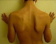
<div class="col-md-6">[[Image:Sca 3.jpg|center|alt=|thumb|220x220px|'''Figure.1''' Neurological Scapular Winging <ref>Scapular Winging. Available from: http://www.maitrise-orthop.com/viewPage_us.do?id=1010</ref>]]</div> | |||
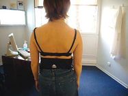
<div class="col-md-6">[[Image:Sca 4.jpg|center|alt=|thumb|239x239px|'''Figure.2''' Musculoskeletal Scapular Winging]]</div> | |||
</div> | |||
== Clinically Relevant Anatomy == | |||
[[File:Scapula bone - Kenhub.png|alt=Scapula bone (highlighted in green) - posterior view|450x450px|'''Figure.3''' Scapula - Posterior View <ref>Scapula Bone (Highlighted in Green) - Posterior View Image - © Kenhub. Available from: https://www.kenhub.com/en/library/anatomy/scapula | |||
</ref>|thumb]]The anatomic structures involved in the winged scapula are: | |||
'''Bones''' | |||
*[[Scapula]] | |||
'''Muscles''' | |||
*[[Trapezius]] | |||
*[[Serratus Anterior]] | |||
*[[Rhomboids]] | |||
*[[Levator Scapulae]] | |||
*[[Pectoralis Minor]] | |||
*[[Latissimus Dorsi Muscle|Latissimus Dorsi]] | |||
'''Nerves''' | |||
*[[Accessory Nerve Cranial Nerve 11|Spinal Accessory Nerve]] | |||
*[[Dorsal Scapular Nerve]] | |||
*[[Long Thoracic Nerve|Thoracicus Longus / Long Thoracic Nerve]] | |||
*[[Brachial Plexus|Brachial Plexus]] | |||
{{#ev:youtube|kA9ISVvc69k}}Structures involving the musculoskeletal winged scapula:<ref name=":2">Ann Cools, Marc Walravens. Oefentherapie bij schouderaandoening. 2005, 104-135. VUB-BIB.fckLREvidence levels : F</ref> | |||
* [[Trapezius]] | |||
*[[Serratus Anterior]] | |||
*[[Rhomboids]] - Rhomboids Major | |||
*[[Levator Scapulae]] | |||
*[[Pectoralis Minor]] | |||
*[[Latissimus Dorsi Muscle|Latissimus Dorsi]] | |||
< | Structures involving the neurological winged scapula:<ref name=":0" /> | ||
* [[Trapezius]] (Pars Descendens) | |||
*[[Serratus Anterior]] | |||
*[[Rhomboids]] | |||
*[[Accessory Nerve Cranial Nerve 11|Spinal Accessory Nerve]] | |||
*[[Dorsal Scapular Nerve]] | |||
*[[Long Thoracic Nerve|Thoracicus Longus / Long Thoracic Nerve]] | |||
*[[Brachial Plexus|Brachial Plexus]] | |||
== | == Etiology == | ||
Scapular winging is rare. Out of which, incidence of scapular winging due to trapezius paralysis is very sparse and difficult to assess. Whereas serratus anterior paralysis due to iatrogenic injury has shown to be the most common reason for scapular winging. Broadly, the etiology of winging are: | |||
* Serratus Anterior Palsy | |||
* Trapezius Palsy | |||
* Rhomboid Palsy<ref name=":5" /> | |||
== Epidemiology == | |||
The causes of all these leading winged scapula are:<ref name=":1" /><ref>C.L. Foo, M. Swann. Isolated paralysis of the serratus anterior, a case report of 20 cases. The journal of bone and joint surgery 1983 (65-B), 552-556.fckLREvidence levels: 3</ref><ref>B. Forthomme, F.C. Wang, J.M. Crielaard, J.L. Croisier. Scapula alata : facteurs musculaires et neurologiques. Épaule neurologique et médecine de réeducation 2009, 21-24.fckLREvidence levels : E</ref> | |||
=== Traumatic === | |||
< | * Acute Traumas, for example a direct shock on the shoulder during a car accident with a sudden traction on the arm. It has also seen amongst professional and amateur athletes of a variety of sports, including archery, ballet, baseball, basketball, body building/weight lifting, bowling, football, golf, gymnastics, hockey, soccer, tennis, and wrestling. | ||
* Micro Traumas, repeated stretching of the neck in later flexion as in tennis (N. Thoracicus longus) or by wearing a heavy backpack (N. Accessories).Occupational injuries in individuals working as car mechanics, navy airmen, scaffolders, welders, carpenters, laborers, and a seamstress has also reported.<ref name=":5" /> | |||
=== Post-infection === | |||
Influenza infection, tonsillitis-bronchitis, [[poliomyelitis]], etc. | |||
== | === Iatrogenic Injury === | ||
* As result of post-surgical complications, like a chest tube placement<ref>W.U. Hassan, N.P. Keaney. Winging of the scapula: an unusual complication of chest tube placement. Journal of accident and Emergency Medicine 1994 (12), 156-157.fckLREvidence levels: A2</ref>. Mastectomies for [[Breast Cancer|breast cancer]] that involve resection of the axillary lymph nodes are at higher risk as the long thoracic nerve lies near the axillae. Neck dissection may lead to injury to the spinal accessory nerve causing trapezius paralysis.<ref name=":6">Park SB, Ramage JL. [https://www.ncbi.nlm.nih.gov/books/NBK541005/ Winging of the Scapula.] StatPearls [Internet]. 2021 Feb 27.</ref> | |||
* Allergic-drug reactions, drug overdose, toxic exposure (herbicides and tetanus antitoxin) | |||
* Consequence of Chiropractic Manipulation | |||
* Use of a Single Axillary Crutch | |||
=== Congenital === | |||
* [[Muscular Dystrophy]]-Fascioscapulohumeral Dystrophy | |||
=== Spontaneous === | |||
* Idiopathic as in the case of [[Parsonage-Turner Syndrome]]. | |||
== Clinical Presentation == | |||
<ref> | * Pain: A severe or excruciating pain, often keeping them awake are mainly caused by a neurological trauma or neuritis. However, winged scapula due to muscular cause are not painful; some may experience moderate pain.<ref name=":0" /> Pain can be the result of the strain and spasm of overcompensating periscapular muscles which could be dull-aching and heaviness feeling. | ||
* Difficulty with elevating the arm above the head and lifting object. Patients couldn't flex their shoulder above 120°.<ref name=":6" /> | |||
* Fatigue was a significant characteristic.<ref name=":5" /> | |||
* On physical exam, | |||
*# Serratus anterior palsy: Health care providers should be able to recognize deformation of the back due to a protrusion of the medial portion of the scapula, which is not anchored against the [[Ribs|rib cage]]. | |||
*# Trapezius palsy: An asymmetrical neckline with drooping of the effected [[Shoulder|shoulder.]] This may be accompanied with lateral displacement and winging of the scapula. | |||
*# Rhomboids palsy: It produces a very subtle winging of the scapula, with the scapula laterally translated and the inferior angle rotated laterally | |||
* Clinical test that providers can use to assess patients are: | |||
*# Serratus anterior palsy: Have the patient face a wall and stand with the affected arm out in front of their body, parallel to the floor. The patient should then be instructed to push against the wall with the palm of their hand on the affected side. A protrusion of the medial portion of the scapula should then be apparent showing serratus anterior palsy. <ref name=":5" />[[File:Sca_2.jpg|right|194x194px]] | |||
*#Trapezius palsy: Typically, winging is minimal so can be easily missed. It is accentuated during arm abduction, with the scapula moving upwards with the superior angle more lateral to the midline than the inferior angle. Winging may disappear during forward flexion of the arm due to the action of the serratus anterior muscle. [[File:Sca_1.jpg|right|187x187px]] | |||
*#Rhomboids palsy: Winging may be accentuated by having the patient extend his or her arm from a fully flexed position, during which the inferior angle of the scapula is pulled laterally and dorsally off the thoracic wall. Weakness of the rhomboids can be tested by having the patient try to bring his or her scapulae together medially, or by having the patient push his or her elbows backwards against resistance with [[Hand Function|hands]] on [[Hip|hips]]. Difficulty with either task suggests weakness of the rhomboids, but this may be masked by trapezius hypertrophy.<ref name=":5" /> | |||
{| class="wikitable" | |||
! colspan="1" rowspan="1" | | |||
! rowspan="1" |Medial winging | |||
! colspan="2" |Lateral winging | |||
|- | |||
| colspan="1" rowspan="1" |Injured nerve | |||
| colspan="1" rowspan="1" |Long thoracic | |||
| colspan="1" rowspan="1" |Spinal accessory | |||
| colspan="1" rowspan="1" |Dorsal scapular | |||
|- | |||
| colspan="1" rowspan="1" |Muscle palsy | |||
| colspan="1" rowspan="1" |Serratus anterior | |||
| colspan="1" rowspan="1" |Trapezius | |||
| colspan="1" rowspan="1" |Rhomboids | |||
|- | |||
| colspan="1" rowspan="1" |Physical exam | |||
| colspan="1" rowspan="1" |Arm flexion; push-up motion against a wall | |||
| colspan="1" rowspan="1" |Arm abduction; external rotation against resistance | |||
| colspan="1" rowspan="1" |Arm extension from full flexion | |||
|- | |||
| colspan="1" rowspan="1" |Position of the scapula compared to normal | |||
| colspan="1" rowspan="1" |Entire scapula displaced more medial and superior | |||
| colspan="1" rowspan="1" |Superior angle more laterally displaced | |||
| colspan="1" rowspan="1" |Inferior angle more laterally displaced | |||
|} | |||
== Diagnostic Procedures == | |||
*Scapular winging can be diagnosed by practitioner with a proper history and physical examination. | |||
*Electrodiagnostic testing can help to establish the underlying neuromuscular pathology. | |||
*Neuromuscular ultrasound can be used to establish the muscular pathology and the neurologic causes of the muscle pathology. <ref name=":6" /> | |||
=== | == Differential Diagnosis == | ||
* [[Rotator Cuff Tendinopathy|Rotator Cuff Disorders]] | |||
* [[Frozen Shoulder]] | |||
* [[Trapezius Myalgia|Over-stressed Trapezius]] | |||
* [[Shoulder Instability|Glenohumeral Instability]] | |||
* [[Thoracic Outlet Syndrome (TOS)|Thoracic Outlet Syndrome]] | |||
* [[Cervical Spondylosis]] | |||
* [[Acromioclavicular Joint Disorders|Acromioclavicular Disorder]]<ref name=":5" /> | |||
== Management == | |||
Currently, no treatment method is considered to be the first line for the resolution of scapular winging. As discussed prior, the recommended treatment for initial treatment is [[Pain Mechanisms|pain]] control and physical therapy. If treatment is not initiated early on in the progression of the condition, patients can develop subsequent issues such as [[Adhesive Capsulitis|adhesive capsulitis]] (or frozen shoulder), [[Subacromial Impingement|subacromial impingement]], and other pathogenesis involving the [[Brachial Plexus|brachial plexus.]]<ref name=":6" /> | |||
=== Medical Management === | |||
== Medical Management | |||
There are surgical treatments with patients being very satisfied about the result. But some studies prefer a non-operative treatment especially in case of older patients who are sedentary and with minimal symptoms.<ref name=":3">Sandro Giannini, MD; Cesare Faldini, MD; Stavroula Pagkrati, MD; Gianluca Grandi, MD;Vitantonio Digennaro, MD; Deianira Luciani, MD; and Luciano Merlini, MD. Fixation of winged scapula in facioscapulahumeral muscular dystrophy. Clinical Medicine and research 2007 (5), 155-162.fckLREvidence levels: A2</ref> | |||
These treatments are: | These treatments are: | ||
* Split | * Split Pectoralis Major Transfer<ref name=":4">Galano GJ, Bigliani LU, Ahmad CS, Levine WN. [https://www.researchgate.net/publication/5655872_Surgical_Treatment_of_Winged_Scapula Surgical treatment of winged scapula]. Clinical orthopaedics and related research. 2008 Mar;466(3):652-60.</ref><ref>John Iceton, W.R. Harris. Treatment of winged scapula by pectoralis major transfer. The journal of bone and joint surgery 1987 (69-B), 108-110.fckLREvidence levels: A2</ref> | ||
* Modified version of the Eden-Lange | * Modified version of the Eden-Lange Procedure<ref name=":4" /> | ||
* | * Scapuloplexy<ref name=":3" /><br> | ||
=== Physical Therapy Management === | |||
Physiotherapy treatment depends upon the etiology of the scapular winging. | |||
=== '''Serratus Anterior Palsy''' === | |||
* Upon diagnosis, patients should be advised to avoid overhead use of the effected extremity and to avoid activities that cause pain . | |||
* [[Range of Motion|Range of motion (ROM)]] exercises in the supine position should also be prescribed. In the supine position, the weight of the body prevents winging by compressing the scapula against the thorax and allows for full shoulder range of motion. | |||
* Special consideration should be taken not to stretch the serratus anterior muscle, as [[stretching]] the denervated muscle can impair the time to and extent of functional recovery. | |||
* A scapular brace may accomplish both tasks of keeping the scapula placed against the thorax and preventing stretching of the serratus anterior muscle, and has been shown to be a generally effective treatment option for compliant individuals. However, the brace tends to be poorly tolerated leading to poor compliance and less functional recovery. | |||
* Watson and Schenkman have identified three stages of long thoracic nerve injury and appropriate treatment at each stage. | |||
** In the acute stage, denervation of the serratus anterior causes pain, and goals of treatment include pain reduction and ROM exercise. Activity modification of the patient is also important to limit further injury to the shoulder. | |||
** In the intermediate stage, the pain has subsided and the nerve is beginning to heal. To maintain full ROM, passive stretching of the rhomboids, [[Levator Scapulae|levator scapulae,]] and pectoralis minor is used to prevent [[Contracture Management for Traumatic Brain Injury|contracture]] of these muscles due to the loss of serratus anterior activity. | |||
** In the third or late stage, the serratus anterior becomes progressively stronger and shoulder mechanics improve. To improve strength and overhead work, [[Strength Training|strengthening exercise]] of all shoulder girdle muscles, including the trapezius, should be implemented and avoidance of overstretching the serratus anterior should be continued.<ref name=":5" /> | |||
=== '''Trapezius Palsy''' === | |||
* Functional recovery of the trapezius muscle due to spinal accessory nerve injury does not consistently benefit from conservative management such as physical therapy, [[Transcutaneous Electrical Nerve Stimulation (TENS)|transcutaneous nerve stimulation]], external support, chiropractic, [[The Influence of NSAIDs on Physiologic Processes and Exercise|NSAIDS,]] and narcotic analgesics. | |||
* A shoulder orthosis has been developed for trapezius muscle paralysis and has been used somewhat successfully in radical neck dissection patients. Within 3 months of using the [[Assistive Devices|orthosis]], 72% of patients were pain free, had improved shoulder girdle muscle function, and increased endurance and function due to loss of pain. However, active abduction only improved by 5–20°. | |||
* The preferred method of treatment for healthy and active patients with isolated chronic trapezius palsy secondary to spinal accessory nerve injury is the Eden-Lange muscle transfer procedure.<ref name=":5" /> | |||
== | === '''Rhomboids Palsy''' === | ||
* Injury to the dorsal scapular nerve or the rhomboid muscles is usually treated conservatively with cervical spine stabilization (collar or cervical traction), muscle relaxants, anti-inflammatories, and physical therapy. | |||
* Strengthening of the trapezius is the primary objective of physical therapy, as the middle portion of the trapezius can compensate for rhomboid weakness or paralysis.<ref name=":5" /> | |||
==== Revalidation Program ==== | |||
{| width="100%" cellspacing="1" cellpadding="1" | {| width="100%" cellspacing="1" cellpadding="1" | ||
|- | |- | ||
| {{#ev:youtube| | | {{#ev:youtube|FBJHyT_MtzA|412}} | ||
| {{#ev:youtube|1Qw77UR7LTE|412}} | | {{#ev:youtube|1Qw77UR7LTE|412}} | ||
|} | |}In the revalidation we have to give attention to diverse monitoring parameters. Timing, muscle activity, muscular balance, endurance and power of the scapular muscles are important in the revalidation program.<ref name=":2" /> | ||
* '''Conscious muscle activation of scapular muscles:<ref name=":2" />''' | |||
In the revalidation we have to give attention to diverse monitoring parameters. Timing, muscle activity, muscular balance, endurance and power of the scapular muscles are important in the revalidation program.<ref | |||
* '''Conscious muscle activation of scapular muscles:<ref | |||
** Correction scapular position with tactile feedback on the level of the angulus inferior and ask the patient to move the scapula downwards and inwards. | ** Correction scapular position with tactile feedback on the level of the angulus inferior and ask the patient to move the scapula downwards and inwards. | ||
** Scapulothoracal feedback of muscle control with feedback on the level of the sternum | ** Scapulothoracal feedback of muscle control with feedback on the level of the sternum | ||
| Line 97: | Line 187: | ||
** Myofeedback | ** Myofeedback | ||
*'''Automate of the scapular muscle control:<ref | *'''Automate of the scapular muscle control:<ref name=":2" />''' | ||
**Rhythmic stabilisation in lateral position, hand supported on 90° anteflexion | **Rhythmic stabilisation in lateral position, hand supported on 90° anteflexion | ||
**Rhythmic stabilisation in stand, hand supports on the ball against the wall | **Rhythmic stabilisation in stand, hand supports on the ball against the wall | ||
**Rhythmic stabilisation in prone, the shoulder girdle in retraction, shoulder in | **Rhythmic stabilisation in prone, the shoulder girdle in retraction, shoulder in external rotation | ||
**Rhythmic stabilisation in prone, shoulder girdle in protraction, arm in submaximal elevation | **Rhythmic stabilisation in prone, shoulder girdle in protraction, arm in submaximal elevation | ||
*'''Dynamic scapulothoracal muscle training:<ref | *'''Dynamic scapulothoracal muscle training:<ref name=":2" />''' | ||
**Push-up with a plus (additional protraction of the scapula) | **Push-up with a plus (additional protraction of the scapula) | ||
**Elevation in the scapular flat in exorotation | **Elevation in the scapular flat in exorotation | ||
| Line 116: | Line 206: | ||
== Clinical Bottom Line == | == Clinical Bottom Line == | ||
* Winged scapula can have a neurological or a musculoskeletal cause<ref name=":0" /><ref | * Winged scapula can have a neurological or a musculoskeletal cause<ref name=":0" /><ref name=":2" /> | ||
* Serratus anterior palsy is the most common cause of scapular winging.<ref name=":6" /> | |||
* It can arise often after an acute trauma or micro-trauma<ref name=":0" /> | * It can arise often after an acute trauma or micro-trauma<ref name=":0" /> | ||
* Physical exercises for the scapula. First conscious muscles activation of the scapula, then automate exercises for the scapula muscles and at last dynamic exercises for the muscles of the scapula.<ref> | * Physical exercises for the scapula. First conscious muscles activation of the scapula, then automate exercises for the scapula muscles and at last dynamic exercises for the muscles of the scapula.<ref name=":2" /> <br> | ||
== Resources == | |||
This 3 minute video is a good summary of the movement of the scapula bone.<ref> Scapula bone video - © Kenhub https://www.kenhub.com/en/library/anatomy/scapula</ref>{{#ev:youtube|fEXrPsGySKI}} | |||
== References == | == References == | ||
Latest revision as of 18:59, 8 March 2024
Original Editors - Derick Glenn
Top Contributors - Derick Glenn, Kim Jackson, Manisha Shrestha, Joao Costa, Admin, Wendy Walker, Naomi O'Reilly, George Prudden, Simisola Ajeyalemi, Oyemi Sillo, Daphne Jackson, WikiSysop and Wanda van Niekerk
Definition/Description[edit | edit source]
The term ‘winged scapula’ (also scapula alata) is used when the muscles of the scapula are too weak or paralyzed, resulting in a limited ability to stabilize the scapula. As a result, the medial or lateral borders of the scapula protrudes from back, like wings. The main reasons for this condition are musculoskeletal- and neurological-related.[1][2]Winging of scapula disturbs scapulohumeral rhythm; contributes to loss of power and limited flexion and abduction of the upper extremity and can be a source of considerable pain. This debilitating condition that can affect the ability to lift, pull, and push heavy objects, as well as to perform daily activities of living, such as brushing one’s hair and teeth and carrying grocery bags.[3]


Clinically Relevant Anatomy[edit | edit source]

The anatomic structures involved in the winged scapula are:
Bones
Muscles
Nerves
- Spinal Accessory Nerve
- Dorsal Scapular Nerve
- Thoracicus Longus / Long Thoracic Nerve
- Brachial Plexus
Structures involving the musculoskeletal winged scapula:[6]
- Trapezius
- Serratus Anterior
- Rhomboids - Rhomboids Major
- Levator Scapulae
- Pectoralis Minor
- Latissimus Dorsi
Structures involving the neurological winged scapula:[1]
- Trapezius (Pars Descendens)
- Serratus Anterior
- Rhomboids
- Spinal Accessory Nerve
- Dorsal Scapular Nerve
- Thoracicus Longus / Long Thoracic Nerve
- Brachial Plexus
Etiology[edit | edit source]
Scapular winging is rare. Out of which, incidence of scapular winging due to trapezius paralysis is very sparse and difficult to assess. Whereas serratus anterior paralysis due to iatrogenic injury has shown to be the most common reason for scapular winging. Broadly, the etiology of winging are:
- Serratus Anterior Palsy
- Trapezius Palsy
- Rhomboid Palsy[3]
Epidemiology[edit | edit source]
The causes of all these leading winged scapula are:[2][7][8]
Traumatic[edit | edit source]
- Acute Traumas, for example a direct shock on the shoulder during a car accident with a sudden traction on the arm. It has also seen amongst professional and amateur athletes of a variety of sports, including archery, ballet, baseball, basketball, body building/weight lifting, bowling, football, golf, gymnastics, hockey, soccer, tennis, and wrestling.
- Micro Traumas, repeated stretching of the neck in later flexion as in tennis (N. Thoracicus longus) or by wearing a heavy backpack (N. Accessories).Occupational injuries in individuals working as car mechanics, navy airmen, scaffolders, welders, carpenters, laborers, and a seamstress has also reported.[3]
Post-infection[edit | edit source]
Influenza infection, tonsillitis-bronchitis, poliomyelitis, etc.
Iatrogenic Injury[edit | edit source]
- As result of post-surgical complications, like a chest tube placement[9]. Mastectomies for breast cancer that involve resection of the axillary lymph nodes are at higher risk as the long thoracic nerve lies near the axillae. Neck dissection may lead to injury to the spinal accessory nerve causing trapezius paralysis.[10]
- Allergic-drug reactions, drug overdose, toxic exposure (herbicides and tetanus antitoxin)
- Consequence of Chiropractic Manipulation
- Use of a Single Axillary Crutch
Congenital[edit | edit source]
- Muscular Dystrophy-Fascioscapulohumeral Dystrophy
Spontaneous[edit | edit source]
- Idiopathic as in the case of Parsonage-Turner Syndrome.
Clinical Presentation[edit | edit source]
- Pain: A severe or excruciating pain, often keeping them awake are mainly caused by a neurological trauma or neuritis. However, winged scapula due to muscular cause are not painful; some may experience moderate pain.[1] Pain can be the result of the strain and spasm of overcompensating periscapular muscles which could be dull-aching and heaviness feeling.
- Difficulty with elevating the arm above the head and lifting object. Patients couldn't flex their shoulder above 120°.[10]
- Fatigue was a significant characteristic.[3]
- On physical exam,
- Serratus anterior palsy: Health care providers should be able to recognize deformation of the back due to a protrusion of the medial portion of the scapula, which is not anchored against the rib cage.
- Trapezius palsy: An asymmetrical neckline with drooping of the effected shoulder. This may be accompanied with lateral displacement and winging of the scapula.
- Rhomboids palsy: It produces a very subtle winging of the scapula, with the scapula laterally translated and the inferior angle rotated laterally
- Clinical test that providers can use to assess patients are:
- Serratus anterior palsy: Have the patient face a wall and stand with the affected arm out in front of their body, parallel to the floor. The patient should then be instructed to push against the wall with the palm of their hand on the affected side. A protrusion of the medial portion of the scapula should then be apparent showing serratus anterior palsy. [3]
- Trapezius palsy: Typically, winging is minimal so can be easily missed. It is accentuated during arm abduction, with the scapula moving upwards with the superior angle more lateral to the midline than the inferior angle. Winging may disappear during forward flexion of the arm due to the action of the serratus anterior muscle.
- Rhomboids palsy: Winging may be accentuated by having the patient extend his or her arm from a fully flexed position, during which the inferior angle of the scapula is pulled laterally and dorsally off the thoracic wall. Weakness of the rhomboids can be tested by having the patient try to bring his or her scapulae together medially, or by having the patient push his or her elbows backwards against resistance with hands on hips. Difficulty with either task suggests weakness of the rhomboids, but this may be masked by trapezius hypertrophy.[3]
- Serratus anterior palsy: Have the patient face a wall and stand with the affected arm out in front of their body, parallel to the floor. The patient should then be instructed to push against the wall with the palm of their hand on the affected side. A protrusion of the medial portion of the scapula should then be apparent showing serratus anterior palsy. [3]


| Medial winging | Lateral winging | ||
|---|---|---|---|
| Injured nerve | Long thoracic | Spinal accessory | Dorsal scapular |
| Muscle palsy | Serratus anterior | Trapezius | Rhomboids |
| Physical exam | Arm flexion; push-up motion against a wall | Arm abduction; external rotation against resistance | Arm extension from full flexion |
| Position of the scapula compared to normal | Entire scapula displaced more medial and superior | Superior angle more laterally displaced | Inferior angle more laterally displaced |
Diagnostic Procedures[edit | edit source]
- Scapular winging can be diagnosed by practitioner with a proper history and physical examination.
- Electrodiagnostic testing can help to establish the underlying neuromuscular pathology.
- Neuromuscular ultrasound can be used to establish the muscular pathology and the neurologic causes of the muscle pathology. [10]
Differential Diagnosis[edit | edit source]
- Rotator Cuff Disorders
- Frozen Shoulder
- Over-stressed Trapezius
- Glenohumeral Instability
- Thoracic Outlet Syndrome
- Cervical Spondylosis
- Acromioclavicular Disorder[3]
Management[edit | edit source]
Currently, no treatment method is considered to be the first line for the resolution of scapular winging. As discussed prior, the recommended treatment for initial treatment is pain control and physical therapy. If treatment is not initiated early on in the progression of the condition, patients can develop subsequent issues such as adhesive capsulitis (or frozen shoulder), subacromial impingement, and other pathogenesis involving the brachial plexus.[10]
Medical Management[edit | edit source]
There are surgical treatments with patients being very satisfied about the result. But some studies prefer a non-operative treatment especially in case of older patients who are sedentary and with minimal symptoms.[11]
These treatments are:
- Split Pectoralis Major Transfer[12][13]
- Modified version of the Eden-Lange Procedure[12]
- Scapuloplexy[11]
Physical Therapy Management[edit | edit source]
Physiotherapy treatment depends upon the etiology of the scapular winging.
Serratus Anterior Palsy[edit | edit source]
- Upon diagnosis, patients should be advised to avoid overhead use of the effected extremity and to avoid activities that cause pain .
- Range of motion (ROM) exercises in the supine position should also be prescribed. In the supine position, the weight of the body prevents winging by compressing the scapula against the thorax and allows for full shoulder range of motion.
- Special consideration should be taken not to stretch the serratus anterior muscle, as stretching the denervated muscle can impair the time to and extent of functional recovery.
- A scapular brace may accomplish both tasks of keeping the scapula placed against the thorax and preventing stretching of the serratus anterior muscle, and has been shown to be a generally effective treatment option for compliant individuals. However, the brace tends to be poorly tolerated leading to poor compliance and less functional recovery.
- Watson and Schenkman have identified three stages of long thoracic nerve injury and appropriate treatment at each stage.
- In the acute stage, denervation of the serratus anterior causes pain, and goals of treatment include pain reduction and ROM exercise. Activity modification of the patient is also important to limit further injury to the shoulder.
- In the intermediate stage, the pain has subsided and the nerve is beginning to heal. To maintain full ROM, passive stretching of the rhomboids, levator scapulae, and pectoralis minor is used to prevent contracture of these muscles due to the loss of serratus anterior activity.
- In the third or late stage, the serratus anterior becomes progressively stronger and shoulder mechanics improve. To improve strength and overhead work, strengthening exercise of all shoulder girdle muscles, including the trapezius, should be implemented and avoidance of overstretching the serratus anterior should be continued.[3]
Trapezius Palsy[edit | edit source]
- Functional recovery of the trapezius muscle due to spinal accessory nerve injury does not consistently benefit from conservative management such as physical therapy, transcutaneous nerve stimulation, external support, chiropractic, NSAIDS, and narcotic analgesics.
- A shoulder orthosis has been developed for trapezius muscle paralysis and has been used somewhat successfully in radical neck dissection patients. Within 3 months of using the orthosis, 72% of patients were pain free, had improved shoulder girdle muscle function, and increased endurance and function due to loss of pain. However, active abduction only improved by 5–20°.
- The preferred method of treatment for healthy and active patients with isolated chronic trapezius palsy secondary to spinal accessory nerve injury is the Eden-Lange muscle transfer procedure.[3]
Rhomboids Palsy[edit | edit source]
- Injury to the dorsal scapular nerve or the rhomboid muscles is usually treated conservatively with cervical spine stabilization (collar or cervical traction), muscle relaxants, anti-inflammatories, and physical therapy.
- Strengthening of the trapezius is the primary objective of physical therapy, as the middle portion of the trapezius can compensate for rhomboid weakness or paralysis.[3]
Revalidation Program[edit | edit source]
In the revalidation we have to give attention to diverse monitoring parameters. Timing, muscle activity, muscular balance, endurance and power of the scapular muscles are important in the revalidation program.[6]
- Conscious muscle activation of scapular muscles:[6]
- Correction scapular position with tactile feedback on the level of the angulus inferior and ask the patient to move the scapula downwards and inwards.
- Scapulothoracal feedback of muscle control with feedback on the level of the sternum
- The patient put his fingers on the processus coracoideus, after this the patient moves his scapula backwards (medio-cranial) with his fingers on the processus coracoideus.
- Myofeedback
- Automate of the scapular muscle control:[6]
- Rhythmic stabilisation in lateral position, hand supported on 90° anteflexion
- Rhythmic stabilisation in stand, hand supports on the ball against the wall
- Rhythmic stabilisation in prone, the shoulder girdle in retraction, shoulder in external rotation
- Rhythmic stabilisation in prone, shoulder girdle in protraction, arm in submaximal elevation
- Dynamic scapulothoracal muscle training:[6]
- Push-up with a plus (additional protraction of the scapula)
- Elevation in the scapular flat in exorotation
- Elbow push-up
- Press-up
- Low rowing
- Horizontal abduction
- Retroflexion against resistance
- Serratus punch upright, prone and fitter
- Dynamic hug
- Elbow in the back pocket’-exercise
Clinical Bottom Line[edit | edit source]
- Winged scapula can have a neurological or a musculoskeletal cause[1][6]
- Serratus anterior palsy is the most common cause of scapular winging.[10]
- It can arise often after an acute trauma or micro-trauma[1]
- Physical exercises for the scapula. First conscious muscles activation of the scapula, then automate exercises for the scapula muscles and at last dynamic exercises for the muscles of the scapula.[6]
Resources[edit | edit source]
This 3 minute video is a good summary of the movement of the scapula bone.[14]
References[edit | edit source]
- ↑ 1.0 1.1 1.2 1.3 1.4 Dr Jacques Vanderstraeten, médecin généraliste et du sport. Scapula alata. La revue de la Médecine Générale 2010 (269), 32-33.fckLREvidence levels : A1
- ↑ 2.0 2.1 Grethe Aalkjӕr, Lisbeth Rejsenhus. Scapula alata or winging scapula. 2006, 1-7.fckLREvidence levels :A2
- ↑ 3.0 3.1 3.2 3.3 3.4 3.5 3.6 3.7 3.8 3.9 Martin RM, Fish DE. Scapular winging: anatomical review, diagnosis, and treatments. Current reviews in musculoskeletal medicine. 2008 Mar 1;1(1):1-1.
- ↑ Scapular Winging. Available from: http://www.maitrise-orthop.com/viewPage_us.do?id=1010
- ↑ Scapula Bone (Highlighted in Green) - Posterior View Image - © Kenhub. Available from: https://www.kenhub.com/en/library/anatomy/scapula
- ↑ 6.0 6.1 6.2 6.3 6.4 6.5 6.6 Ann Cools, Marc Walravens. Oefentherapie bij schouderaandoening. 2005, 104-135. VUB-BIB.fckLREvidence levels : F
- ↑ C.L. Foo, M. Swann. Isolated paralysis of the serratus anterior, a case report of 20 cases. The journal of bone and joint surgery 1983 (65-B), 552-556.fckLREvidence levels: 3
- ↑ B. Forthomme, F.C. Wang, J.M. Crielaard, J.L. Croisier. Scapula alata : facteurs musculaires et neurologiques. Épaule neurologique et médecine de réeducation 2009, 21-24.fckLREvidence levels : E
- ↑ W.U. Hassan, N.P. Keaney. Winging of the scapula: an unusual complication of chest tube placement. Journal of accident and Emergency Medicine 1994 (12), 156-157.fckLREvidence levels: A2
- ↑ 10.0 10.1 10.2 10.3 10.4 Park SB, Ramage JL. Winging of the Scapula. StatPearls [Internet]. 2021 Feb 27.
- ↑ 11.0 11.1 Sandro Giannini, MD; Cesare Faldini, MD; Stavroula Pagkrati, MD; Gianluca Grandi, MD;Vitantonio Digennaro, MD; Deianira Luciani, MD; and Luciano Merlini, MD. Fixation of winged scapula in facioscapulahumeral muscular dystrophy. Clinical Medicine and research 2007 (5), 155-162.fckLREvidence levels: A2
- ↑ 12.0 12.1 Galano GJ, Bigliani LU, Ahmad CS, Levine WN. Surgical treatment of winged scapula. Clinical orthopaedics and related research. 2008 Mar;466(3):652-60.
- ↑ John Iceton, W.R. Harris. Treatment of winged scapula by pectoralis major transfer. The journal of bone and joint surgery 1987 (69-B), 108-110.fckLREvidence levels: A2
- ↑ Scapula bone video - © Kenhub https://www.kenhub.com/en/library/anatomy/scapula

