Midbrain: Difference between revisions
No edit summary |
No edit summary |
||
| Line 12: | Line 12: | ||
When viewed in cross-section, the midbrain can be divided into three portions: | When viewed in cross-section, the midbrain can be divided into three portions: | ||
Tectum (posterior) | # Tectum (posterior) | ||
# Tegmentum | |||
Tegmentum | # Cerebral peduncles (anterior)<ref>Radiopedia Midbrain Available: https://radiopaedia.org/articles/midbrain<nowiki/>(accessed 23.4.2022)</ref> | ||
Cerebral peduncles (anterior)<ref>Radiopedia Midbrain Available: https://radiopaedia.org/articles/midbrain<nowiki/>(accessed 23.4.2022)</ref> | |||
== Tectum == | == Tectum == | ||
| Line 22: | Line 20: | ||
== Tegmentum == | == Tegmentum == | ||
The tegmentum is the phylogenetically-old part of the brainstem and runs through the pons and medulla oblongata. The tegmentum contains a variety of ascending and descending tracts that pass through the midbrain (eg the medial lemniscus and the anterolateral | The tegmentum is the phylogenetically-old part of the brainstem and runs through the pons and medulla oblongata. The tegmentum contains a variety of ascending and descending tracts that pass through the midbrain (eg the [[Dorsal Column Medial Lemniscal Pathway|medial lemniscus]] and the [[Spinothalamic tract|anterolateral tract]]<nowiki/>s). | ||
* Fibers from the superior cerebellar peduncles, the major efferent pathway from the cerebellum, decussate in the midbrain. | * Fibers from the superior cerebellar peduncles, the major efferent pathway from the [[cerebellum]], decussate in the midbrain. | ||
* Some of these fibers project to a midbrain nucleus called the red nucleus, which is thought to play an important role in motor coordination. | * Some of these fibers project to a midbrain nucleus called the [[Rubrospinal Tract|red nucleus]], which is thought to play an important role in motor coordination. | ||
* There are several other important nuclei and neuronal clusters in the midbrain tegmentum. eg nuclei for two cranial nerves: cranial nerve III (oculomotor nerve) and cranial nerve IV (trochlear nerve). | * There are several other important nuclei and neuronal clusters in the midbrain tegmentum. eg nuclei for two [[Cranial Nerves|cranial nerves]]: cranial nerve III ([[Oculomotor Nerve|oculomotor nerve]]) and cranial nerve IV ([[Trochlear Nerve|trochlear nerve]]). | ||
* It also contains neurons that are part of the raphe nuclei (clusters of serotonin-producing neurons found in the brainstem that send serotonin throughout the central nervous system); | * It also contains neurons that are part of the raphe nuclei (clusters of serotonin-producing neurons found in the brainstem that send serotonin throughout the central nervous system); | ||
* Also contains one of the largest collections of dopamine-producing neurons in the brain, the ventral tegmental area. | * Also contains one of the largest collections of [[dopamine]]-producing [[Neurone|neurons]] in the brain, the ventral tegmental area. | ||
== Cerebral Peduncles == | == Cerebral Peduncles == | ||
| Line 34: | Line 32: | ||
# The cerebral peduncles contain the substantia nigrae, which (like the ventral tegmental area) contain large collections of dopamine-producing neurons. | # The cerebral peduncles contain the substantia nigrae, which (like the ventral tegmental area) contain large collections of dopamine-producing neurons. | ||
# The area surrounding the cerebral aqueduct is called the periaqueductal gray, or PAG, long been recognized for its role in pain inhibition, although it is also thought to be involved in many other functions ranging from emotional responses to the production of vocalizations<ref name=":0" />. | # The area surrounding the cerebral aqueduct is called the periaqueductal gray, or PAG, long been recognized for its role in [[Pain-Modulation|pain inhibition]], although it is also thought to be involved in many other functions ranging from emotional responses to the production of vocalizations<ref name=":0" />. | ||
== Sub Heading 3 == | == Sub Heading 3 == | ||
Revision as of 02:51, 23 April 2022
Original Editor - Lucinda hampton
Top Contributors - Lucinda hampton, Uchechukwu Chukwuemeka and Kim Jackson
Introduction[edit | edit source]
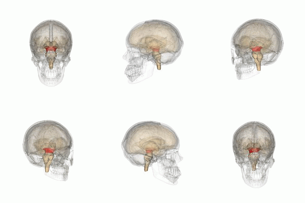
The midbrain (derived from the mesencephalon of the neural tube) is a part of the central nervous system, located below the cerebral cortex and at the topmost part of the brainstem. This small but important structure plays a crucial role in processing information related to hearing, vision, movement, pain, sleep, and arousal.
Divisions[edit | edit source]
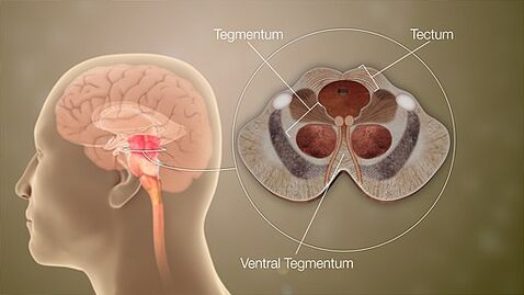
When viewed in cross-section, the midbrain can be divided into three portions:
- Tectum (posterior)
- Tegmentum
- Cerebral peduncles (anterior)[1]
Tectum[edit | edit source]
Sitting posteriorly, the tectum (Latin for "roof" or "covering") is composed of the tectal (quadrigeminal) plate and superior and inferior colliculi. The tectum is unique to the midbrain and does not have a counterpart in the rest of the brainstem.
Tegmentum[edit | edit source]
The tegmentum is the phylogenetically-old part of the brainstem and runs through the pons and medulla oblongata. The tegmentum contains a variety of ascending and descending tracts that pass through the midbrain (eg the medial lemniscus and the anterolateral tracts).
- Fibers from the superior cerebellar peduncles, the major efferent pathway from the cerebellum, decussate in the midbrain.
- Some of these fibers project to a midbrain nucleus called the red nucleus, which is thought to play an important role in motor coordination.
- There are several other important nuclei and neuronal clusters in the midbrain tegmentum. eg nuclei for two cranial nerves: cranial nerve III (oculomotor nerve) and cranial nerve IV (trochlear nerve).
- It also contains neurons that are part of the raphe nuclei (clusters of serotonin-producing neurons found in the brainstem that send serotonin throughout the central nervous system);
- Also contains one of the largest collections of dopamine-producing neurons in the brain, the ventral tegmental area.
Cerebral Peduncles[edit | edit source]
Cerebral peduncles: Anterior to the tegmentum are the cerebral peduncles which are composed of the large ascending and descending tracts that run to and from the cerebrum[2].
- The cerebral peduncles contain the substantia nigrae, which (like the ventral tegmental area) contain large collections of dopamine-producing neurons.
- The area surrounding the cerebral aqueduct is called the periaqueductal gray, or PAG, long been recognized for its role in pain inhibition, although it is also thought to be involved in many other functions ranging from emotional responses to the production of vocalizations[2].
Sub Heading 3[edit | edit source]
Resources[edit | edit source]
- bulleted list
- x
or
- numbered list
- x
References[edit | edit source]
- ↑ Radiopedia Midbrain Available: https://radiopaedia.org/articles/midbrain(accessed 23.4.2022)
- ↑ 2.0 2.1 Neuroscientifically challenged Know your midbrain Available:https://neuroscientificallychallenged.com/posts/know-your-brain-midbrain (accessed 23.4.2022)


