Hip Adductors: Difference between revisions
No edit summary |
Kim Jackson (talk | contribs) No edit summary |
||
| Line 6: | Line 6: | ||
== Introduction == | == Introduction == | ||
[[File:Adductor magnus.png|thumb|Hip Adductors (and others).]] | [[File:Adductor magnus.png|thumb|Hip Adductors (and others).]] | ||
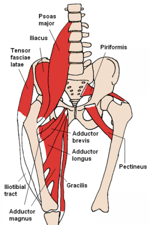
The [[Muscle Cells (Myocyte)|muscles]] in the medial compartment of the thigh are collectively known as the [[Hip Anatomy|hip]] adductors. There are four muscles in this group; [[Adductor Longus|adductor longus]], [[Adductor Brevis|adductor brevis]], [[Adductor Magnus|adductor magnus]], and [[Gracilis|gracillis]]. | The [[Muscle Cells (Myocyte)|muscles]] in the medial compartment of the thigh are collectively known as the [[Hip Anatomy|hip]] adductors. There are four primary muscles in this group; [[Adductor Longus|adductor longus]], [[Adductor Brevis|adductor brevis]], [[Adductor Magnus|adductor magnus]], and [[Gracilis|gracillis]]. These muscles primarily function to move the thigh/lower extremity closer to the body's central axis. Additionally, the Pectineus muscle, while not a primary hip adductor, assists in this movement and also plays a role in hip flexion.<ref name=":1" /> | ||
* All the hip adductors are innervated by the [[Obturator Nerve|obturator nerve]], which arises from the [[Lumbar Plexus|lumbar plexus]]<ref name=":0">Teach me anatomy Available: https://teachmeanatomy.info/lower-limb/muscles/thigh/medial-compartment/<nowiki/>(accessed 19.1.22)</ref><ref name=":1">Attum B, Varacallo M[https://www.statpearls.com/articlelibrary/viewarticle/24169/ . Anatomy, bony pelvis and lower limb, thigh muscles].Available:https://www.statpearls.com/articlelibrary/viewarticle/24169/ (accessed 19.1.2022)</ref>. | * All the hip adductors are innervated by the [[Obturator Nerve|obturator nerve]], which arises from the [[Lumbar Plexus|lumbar plexus]]<ref name=":0">Teach me anatomy Available: https://teachmeanatomy.info/lower-limb/muscles/thigh/medial-compartment/<nowiki/>(accessed 19.1.22)</ref><ref name=":1">Attum B, Varacallo M[https://www.statpearls.com/articlelibrary/viewarticle/24169/ . Anatomy, bony pelvis and lower limb, thigh muscles].Available:https://www.statpearls.com/articlelibrary/viewarticle/24169/ (accessed 19.1.2022)</ref>. | ||
Latest revision as of 13:17, 3 October 2023
Original Editor - Lucinda hampton
Top Contributors - Lucinda hampton and Kim Jackson
Introduction[edit | edit source]
The muscles in the medial compartment of the thigh are collectively known as the hip adductors. There are four primary muscles in this group; adductor longus, adductor brevis, adductor magnus, and gracillis. These muscles primarily function to move the thigh/lower extremity closer to the body's central axis. Additionally, the Pectineus muscle, while not a primary hip adductor, assists in this movement and also plays a role in hip flexion.[1]
- All the hip adductors are innervated by the obturator nerve, which arises from the lumbar plexus[2][1].
- In closed chain activation, the hip adductors help stabilize the pelvis and lower extremity during the stance phase of gait, and assist in postural control.
- They also have secondary roles including hip flexion and rotation.
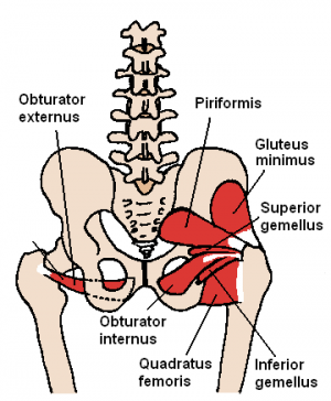
The Obturator Externus, pectineus, gemelli (superior and inferior ) and quadratus femoris may also be included by some authorities.[3][2][4]
Muscles[edit | edit source]
The adductor longus, adductor brevis, adductor magnus and gracilis primarily provide adduction of the thigh.
- The adductor magnus is the largest muscle in the medial compartment. It lies posteriorly to the other muscles, and is the most commonly injured.
- The adductor longus is a large, flat muscle. It partially covers the adductor brevis and magnus, and forms the medial border of the femoral triangle. Adductor longus provides some medial rotation..
- The adductor brevis is a short muscle that lies underneath the adductor longus.
- The gracilis is the most superficial and medial of hip adductors. It crosses at both the hip and knee joints, and adducts the thigh at the hip, and flexion of the leg at the knee.[5]
Physiotherapy Relevance[edit | edit source]
Strain of the adductor muscles is the underlying cause of a ‘groin strain‘/ Adductor Tendinopathy, among the most common sport injuries.[3] The proximal part of the muscle is most commonly affected, tearing near their bony attachments in the pelvis[2]. See also Assessment of Athletes with Groin Pain
The adductor squeeze test is used in the diagnosis of groin injuries and for the measurement of adductor muscles strength
Adductor tenotomy (cutting the origin tendons of the adductor muscles of the thigh) is sometimes used for infants with spastic quadriparesis (cerebral palsy).[6]Due to the spasticity in the hip adductors the affected children walk (if possible) with an adducted, flexed and internally rotated legs (scissor gait)[3].
References[edit | edit source]
- ↑ 1.0 1.1 Attum B, Varacallo M. Anatomy, bony pelvis and lower limb, thigh muscles.Available:https://www.statpearls.com/articlelibrary/viewarticle/24169/ (accessed 19.1.2022)
- ↑ 2.0 2.1 2.2 Teach me anatomy Available: https://teachmeanatomy.info/lower-limb/muscles/thigh/medial-compartment/(accessed 19.1.22)
- ↑ 3.0 3.1 3.2 Ken Hub Thigh Adductors Available: https://www.kenhub.com/en/library/anatomy/the-hip-adductors(accessed 19.1.2022)
- ↑ Radiopedia Hip Muscles https://radiopaedia.org/articles/hip-muscles-1?lang=gb (accessed 19.1.2022)
- ↑ Kiel J, Kaiser K. Adductor Strain. Available: https://www.ncbi.nlm.nih.gov/books/NBK493166/(accessed 19.1.2022)
- ↑ Orthobullets Cerebral Palsy - Hip Conditions Available: https://www.orthobullets.com/pediatrics/4130/cerebral-palsy--hip-conditions(accessed 19.1.2022)


