Lumbar Fusion Rehabilitation: Difference between revisions

No edit summary
mNo edit summary
 
(52 intermediate revisions by 6 users not shown)
Line 1: Line 1:
<div class="editorbox">Original Editor - [[User:Mariam Hashem|Mariam Hashem]]


== Indications ==
Top Contributors - {{Special:Contributors/{{FULLPAGENAME}}}}
</div>
-Severe, disabling back or leg pain. 


-Posttraumatic cases of segmental instability or potential neurologic injury 


-Degenerative spinal pathology with failure of conservative treatment. 


== Degenerative cascade ==


[[Degenerative Disc Disease]]
==Introduction==
[[Lumbar]] Spine Fusion is an effective treatment option to stabilise the painful motion segment, and may provide indirect decompression of the neural elements, restore lordosis and correct deformity<ref name=":1">Mobbs, R. J., Phan, K., Malham, G., Seex, K., & Rao, P. J. (2015). [https://scholar.google.com/scholar_url?url=https://www.ncbi.nlm.nih.gov/pmc/articles/PMC5039869/&hl=en&sa=T&oi=gsb-ggp&ct=res&cd=0&d=13017676127347181189&ei=GZ2uY_yfMYa8yATUzZGgCg&scisig=AAGBfm03lVzUHVhRGS8YC8pG4Qkj11Oclg Lumbar interbody fusion] : techniques , indications and comparison of interbody fusion options including PLIF , TLIF , MI-TLIF , OLIF / ATP , LLIF and ALIF, ''3''(1), 2–18. <nowiki>https://doi.org/10.3978/j.issn.2414-469X.2015.10.05</nowiki></ref>.


== Diagnosis: ==
The [[Surgery and General Anaesthetic|surgery]] involves placement of an implant (cage, spacer or structural graft) within the intervertebral space after discectomy<ref name=":1" /> and successful union of two or more [[Lumbar Vertebrae|vertebra]]<ref name=":0" />.


==== Spinal radiographs showing:           ====
==Indications==
-Osteophytes and segmental disc space narrowing in patients with degenerative spondylosis.
* Discogenic/facetogenic low back pain<ref name=":1" />.
* Post traumatic cases of segmental instability or potential neurologic injury<ref name=":1" />.


-A defect in the pars interarticularis  
* Degenerative spinal pathology with failure of conservative treatment<ref name=":0">Maxey, L. and Magnusson, J. (n.d.). [https://scholar.google.com/scholar_url?url=https://books.google.com/books%3Fhl%3Den%26lr%3D%26id%3DnxO7kIdEsPMC%26oi%3Dfnd%26pg%3DPP1%26dq%3DMaxey,%2BL.%2Band%2BMagnusson,%2BJ.%2B(n.d.).%25C2%25A0Rehabilitation%2Bfor%2Bthe%2Bpostsurgical%2Borthopedic%2Bpatient.%26ots%3DSR6thhGWWS%26sig%3Dklqy-qyNuMYlSy0rQ8DGa_bGFtg&hl=en&sa=T&oi=gsb&ct=res&cd=0&d=735113821912823632&ei=Rp2uY4zkK86vywS1g6foAw&scisig=AAGBfm1tw_Lpmaw0JpgOf8sR51piVaVw_A Rehabilitation for the postsurgical orthopedic patient.]</ref>.
* Neurogenic claudication<ref name=":1" />.  
* [[Radiculopathy]] due to foraminal stenosis<ref name=":1" />.
* Lumbar degenerative spinal deformity including symptomatic [[spondylolisthesis]] and degenerative [[scoliosis]]<ref name=":1" />.


-Anterolisthesis, or a forward slippage of one vertebra on the next, is the hallmark radiographic finding in spondylolisthesis.
==Degenerative cascade==


- Flexion and extension films can help to detect hypermobility or excessive motion in degenerative lumbar conditions
[[Degenerative Disc Disease]]
 
==== Computed tomography (CT) reliably evaluates the bone or spondylosis compression against the nerves. ====
 
==== Confirmatory diagnostic testing often includes MRI scanning and discography for equivocal cases. ====
 
== Types of lumbar fusion: ==


*The goal of a lumbar arthrodesis is the successful union of two or more vertebra
==Diagnosis==
*Instrumentation can be used to immobilize the moving segments while the fusion becomes solid.
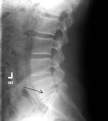
[[File:Narrow disc space.jpg|thumb]]
*Today, most spine surgeons use pedicle screw constructs to immobilize the vertebrae rigidly while preserving the normal lumbar lordosis 2
* Confirmatory diagnostic testing often includes [[MRI Scans|MRI]] scanning and discography for equivocal cases.<ref name=":0" />
* Spinal radiographs showing:
** [[Osteophyte|Osteophytes]] and segmental disc space narrowing in patients with degenerative spondylosis.
** A defect in the pars interarticularis.
** Anterolisthesis, or a forward slippage of one vertebra on the next, is the hallmark radiographic finding in spondylolisthesis.
** Flexion and extension films can help to detect hypermobility or excessive motion in degenerative lumbar conditions.


==== Posterolateral Lumbar Fusion: ====
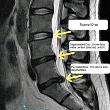
[[File:Degenerative disc diseaseMRI.jpg|thumb]]
-A midline posterior incision, with a laminectomy if necessary.  
-Transverse process, pars interarticularis, and if needed, the sacral alae are decorticated (posterior fusion). then a bone graft is placed on the decorticated surfaces. 


Pedicle screws and rods or plates may be placed to immobilize the motion segments rigidly and augment the formation of a solid union.
==Lumbar Fusion Approaches==


- In routine cases of posterolateral fusions the disc is not radically resected. Biomechanical studies have shown that people bear load through the middle and posterior thirds of the disc. Several reports describe a persistently painful disc under a solid posterior fusion.14
Today, most spine surgeons use pedicle screw constructs to immobilise the vertebrae rigidly while preserving the normal lumbar lordosis.<ref>Burkus K, et al: [https://pubmed.ncbi.nlm.nih.gov/19411467/ Six-year outcomes of anterior lumbar interbody arthrodesis with use of interbody fusion cages and recombinant human bone morphogenic protein-2.] J Bone Joint Surg 91:1181-1189, 2009.
</ref>


==== Interbody Fusion: ====
=== Posterolateral Lumbar Fusion ===
A midline posterior incision, with a [[laminectomy]] if necessary. 


===== A-posterior lumbar fusion (PLIF) =====
Transverse process, pars interarticularis, and if needed, the sacral alae are decorticated (posterior fusion). then a bone graft is placed on the decorticated surfaces.
-Associated with a higher incidence of postsurgical nerve injuries.


===== B- Transformational Lumbar Interbody Fusion =====
Pedicle screws and rods or plates may be placed to immobilise the motion segments rigidly and augment the formation of a solid union.


===== C-Anterior Lumbar Interbody Fusion =====
In routine cases of posterolateral fusions the disc is not radically resected. Biomechanical studies have shown that people bear load through the middle and posterior thirds of the disc. Several reports describe a persistently painful disc under a solid posterior fusion.<ref>Ibrahim T, et al: [https://scholar.google.com/scholar_url?url=https://link.springer.com/article/10.1007/s00264-006-0269-6&hl=en&sa=T&oi=gsb&ct=res&cd=0&d=403217471083671544&ei=9p2uY_yAG8iBywTQ4KTQBg&scisig=AAGBfm1wcM6MmVGO41v5s4Dk4dWF4SKhAg Surgical versus non-surgical treatment of chronic low back pain]: A meta-analysis of randomized trials. Int Orthop 32(7):107-113, 2006.
</ref>


===== D-Lateral Interbody Fusion =====
=== Interbody Fusion ===
-Nerve stretch injury reported, with L4 nerve root injury most common.17
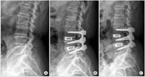
[[File:Posterior lumbar interbody fusion.jpg|thumb]]
* A-posterior lumbar fusion (PLIF): associated with a higher incidence of postsurgical nerve injuries.
* B- Transformational Lumbar Interbody Fusion
* C-Anterior Lumbar Interbody Fusion
* D-Lateral Interbody Fusion: nerve stretch injury reported, with L4 nerve root injury most common.<ref>Knight RQ, et al: [https://scholar.google.com/scholar_url?url=https://journals.lww.com/jspinaldisorders/fulltext/2009/02000/Direct_Lateral_Lumbar_Interbody_Fusion_for.6.aspx&hl=en&sa=T&oi=gsb&ct=res&cd=0&d=12035724177052866777&ei=G56uY8LKOs6TywSl1LXoCA&scisig=AAGBfm2TE8ro7OlZOPlqajVOaxFcK8GCAg Direct lateral lumbar interbody fusion for degenerative conditions]: Early complication profile. J Spinal Disord Tech 22(1):34-37, 2009.
</ref>
* E-Interbody Cages: hollow cylinders made of titanium, carbon ,or bone filled with autogenous bone graft or a bone graft substitute and inserted between the vertebral bodies.<ref name=":0" />


===== E-Interbody Cages =====
{{#ev:youtube|N-2Gy3iIeu8}}
-Hollow cylinders made of titanium, carbon ,or bone filled with autogenous bone graft or a bone graft substitute and inserted between the vertebral bodies.


== '''SURGICAL PROCEDURE''' == 


== '''Rehabilittion:''' ==
==Rehabilitation==


=== '''Preoperative phase''' ===
===Preoperative phase:===
This is the proper timing for assessing functional outcomes and psychological support. this is possible through patient education, which includes:
This is the proper timing for assessing functional outcomes and psychological support. This is possible through patient education, which includes:
* stabilization-based exercises  
* [[CPR for Lumbar Stabilisation|Stabilisation-based exercises]]
* Postoperative precautions
* Bed mobility and transfers
* Initial postoperative exercises
* Gait training with any necessary assistive devices
* [[Gait training]] with any necessary [[Assistive Devices|assistive devices]]
* Donning and doffing any required braces
* Wound care
* [[Wound Care Basics: Objective Assessment|Wound care]]
* General overview and prognosis of the postoperative rehabilitation process


'''SURGICAL PROCEDURE'''
A tour of the facility and operating room along with meeting with individuals who have already undergone such a procedure may also help to decrease patient anxiety surrounding the surgery and hospital experience.<ref>Kisner C, Colby LA: [https://scholar.google.com/scholar_url?url=https://books.google.com/books%3Fhl%3Den%26lr%3D%26id%3DyZc6DwAAQBAJ%26oi%3Dfnd%26pg%3DPR1%26dq%3DKisner%2BC,%2BColby%2BLA%2B%2BTherapeutic%2Bexercise%2B%2BFoundations%2Band%2Btechniques,%2Bed%2B5,%2BPhiladelphia,%2B2007,%2BFA%2BDavis.%26ots%3DNfw0F2hDUe%26sig%3D10CnqTfr7cQUruuwAq0Z_ABIgUw&hl=en&sa=T&oi=gsb&ct=res&cd=0&d=16766534394545827191&ei=RZ6uY_z3K86TywSl1LXoCA&scisig=AAGBfm3-Xke7HiSACVwH3GheEawiwvbxgg Therapeutic exercise: Foundations and techniques], ed 5, Philadelphia, 2007, FA Davis.
 
</ref>
'''Video''' 
 
'''Rehabilittion:'''  
 
-individuals with more chronic pain symptoms will most likely exhibit altered pain processing, which may be addressed through including cognitive-behavioral interventions during the recovery process.
 
'''Preoperative phase'''
 
-conservative measures have not had a significant impact on the patient’s condition and that they have gone through an extensive therapy program.
 
- taught stabilization-based exercises and has begun to address other relevant physical and cognitive dysfunctions.
 
-preoperative management may be very useful in determining functionally relevant outcomes along with realistic goals.21 This is also the time to start on patient education regarding issues such as:
 
• Postoperative precautions
 
• Bed mobility and transfers
 
• Initial postoperative exercises
 
• Gait training with any necessary assistive devices
 
• Donning and doffing any required braces
 
• Wound care
 
• General overview and prognosis of the postoperative rehabilitation process
 
-A tour of the facility and operating room along with meeting with individuals who have already undergone such a procedure may also help to decrease patient anxiety surrounding the surgery and hospital experience.21
 
-Therapist must have a complete picture about the procedure, surgical approach and fused levels.
 
<nowiki>*</nowiki>clinical note: During a posterior fusion, the multifidi are retracted from the spine. This partially tears the dorsal divisions of the spinal nerves, resulting in partial denervation of the multifidi. If an anterior fusion also has been performed, then a midline skin incision will be apparent and the abdominal muscular incision is lateral. The incision passes through the obliques, also partially denervating them. For this reason the therapist should teach the patient the proper way to recruit the transverse abdominis (TA), multifidi, and pelvic floor muscles and watch for any substitution patterns to promote proper spinal stabilization.
 
=== '''Phase I''' ===
-1 to 5 days after surgery (inpatient) and up to 6 Weeks
 
-Most patients are referred for physical therapy anywhere between 4 to 7 weeks after their discharge from the hospital.
 
-Physical therapy management during this phase consists of teaching patients the proper way to get in and out of bed, dress and perform other self-care activities, and walk (perhaps with a walker for the first 1 or 2 days).
 
-Light TA and pelvic floor contractions to begin to practice them in different positions.
 
.-The therapist also can teach basic and simple neural mobilization for the nerves involving the lumbosacral plexus.
 
-Patients and their family should leave the hospital with an understanding of the home care required until they begin their outpatient physical therapy,
 
=== '''Phase II''' ===
'''TIME:''' 6 to 10 weeks after surgery
 
'''GOALS:''' Increased activity, tissue remodeling, stabilization, and reconditioning
 
-During phase II, patients gradually increase their activity level. While taking soft tissue healing into account, the PT can safely begin to influence the direction of tissue modeling through carefully applied stress. Patients should begin to approximate normal activities while the therapist controls
 
-the intensity of movement and exercise. Also toward the end of this phase.
 
-patients should be slowly working up to 30 minutes of exercise and physical activity at least 5 days a week as recommended by the American College of Sports Medicine.23
 
-They can begin a light weight-training program, avoiding exercises that inappropriately load the lumbar spine but making sure to include some exercise for the lumbar paraspinals and other muscles that attach to the thoracodorsal fascia.
 
'''-Common restrictions are no lifting greater than 10╯lb'''
 
'''and no overhead lifting. Examples of exercises for this'''
 
'''phase are listed in the following sections.''' 
 
'''Evaluation'''
 
-assess the patient’s status and help to create an individualized program. The examination should include relevant tests and measures, such as posture, gait, range of motion (ROM), strength, balance, body mechanics, and specific functional tasks while making sure not to overload the lumbar spine. The therapist and patient can then begin to collaborate on and establish goals for treatment.
 
'''-This evaluation should include ROM for the LEs and upper extremities (UEs) but not for the lumbar spine.''' A complete neurologic examination should be performed to establish a baseline and should include neural tension testing.
 
'''-The therapist can perform strength testing for the Les with the exception of testing hip flexor strength.'''
 
'''-''' He or she also can check the patient’s ability to stabilize or brace the lumbar spine isometrically, which is a test of the patient’s ability to recruit the core trunk muscles to control the spine.
 
The goals of phase II are as follows:
 
• Demonstrate good body mechanics for activities of daily
 
living (ADL)
 
• Protect the surgical site from infection and mechanical
 
stress
 
• Maintain nerve root mobility at the involved levels
 
• Control pain and inflammation
 
• Minimize patient fear and apprehension
 
• Begin a stabilization and reconditioning program
 
• Improve scar and surrounding soft tissue mobility
 
• Treat restrictions of thoracic, UEs, and LEs that can lead
 
to more strain on the lumbar spine
 
• Education to minimize sitting time and maximize walking
 
time
 
'''Body Mechanics Training'''
 
The therapist should go through the entire program, which is as follows:
 
• In and out of bed (Fig. 16-4)
 
• In and out of a chair (Fig. 16-5)
 
• Up and down from the floor (Fig. 16-6)
 
• Lying postures (Fig. 16-7)
 
• Sitting (Fig. 16-8)
 
• Standing
 
• Dressing
 
• Bending (Fig. 16-9)
 
• Reaching
 
• Pushing and pulling (Fig. 16-10)
 
• Lifting (Fig. 16-11)
 
• Carrying (Fig. 16-12)
 
-motion if they move their hips rather than
 
the spine.
 
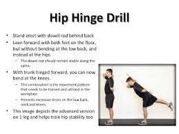
'''Instead of flexing the lumbar spine, they can “hip'''
 
'''hinge”''' (see Fig. 16-9). Rather than twist in the lumbar spine,
 
they can pivot on another body part (e.g., knees, elbows,
 
hips). When teaching a hip hinge, the PT should point out
 
that the hips should move back rather than down. After
 
surgery, patients tend to guard and move cautiously. Showing
 
them the way to use their momentum safely in many
 
'''Nerve Root Gliding.╇'''
 
'''-''' Patients should extend the knee while lying supine with the spine in a neutral position and the hip flexed to a 90° angle.
 
When tension is encountered,
 
-the therapist helps the patient work the knee or ankle gently back and forth, gradually increasing the ROM (Fig. 16-13).
 
-This stretch may cause increased symptoms during the stretch, which should resolve immediately on relaxing.
 
-Local inflammation occurs after lumbar spine surgery.
 
Because the body forms scar tissue in response to inflammation,
 
the nerve root can become adherent to the neural
 
foramen or lose elasticity. It is theorized that a nerve root
 
that is kept moving within its sheath cannot develop adhesions.
 
-the patient must keep the spine stabilized while moving the leg.
 
'''-Ultrasound should not be applied over a healing bony fusion.''' Patients with severe pain problems can try using a home transcutaneous electrical nerve stimulation (TENS) unit or interferential unit.
 
-Incisional pain can be expected to decrease gradually over 6 to 8 weeks.
 
The therapist should teach patients to manage flare-ups using ice, rest, and resumption of previous activities within 1 or 2 days. 
 
'''Minimizing Patient Fear and Apprehension.╇'''   
 
-Psychosocial variables have been shown to have a large influence on disability and function in individuals with chronic back pain, so ignoring these concepts could be a large detriment to the patient’s functional improvement.11
 
.
 
'''Stabilization, Strength, and Reconditioning.╇'''
 
• Cocontraction of the TA, multifidus, and pelvic floor muscles with and without using pressure biofeedback (BFB) (Fig. 16-14)
 
• Abdominal breathing
 
• Abdominal bracing with appropriate progression
 
(Fig. 16-15)
 
Abdominal bracing and supine marching are good exercises
 
to begin strengthening the trunk. Before bracing is
 
initiated, it is best to make sure the patient can isometrically
 
contract the TA, multifidi, and pelvic floor muscles.6,9,28-31
 
After the patient is able to do such, it is important to progress
 
those stabilization exercises, eventually working toward
 
functional goals that have been established. The patient
 
should be able to contract the appropriate stabilization
 
muscles in different postures and positions, so it is recommended
 
that these be practiced also in sitting, standing, and
 
quadruped.
 
In quadruped (four-point kneeling) the patient should be
 
able to more easily work on contracting TA while keeping
 
other global muscles relaxed
 
Adding bracing along with arm
 
and leg movements in this quadruped position is also a great
 
way to activate the multifidus and lumbar spine paraspinals
 
without placing the lumbar spine under undue axial load32,33
 
General balance activities would
 
also help with this type of challenge. Examples of these types
 
of exercises may include:
 
• UE or LE activities while sitting on an exercise ball (Fig.
 
16-17)
 
• Supine/hooklying activities laying vertical on a foam roll
 
(Fig. 16-18)
 
• Standing activities on a disc or rocker board
 
(Fig. 16-19)
 
• Trunk or hip perturbations in sitting or standing
 
(Fig. 16-20)
 
General strength and conditioning exercises should also
 
be initiated during this phase of rehabilitation after it is
 
cleared by the physician and the patient demonstrates appropriate
 
stabilization. Examples of exercises would include:
 
• Wall squats and sit to stand
 
• Half lunges
 
• Step ups and step downs
 
• Walking
 
• Cardiovascular reconditioning (using stair climber,
 
brisk walking, and pool exercises once the incision is
 
closed)
 
-Care should be taken when starting more vigorous strengthening activities, because it is recommended that the patient be able to use the appropriate stabilization muscles during components of the exercise before doing the full exercise.
 
'''Maintaining Scar and Soft Tissue Mobility.╇'''
 
'''-'''The therapist should use soft tissue techniques to maintain good scar and soft tissue mobility without disrupting the healing of these tissues (Fig. 16-21). Scar tissue tends to contract
 
while healing. This can create a “tight” scar that restricts
 
mobility.35 In cases of prolonged incisional pain it may be
 
beneficial to use techniques to desensitize the tissue starting
 
with very soft and gentle surfaces progressing to more firm
 
and vigorous materials.
 
'''Assessment and Treatment for Restrictions of'''
 
'''Thoracic, Shoulder, and Hip Mobility'''
 
The following steps will help ease restrictions of the thoracic spine and hip:
 
• Manual therapy for thoracic motion restrictions
 
• LE and UE stretches for soft tissue restrictions
 
• Hamstring stretches
 
• Hip flexor stretches (Fig. 16-22) can be initiated in later
 
stages with permission from the surgeon
 
Quadriceps stretches (begin with prone knee flexion
 
before progressing )
 
• Lumbar flexion stretch (Fig. 16-23) with surgeon approval
 
'''When initiating this stretch, the therapist must not be overly aggressive, obtaining ROM at the expense of compromising the fusion site'''
 
Up and down from the floor (Fig. 16-24)
 
• Hip rotator stretches (Fig. 16-25)
 
• Latissimus dorsi stretches (Fig. 16-26)
 
''*One of the most stressful motions in the lumbar spine is rotation,''
 
''which causes a shearing effect across the disc. Since the''
 
''thoracic spine is designed to allow more rotation, limited''
 
''motion here may increase strain on the lumbar spine during''
 
''twisting motions.'' The PT can use manual mobilization techniques
 
to increase thoracic spine mobility
 
'''Stretching throughout phase II should be'''
 
'''very gentle and only pushed to the point the patient can'''
 
'''brace to prevent lumbar motion'''.
 
<nowiki>*</nowiki>Iliopsoas stretching is initiated in a later phase with the permission of the physician. The aggressiveness of any hip stretching is dictated by the patient’s ability to control the spine while stretching
 
Examples of other exercises (performed while bracing)
 
initiated in the later stages of phase II include the following:
 
• Bridging
 
• Heel lifts
 
• Superman (avoiding lumbar extension)
 
• Lateral pulls (light resistance with approval of surgeon)
 
(Fig. 16-27)
 
• Seated upright rowing machine
 
• Scapular depression (avoid resisting more than 40% of
 
body weight)
 
• Push-ups standing and leaning into the wall
 
• Stair climber
 
• Upper body ergometer (UBE)
 
What the therapist is attempting to
 
develop at this stage is not so much muscle power as kinesthetic
 
sense for the muscles and their role in protecting the
 
spine. Therefore the proper form of each exercise should be
 
emphasiz
 
'''Phase III'''
 
'''TIME:''' 11 to 19 weeks after surgery
 
'''GOALS:''' Return to work, continue to advance/progress
 
exercise program, practice specific skills program,
 
initiate resistance training program
 
-They often return to work with modified duties or on a part-time schedule.
 
At this time they should be independent with self-care duties and also with a moderately challenging home exercise program.
 
The use of proper mechanics should be becoming a habit but will need
 
to be continually reinforced during specific activities. Exercises
 
that address functional movement may be a great time
 
to reinforce those principles. More strenuous stabilization
 
activities, such as half and full front and side planks could
 
be added.
 
The early development of these
 
muscles in their role as spinal stabilizers rather than spinal
 
movers is a crucial component of this phase.
 
The previous
 
trunk stabilization activities should be progressed within the
 
patient tolerance by modifying, for example, the number of
 
repetitions, adding Thera-Band resistance, or performing the
 
exercise on a more challenging surface.
 
It is not advised to do complex weight lifting
 
tasks, but to focus on light free weight activity and machinebased
 
exercises that allow the patient to perform them with
 
proper posture, technique, and bracing. Patients with a poor
 
tolerance for any one position may do better on a circuittraining
 
program.
 
'''*Patients should be extremely careful with overhead lifting because of the axial load and compressive forces placed on the spine. -'''Endurance and cardiovascular exercises should also be progressed at this stage and start to progress gradually.
 
-For some individuals it may be advised to do more cardiovascular or resisted exercises in an aquatic rehabilitation environment
 
The buoyancy of the water may help to unload the spine but allow the patient to do partial weight-bearing exercises along with core and resisted extremity activity.
 
<nowiki>*</nowiki>At this stage the expectation is that pain continues to decrease and be at a minimal level. Those patients that continue to have an unexpected degree of pain may need to be  by the PT or by the surgeon In the absence of any physical explanation of the pain, the rehabilitation team
 
needs to reinforce the functional improvements and minimize
 
the importance of pain as a marker of improvement.
 
'''Phase IV'''
 
'''TIME:''' 20 weeks to 1 year after surgery
 
'''GOALS:''' Restore preinjury status, continue home
 
program of conditioning and stabilization
 
Patients should be progressing to full restoration of their preinjury
 
level of function and be independent with conducting their previous home and gym program.
 
-They should have a good grasp of not only the exercises and physical activity required to reach their goals but also ways to modify those activities, because at this stage it might be expected that the patient
 
may be finishing with outpatient therapy. Proper
 
''Patients with fusions frequently develop''
 
''problems at the level above or below the fusion.'' For these
 
reasons, the patient should learn that spinal care is now
 
a lifetime habit and must be maintained with regular
 
exercise and good mechanics during all daily activities (not
 
just those the patient perceives as stressful).
 
-Patients returning to a more strenuous job or sports
 
are now developing the extra degree of strength and skill to do so.
 
- ''Later in this phase (and with clearance from the'' ''surgeon)'' they may begin agility and sport-specific drills, such as running, cutting, and jumping. If a more comprehensive weight training program is called for it should be again geared to the specific activity faced by the patient
 
The patient must demonstrate good trunk strength and control and good LE strength and flexibility before initiating agility drills. Although all therapists would like to relieve pain, some suffering is beyond the ability of current medical science to alleviate. This is a difficult concept for some patients to understand, and they may not be willing to accept it. Focusshould again be on improving function and less on pain abatement.
 
-Cognitive-behavioral interventions can continue to help with pain-related fear, social adjustments, and coping strategies that may still be difficult for patients during these later stages. Therapists should make every effort to help patients accept this reality and learn to care for themselves without seeking constant medical intervention.
 
reference:
 
1-the book
 
2-Burkus K, et al: Six-year outcomes of anterior lumbar interbody arthrodesis
 
with use of interbody fusion cages and recombinant human bone
 
morphogenic protein-2. J Bone Joint Surg 91:1181-1189, 2009.
 
Before beginning a rehabilitation program, the therapist must know whether the patient has had a fusion with or without instrumentation.
 
-The therapist also must know the surgical approachand the levels fused. *clinical note:After a motion segment is fused, increased stress is placed on the levels above and below thefusion. This creates risk for acceleration of the degenerative cascade at the adjacent levels. Obviously the more levels that have been fused, the greater the stress placed on the remaining segments. When the fusion includes the L5-S1 motion segment, abnormal forces are then translated to the sacroiliac joints. To minimize these forces, the therapist must be sure that normal motion exists at all remaining segments, including the thoracic spine, shoulders, and lower extremities (LEs).
 
<nowiki>*</nowiki>clinical note: During a posterior fusion, the multifidi are retracted from the spine. This partially tears the dorsal divisions of the spinal nerves, resulting in partial denervation of the multifidi.
 
5,22 If an anterior fusion also has been performed, then a midline skin incision will be apparent and the abdominal muscular incision is lateral. The incision passes through the obliques, also partially denervating them. For this reason the therapist should teach the patient the proper way to recruit the transverse abdominis (TA), multifidi, and pelvic floormuscles and watch for any substitution patterns to promote
 
proper spinal stabilization.
 
'''Phase I'''
 
'''TIME:''' 1 to 5 days after surgery (inpatient) and up to 6
 
Weeks
 
-Most patients are referred for physical therapy anywhere between 4 to 7 weeks after their discharge from the hospital.
 
'''GOALS:''' Patient education about daily movements,
 
abdominal stabilization, neural mobilization, and
 
home care principles
 
inpatients:
 
-Most patients remain in the hospital for several days after fusion surgery
 
-Physical therapy management during this phase consists of teaching patients the proper way to get in and out of bed, dress and perform other self-care activities, and walk (perhaps with a walker for the first 1 or 2 days).
 
-Strenuous abdominal stabilization exercises are ''not'' recommended at this time; however, attempts should be made to perform light TA and pelvic floor contractions to begin to practice them in different positions. The patient may use a large “sigh” or more forceful exhalation such as “blowing out a candle” to start to facilitate other abdominal muscles that
 
assist with bracing.
 
-The therapist also can teach basic and simple neural mobilization for the nerves involving the lumbosacral plexus.
 
-Patients and their family should leave the hospital with an understanding of the home care required until they begin their outpatient physical therapy, especially in the absence of home PT during the interim.
 
'''-Patients will be given instructions from the physician to'''
 
'''avoid driving, prolonged sitting, lifting, bending, and twisting.'''
 
'''Phase II'''
 
'''TIME:''' 6 to 10 weeks after surgery
 
'''GOALS:''' Increased activity, tissue remodeling, stabilization, and reconditioning
 
-During phase II, patients gradually increase their activity level. While taking soft tissue healing into account, the PT can safely begin to influence the direction of tissue modeling through carefully applied stress. Patients should begin to approximate normal activities while the therapist controls
 
-the intensity of movement and exercise. Also toward the end of this phase.
 
-patients should be slowly working up to 30 minutes of exercise and physical activity at least 5 days a week as recommended by the American College of Sports Medicine.23
 
-They can begin a light weight-training program, avoiding exercises that inappropriately load the lumbar spine but making sure to include some exercise for the lumbar paraspinals and other muscles that attach to the thoracodorsal fascia.
 
'''-Common restrictions are no lifting greater than 10╯lb'''
 
'''and no overhead lifting. Examples of exercises for this'''
 
'''phase are listed in the following sections.''' 
 
'''Evaluation'''
 
-assess the patient’s status and help to create an individualized program. The examination should include relevant tests and measures, such as posture, gait, range of motion (ROM), strength, balance, body mechanics, and specific functional tasks while making sure not to overload the lumbar spine. The therapist and patient can then begin to collaborate on and establish goals for treatment.
 
'''-This evaluation should include ROM for the LEs and upper extremities (UEs) but not for the lumbar spine.''' A complete neurologic examination should be performed to establish a baseline and should include neural tension testing.
 
'''-The therapist can perform strength testing for the Les with the exception of testing hip flexor strength.'''
 
'''-''' He or she also can check the patient’s ability to stabilize or brace the lumbar spine isometrically, which is a test of the patient’s ability to recruit the core trunk muscles to control the spine.
 
The goals of phase II are as follows:
 
• Demonstrate good body mechanics for activities of daily
 
living (ADL)
 
• Protect the surgical site from infection and mechanical
 
stress
 
• Maintain nerve root mobility at the involved levels
 
• Control pain and inflammation
 
• Minimize patient fear and apprehension
 
• Begin a stabilization and reconditioning program
 
• Improve scar and surrounding soft tissue mobility
 
• Treat restrictions of thoracic, UEs, and LEs that can lead
 
to more strain on the lumbar spine
 
• Education to minimize sitting time and maximize walking
 
time
 
'''Body Mechanics Training'''
 
The therapist should go through the entire program, which is as follows:
 
• In and out of bed (Fig. 16-4)
 
• In and out of a chair (Fig. 16-5)
 
• Up and down from the floor (Fig. 16-6)
 
• Lying postures (Fig. 16-7)
 
• Sitting (Fig. 16-8)
 
• Standing
 
• Dressing
 
• Bending (Fig. 16-9)
 
• Reaching
 
• Pushing and pulling (Fig. 16-10)
 
• Lifting (Fig. 16-11)
 
• Carrying (Fig. 16-12)
 
-motion if they move their hips rather than
 
the spine.
 
'''Instead of flexing the lumbar spine, they can “hip'''
 
'''hinge”''' (see Fig. 16-9). Rather than twist in the lumbar spine,
 
they can pivot on another body part (e.g., knees, elbows,
 
hips). When teaching a hip hinge, the PT should point out
 
that the hips should move back rather than down. After
 
surgery, patients tend to guard and move cautiously. Showing
 
them the way to use their momentum safely in many
 
'''Nerve Root Gliding.╇'''
 
'''-''' Patients should extend the knee while lying supine with the spine in a neutral position and the hip flexed to a 90° angle.
 
When tension is encountered,
 
-the therapist helps the patient work the knee or ankle gently back and forth, gradually increasing the ROM (Fig. 16-13).
 
-This stretch may cause increased symptoms during the stretch, which should resolve immediately on relaxing.
 
-Local inflammation occurs after lumbar spine surgery.
 
Because the body forms scar tissue in response to inflammation,
 
the nerve root can become adherent to the neural
 
foramen or lose elasticity. It is theorized that a nerve root
 
that is kept moving within its sheath cannot develop adhesions.
 
-the patient must keep the spine stabilized while moving the leg.
 
'''-Ultrasound should not be applied over a healing bony fusion.''' Patients with severe pain problems can try using a home transcutaneous electrical nerve stimulation (TENS) unit or interferential unit.
 
-Incisional pain can be expected to decrease gradually over 6 to 8 weeks.
 
The therapist should teach patients to manage flare-ups using ice, rest, and resumption of previous activities within 1 or 2 days. 
 
'''Minimizing Patient Fear and Apprehension.╇'''   
 
-Psychosocial variables have been shown to have a large influence on disability and function in individuals with chronic back pain, so ignoring these concepts could be a large detriment to the patient’s functional improvement.11
 
.
 
'''Stabilization, Strength, and Reconditioning.╇'''
 
• Cocontraction of the TA, multifidus, and pelvic floor muscles with and without using pressure biofeedback (BFB) (Fig. 16-14)
 
• Abdominal breathing
 
• Abdominal bracing with appropriate progression
 
(Fig. 16-15)
 
Abdominal bracing and supine marching are good exercises
 
to begin strengthening the trunk. Before bracing is
 
initiated, it is best to make sure the patient can isometrically
 
contract the TA, multifidi, and pelvic floor muscles.6,9,28-31
 
After the patient is able to do such, it is important to progress
 
those stabilization exercises, eventually working toward
 
functional goals that have been established. The patient
 
should be able to contract the appropriate stabilization
 
muscles in different postures and positions, so it is recommended
 
that these be practiced also in sitting, standing, and
 
quadruped.
 
In quadruped (four-point kneeling) the patient should be
 
able to more easily work on contracting TA while keeping
 
other global muscles relaxed
 
Adding bracing along with arm
 
and leg movements in this quadruped position is also a great
 
way to activate the multifidus and lumbar spine paraspinals
 
without placing the lumbar spine under undue axial load32,33
 
General balance activities would
 
also help with this type of challenge. Examples of these types
 
of exercises may include:
 
• UE or LE activities while sitting on an exercise ball (Fig.
 
16-17)
 
• Supine/hooklying activities laying vertical on a foam roll
 
(Fig. 16-18)
 
• Standing activities on a disc or rocker board
 
(Fig. 16-19)
 
• Trunk or hip perturbations in sitting or standing
 
(Fig. 16-20)
 
General strength and conditioning exercises should also
 
be initiated during this phase of rehabilitation after it is
 
cleared by the physician and the patient demonstrates appropriate
 
stabilization. Examples of exercises would include:
 
• Wall squats and sit to stand
 
• Half lunges
 
• Step ups and step downs
 
• Walking
 
• Cardiovascular reconditioning (using stair climber,
 
brisk walking, and pool exercises once the incision is
 
closed)
 
-Care should be taken when starting more vigorous strengthening activities, because it is recommended that the patient be able to use the appropriate stabilization muscles during components of the exercise before doing the full exercise.
 
'''Maintaining Scar and Soft Tissue Mobility.╇'''
 
'''-'''The therapist should use soft tissue techniques to maintain good scar and soft tissue mobility without disrupting the healing of these tissues (Fig. 16-21). Scar tissue tends to contract
 
while healing. This can create a “tight” scar that restricts
 
mobility.35 In cases of prolonged incisional pain it may be
 
beneficial to use techniques to desensitize the tissue starting
 
with very soft and gentle surfaces progressing to more firm
 
and vigorous materials.
 
'''Assessment and Treatment for Restrictions of'''
 
'''Thoracic, Shoulder, and Hip Mobility'''
 
The following steps will help ease restrictions of the thoracic spine and hip:
 
• Manual therapy for thoracic motion restrictions
 
• LE and UE stretches for soft tissue restrictions
 
• Hamstring stretches
 
• Hip flexor stretches (Fig. 16-22) can be initiated in later
 
stages with permission from the surgeon
 
Quadriceps stretches (begin with prone knee flexion
 
before progressing )
 
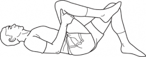
• Lumbar flexion stretch (Fig. 16-23) with surgeon approval
 
'''When initiating this stretch, the therapist must not be overly aggressive, obtaining ROM at the expense of compromising the fusion site'''
 
Up and down from the floor (Fig. 16-24)
 
• Hip rotator stretches (Fig. 16-25)
 
• Latissimus dorsi stretches (Fig. 16-26)
 
''*One of the most stressful motions in the lumbar spine is rotation,''
 
''which causes a shearing effect across the disc. Since the''
 
''thoracic spine is designed to allow more rotation, limited''
 
''motion here may increase strain on the lumbar spine during''
 
''twisting motions.'' The PT can use manual mobilization techniques
 
to increase thoracic spine mobility
 
'''Stretching throughout phase II should be'''
 
'''very gentle and only pushed to the point the patient can'''
 
'''brace to prevent lumbar motion'''.
 
<nowiki>*</nowiki>Iliopsoas stretching is initiated in a later phase with the permission of the physician. The aggressiveness of any hip stretching is dictated by the patient’s ability to control the spine while stretching
 
Examples of other exercises (performed while bracing)
 
initiated in the later stages of phase II include the following:
 
• Bridging
 
• Heel lifts
 
• Superman (avoiding lumbar extension)
 
• Lateral pulls (light resistance with approval of surgeon)
 
(Fig. 16-27)
 
• Seated upright rowing machine
 
• Scapular depression (avoid resisting more than 40% of
 
body weight)
 
• Push-ups standing and leaning into the wall
 
• Stair climber
 
• Upper body ergometer (UBE)
 
What the therapist is attempting to
 
develop at this stage is not so much muscle power as kinesthetic
 
sense for the muscles and their role in protecting the
 
spine. Therefore the proper form of each exercise should be
 
emphasiz
 
'''Phase III'''
 
'''TIME:''' 11 to 19 weeks after surgery
 
'''GOALS:''' Return to work, continue to advance/progress
 
exercise program, practice specific skills program,
 
initiate resistance training program
 
-They often return to work with modified duties or on a part-time schedule.
 
At this time they should be independent with self-care duties and also with a moderately challenging home exercise program.
 
The use of proper mechanics should be becoming a habit but will need
 
to be continually reinforced during specific activities. Exercises
 
that address functional movement may be a great time
 
to reinforce those principles. More strenuous stabilization
 
activities, such as half and full front and side planks could
 
be added.
 
The early development of these
 
muscles in their role as spinal stabilizers rather than spinal
 
movers is a crucial component of this phase.
 
The previous
 
trunk stabilization activities should be progressed within the
 
patient tolerance by modifying, for example, the number of
 
repetitions, adding Thera-Band resistance, or performing the
 
exercise on a more challenging surface.
 
It is not advised to do complex weight lifting
 
tasks, but to focus on light free weight activity and machinebased
 
exercises that allow the patient to perform them with
 
proper posture, technique, and bracing. Patients with a poor
 
tolerance for any one position may do better on a circuittraining
 
program.
 
'''*Patients should be extremely careful with overhead lifting because of the axial load and compressive forces placed on the spine. -'''Endurance and cardiovascular exercises should also be progressed at this stage and start to progress gradually.
 
-For some individuals it may be advised to do more cardiovascular or resisted exercises in an aquatic rehabilitation environment
 
The buoyancy of the water may help to unload the spine but allow the patient to do partial weight-bearing exercises along with core and resisted extremity activity.
 
<nowiki>*</nowiki>At this stage the expectation is that pain continues to decrease and be at a minimal level. Those patients that continue to have an unexpected degree of pain may need to be  by the PT or by the surgeon In the absence of any physical explanation of the pain, the rehabilitation team
 
needs to reinforce the functional improvements and minimize
 
the importance of pain as a marker of improvement.
 
'''Phase IV'''


'''TIME:''' 20 weeks to 1 year after surgery
Therapist must have a complete picture about the procedure, surgical approach and fused levels.


'''GOALS:''' Restore preinjury status, continue home
Note: During a posterior fusion, the multifidi are retracted from the spine. This partially tears the dorsal divisions of the spinal nerves, resulting in partial denervation of the multifidi. If an anterior fusion also has been performed, then a midline skin incision will be apparent and the abdominal muscular incision is lateral. The incision passes through the obliques, also partially denervating them. For this reason the therapist should teach the patient the proper way to recruit the transverse abdominis (TA), multifidi, and pelvic floor muscles and watch for any substitution patterns to promote proper spinal stabilisation.<ref name=":0" />
* [[Range of Motion|ROM]]
* [[Posture]]
* [[Balance]]
* Gait
* Strength
* Body mechanics
* Specific functional tasks
* Specific [[Neurological Assessment|Neurological examination]]
* The evaluation shouldn't include Lumbar ROM or hip flexor mobility especially at first stages of rehabilitation.<ref name=":0" />


program of conditioning and stabilization
===Phase I:===
* 1 to 5 days after surgery (inpatient) and up to 6 Weeks
* Most patients are referred for physiotherapy anywhere between 4 to 7 weeks after their discharge from the hospital.
* Physical therapy management during this phase consists of teaching patients the proper way to get in and out of bed, dress and perform other self-care activities, and walk (perhaps with a walker for the first 1 or 2 days).
* Light TA and pelvic floor contractions to begin to practice them in different positions.
* The therapist also can teach basic and simple neural mobilisation for the nerves involving the lumbosacral plexus.
* Patients and their family should leave the hospital with an understanding of the home care required until they begin their outpatient physical therapy.<ref name=":0" />


Patients should be progressing to full restoration of their preinjury
===Phase II:===
* 6 to 10 weeks after surgery
* Patients should begin to approximate normal activities while the therapist controls
* Patients should be slowly working up to 30 minutes of exercise and physical activity at least 5 days a week.
* They can begin a light weight-training program, avoiding exercises that inappropriately load the lumbar spine but making sure to include some exercise for the lumbar paraspinals and other muscles that attach to the thoracodorsal fascia.<ref name=":0" />
* '''Common restrictions are no lifting greater than 10lb.''' '''and no overhead lifting.'''<ref>Thompson WR, editor: [https://scholar.google.com/scholar_url?url=https://books.google.com/books%3Fhl%3Den%26lr%3D%26id%3DhhosAwAAQBAJ%26oi%3Dfnd%26pg%3DPP1%26dq%3DThompson%2BWR,%2Beditor%2B%2BACSM%25E2%2580%2599s%2Bguidelines%2Bfor%2Bexercise%2Btesting%2Band%2Bprescription,%2Bed%2B8,%2BAmerican%2BCollege%2Bof%2BSports%2BMedicine,%2BBaltimore,%2B2004,%2BLippincott%2BWilliams%2B%2526%2BWilkins.%26ots%3DlkH41J-YPC%26sig%3D6faYgZ80ZWts_MwOxMeW7XF2-90&hl=en&sa=T&oi=gsb&ct=res&cd=0&d=15677296915985333758&ei=hZ6uY5HJIoa8yATUzZGgCg&scisig=AAGBfm1n3SOrwWUIlSL1Sd4nt23Y4M1vZQ ACSM’s guidelines for exercise testing and prescription], ed 8, American College of Sports Medicine, Baltimore, 2004, Lippincott Williams & Wilkins.
</ref>


level of function and be independent with conducting their previous home and gym program.
==== '''Goals''' ====
* Achieve good body mechanics during ADL
* Protect the surgical site from infection and mechanical stress
* Nerve root mobility at the involved levels
* Control pain and inflammation
* Begin a stabilisation and reconditioning program
* Improve scar and surrounding soft tissue mobility
* Treat restrictions of thoracic, UEs, and LEs that can lead to more strain on the lumbar spine<ref name=":0" />


-They should have a good grasp of not only the exercises and physical activity required to reach their goals but also ways to modify those activities, because at this stage it might be expected that the patient
==== Body mechanics education ====
* Ved mobility
* Pushing and pulling
* [[Lifting]] and carrying
* [[Hip Hinge|Hip hinge technique]]


may be finishing with outpatient therapy. Proper
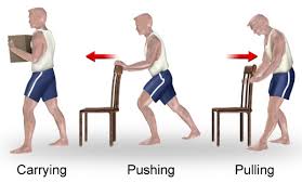
See [[Injury Prevention and Body Mechanics]][[File:Push and pull.jpg|thumb|none]]
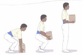
[[File:Lifting mechanics.jpg|thumb|none]][[File:Hip hinge.jpg|thumb|none]]


''Patients with fusions frequently develop''
==== Nerve root gliding techniques ====
[[File:Nerve root gliding.png|thumb]]
Patients should extend the knee while lying supine with the spine in a neutral position and the hip flexed to a 90° angle. When tension is encountered, the therapist helps the patient work the knee or ankle gently back and forth, gradually increasing the ROM.  This stretch may cause increased symptoms during the stretch, which should resolve immediately on relaxing.


''problems at the level above or below the fusion.'' For these
==== Pain management ====
* Prescribed medications and ice.
* Ultrasound shouldn't be applied over a healing bony fusion.
* Patients with severe pain problems can try using a home [https://www.physio-pedia.com/Transcutaneous_Electrical_Nerve_Stimulation_(TENS) transcutaneous electrical nerve stimulation] (TENS) unit or [https://www.physio-pedia.com/Interferential_Therapy interferential unit].
* Incisional pain can be expected to decrease gradually over 6 to 8 weeks.      


reasons, the patient should learn that spinal care is now
==== Strength and Reconditioning ====
* Co-contraction of the TA, multifidus, and pelvic floor muscles with and without using pressure biofeedback (BFB). See [[Spinal Stabilization|lumbar stabilisation]]
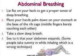
* Abdominal breathing
[[File:Abdominal breathing.jpg|thumb|none]]
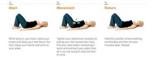
* Abdominal bracing and supine marching are good exercises to begin strengthening the trunk. Before bracing is initiated, it is best to make sure the patient can isometrically contract the TA, multifidi, and pelvic floor muscles.


a lifetime habit and must be maintained with regular
* [[File:Abdominal bracing.jpg|thumb|none]]
* Exercises are recommended to  be practiced in sitting, standing, and quadruped.
* General strength and conditioning exercises should also be initiated during this phase of rehabilitation after it is cleared by the physician and the patient demonstrates appropriate stabilisation. Examples of exercises would include:
** Wall squats and sit to stand
** Half lunges
** Step ups and step downs
** Walking
* Cardiovascular reconditioning (using stair climber, brisk walking, and pool exercises once the incision is closed)
* Care should be taken when starting more vigorous strengthening activities, because it is recommended that the patient be able to use the appropriate stabilisation muscles during components of the exercise before doing the full exercise.
* Hip flexors stretching should only start after the permission of the surgeon. stretching should be very gentle and only pushed to the point the patient can brace to prevent lumbar motion.
* ''one of the most stressful motions in the lumbar spine is rotation,'' ''which causes a shearing effect across the disc. Since the'' ''thoracic spine is designed to allow more rotation, limited'' ''motion here may increase strain on the lumbar spine during'' ''twisting motions.'' The PT can use manual mobilization techniques to increase thoracic spine mobility
* Examples of other exercises (performed while bracing) initiated in the later stages of phase II include the following:


exercise and good mechanics during all daily activities (not
** Bridging
** Heel lifts
** Superman (avoiding lumbar extension)
** Lateral pulls (light resistance with approval of surgeon)
** Seated upright rowing machine
** Scapular depression (avoid resisting more than 40% of body weight)
** Push-ups standing and leaning into the wall
** Stair climber
** Upper body ergometer (UBE)


just those the patient perceives as stressful).  
* What the therapist is attempting to develop at this stage is not so much muscle power as kinesthetic sense for the muscles and their role in protecting the spine. Therefore the proper form of each exercise should be emphasised.


-Patients returning to a more strenuous job or sports
==== Scar and soft tissue mobility ====
Through soft tissue mobilisation techniques.<ref name=":0" />


are now developing the extra degree of strength and skill to do so.
===Phase III===
* 11 to 19 weeks after surgery


- ''Later in this phase (and with clearance from the'' ''surgeon)'' they may begin agility and sport-specific drills, such as running, cutting, and jumping. If a more comprehensive weight training program is called for it should be again geared to the specific activity faced by the patient
==== Goals ====
* Return to work
* continue to advance/progress exercise program,
* practice specific skills program,
* initiate resistance training program


The patient must demonstrate good trunk strength and control and good LE strength and flexibility before initiating agility drills. Although all therapists would like to relieve pain, some suffering is beyond the ability of current medical science to alleviate. This is a difficult concept for some patients to understand, and they may not be willing to accept it. Focusshould again be on improving function and less on pain abatement.
* They often return to work with modified duties or on a part-time schedule.
* patient should be independent with self-care duties and also with a moderately challenging home exercise program.
* More strenuous stabilisation activities, such as half and full front and side planks could be added.
* The previous trunk stabilisation activities should be progressed within the patient tolerance with modifications according to the patient's condition.
* It is not advised to do complex weight lifting tasks, but to focus on light free weight activity and machine based exercises that allow the patient to perform them with proper posture, technique, and bracing.
* '''Patients should be extremely careful with overhead lifting because of the axial load and compressive forces placed on the spine.'''
* Endurance and cardiovascular exercises should also be progressed at this stage and start to progress gradually.  
* For some individuals it may be advised to do more cardiovascular or resisted exercises in an aquatic rehabilitation environment.
* At this stage the expectation is that pain continues to decrease and be at a minimal level.  However, if pain persists and the surgeon reported absence of physical explanation, then the therapist should reinforce the functional improvements and minimise the importance of pain as a marker of improvement.<ref name=":0" />


-Cognitive-behavioral interventions can continue to help with pain-related fear, social adjustments, and coping strategies that may still be difficult for patients during these later stages. Therapists should make every effort to help patients accept this reality and learn to care for themselves without seeking constant medical intervention.
===Phase IV===
* 20 weeks to 1 year after surgery


reference:
==== Goals ====
* Restore pre-injury status
* Continue home program of conditioning and stabilisation
* Patients should be progressing to full restoration of their pre-injury level of function and be independent with conducting their previous home and gym program.
* They should have a good grasp of not only the exercises and physical activity required to reach their goals but also ways to modify those activities.


1-the book
* Patients with fusions frequently develop problems at the level above or below the fusion. For these reasons, the patient should learn that spinal care is now a lifetime habit and must be maintained with regular exercise and good mechanics during all daily activities.
* Later in this phase (and with clearance from the surgeon) they may begin agility and sport-specific drills, such as running, cutting, and jumping. If a more comprehensive weight training program is called for it should be again geared to the specific activity faced by the patient<ref name=":0" />


2-Burkus K, et al: Six-year outcomes of anterior lumbar interbody arthrodesis
==Useful links:==
[[Core Stability|Core stability]]


with use of interbody fusion cages and recombinant human bone
[[Exercises for Lumbar Instability|Exercises for Lumbar Instability]]


morphogenic protein-2. J Bone Joint Surg 91:1181-1189, 2009.
==References:==

Latest revision as of 11:56, 24 April 2024


Introduction[edit | edit source]

Lumbar Spine Fusion is an effective treatment option to stabilise the painful motion segment, and may provide indirect decompression of the neural elements, restore lordosis and correct deformity[1].

The surgery involves placement of an implant (cage, spacer or structural graft) within the intervertebral space after discectomy[1] and successful union of two or more vertebra[2].

Indications[edit | edit source]

  • Discogenic/facetogenic low back pain[1].
  • Post traumatic cases of segmental instability or potential neurologic injury[1].

Degenerative cascade[edit | edit source]

Degenerative Disc Disease

Diagnosis[edit | edit source]

Narrow disc space.jpg
  • Confirmatory diagnostic testing often includes MRI scanning and discography for equivocal cases.[2]
  • Spinal radiographs showing:
    • Osteophytes and segmental disc space narrowing in patients with degenerative spondylosis.
    • A defect in the pars interarticularis.
    • Anterolisthesis, or a forward slippage of one vertebra on the next, is the hallmark radiographic finding in spondylolisthesis.
    • Flexion and extension films can help to detect hypermobility or excessive motion in degenerative lumbar conditions.
Degenerative disc diseaseMRI.jpg

Lumbar Fusion Approaches[edit | edit source]

Today, most spine surgeons use pedicle screw constructs to immobilise the vertebrae rigidly while preserving the normal lumbar lordosis.[3]

Posterolateral Lumbar Fusion[edit | edit source]

A midline posterior incision, with a laminectomy if necessary.

Transverse process, pars interarticularis, and if needed, the sacral alae are decorticated (posterior fusion). then a bone graft is placed on the decorticated surfaces.

Pedicle screws and rods or plates may be placed to immobilise the motion segments rigidly and augment the formation of a solid union.

In routine cases of posterolateral fusions the disc is not radically resected. Biomechanical studies have shown that people bear load through the middle and posterior thirds of the disc. Several reports describe a persistently painful disc under a solid posterior fusion.[4]

Interbody Fusion[edit | edit source]

Posterior lumbar interbody fusion.jpg
  • A-posterior lumbar fusion (PLIF): associated with a higher incidence of postsurgical nerve injuries.
  • B- Transformational Lumbar Interbody Fusion
  • C-Anterior Lumbar Interbody Fusion
  • D-Lateral Interbody Fusion: nerve stretch injury reported, with L4 nerve root injury most common.[5]
  • E-Interbody Cages: hollow cylinders made of titanium, carbon ,or bone filled with autogenous bone graft or a bone graft substitute and inserted between the vertebral bodies.[2]


Rehabilitation[edit | edit source]

Preoperative phase:[edit | edit source]

This is the proper timing for assessing functional outcomes and psychological support. This is possible through patient education, which includes:

A tour of the facility and operating room along with meeting with individuals who have already undergone such a procedure may also help to decrease patient anxiety surrounding the surgery and hospital experience.[6]

Therapist must have a complete picture about the procedure, surgical approach and fused levels.

Note: During a posterior fusion, the multifidi are retracted from the spine. This partially tears the dorsal divisions of the spinal nerves, resulting in partial denervation of the multifidi. If an anterior fusion also has been performed, then a midline skin incision will be apparent and the abdominal muscular incision is lateral. The incision passes through the obliques, also partially denervating them. For this reason the therapist should teach the patient the proper way to recruit the transverse abdominis (TA), multifidi, and pelvic floor muscles and watch for any substitution patterns to promote proper spinal stabilisation.[2]

  • ROM
  • Posture
  • Balance
  • Gait
  • Strength
  • Body mechanics
  • Specific functional tasks
  • Specific Neurological examination
  • The evaluation shouldn't include Lumbar ROM or hip flexor mobility especially at first stages of rehabilitation.[2]

Phase I:[edit | edit source]

  • 1 to 5 days after surgery (inpatient) and up to 6 Weeks
  • Most patients are referred for physiotherapy anywhere between 4 to 7 weeks after their discharge from the hospital.
  • Physical therapy management during this phase consists of teaching patients the proper way to get in and out of bed, dress and perform other self-care activities, and walk (perhaps with a walker for the first 1 or 2 days).
  • Light TA and pelvic floor contractions to begin to practice them in different positions.
  • The therapist also can teach basic and simple neural mobilisation for the nerves involving the lumbosacral plexus.
  • Patients and their family should leave the hospital with an understanding of the home care required until they begin their outpatient physical therapy.[2]

Phase II:[edit | edit source]

  • 6 to 10 weeks after surgery
  • Patients should begin to approximate normal activities while the therapist controls
  • Patients should be slowly working up to 30 minutes of exercise and physical activity at least 5 days a week.
  • They can begin a light weight-training program, avoiding exercises that inappropriately load the lumbar spine but making sure to include some exercise for the lumbar paraspinals and other muscles that attach to the thoracodorsal fascia.[2]
  • Common restrictions are no lifting greater than 10lb. and no overhead lifting.[7]

Goals[edit | edit source]

  • Achieve good body mechanics during ADL
  • Protect the surgical site from infection and mechanical stress
  • Nerve root mobility at the involved levels
  • Control pain and inflammation
  • Begin a stabilisation and reconditioning program
  • Improve scar and surrounding soft tissue mobility
  • Treat restrictions of thoracic, UEs, and LEs that can lead to more strain on the lumbar spine[2]

Body mechanics education[edit | edit source]

See Injury Prevention and Body Mechanics

Push and pull.jpg
Lifting mechanics.jpg
Hip hinge.jpg

Nerve root gliding techniques[edit | edit source]

Nerve root gliding.png

Patients should extend the knee while lying supine with the spine in a neutral position and the hip flexed to a 90° angle. When tension is encountered, the therapist helps the patient work the knee or ankle gently back and forth, gradually increasing the ROM. This stretch may cause increased symptoms during the stretch, which should resolve immediately on relaxing.

Pain management[edit | edit source]

  • Prescribed medications and ice.
  • Ultrasound shouldn't be applied over a healing bony fusion.
  • Patients with severe pain problems can try using a home transcutaneous electrical nerve stimulation (TENS) unit or interferential unit.
  • Incisional pain can be expected to decrease gradually over 6 to 8 weeks.      

Strength and Reconditioning[edit | edit source]

  • Co-contraction of the TA, multifidus, and pelvic floor muscles with and without using pressure biofeedback (BFB). See lumbar stabilisation
  • Abdominal breathing
Abdominal breathing.jpg
  • Abdominal bracing and supine marching are good exercises to begin strengthening the trunk. Before bracing is initiated, it is best to make sure the patient can isometrically contract the TA, multifidi, and pelvic floor muscles.
  • Abdominal bracing.jpg
  • Exercises are recommended to be practiced in sitting, standing, and quadruped.
  • General strength and conditioning exercises should also be initiated during this phase of rehabilitation after it is cleared by the physician and the patient demonstrates appropriate stabilisation. Examples of exercises would include:
    • Wall squats and sit to stand
    • Half lunges
    • Step ups and step downs
    • Walking
  • Cardiovascular reconditioning (using stair climber, brisk walking, and pool exercises once the incision is closed)
  • Care should be taken when starting more vigorous strengthening activities, because it is recommended that the patient be able to use the appropriate stabilisation muscles during components of the exercise before doing the full exercise.
  • Hip flexors stretching should only start after the permission of the surgeon. stretching should be very gentle and only pushed to the point the patient can brace to prevent lumbar motion.
  • one of the most stressful motions in the lumbar spine is rotation, which causes a shearing effect across the disc. Since the thoracic spine is designed to allow more rotation, limited motion here may increase strain on the lumbar spine during twisting motions. The PT can use manual mobilization techniques to increase thoracic spine mobility
  • Examples of other exercises (performed while bracing) initiated in the later stages of phase II include the following:
    • Bridging
    • Heel lifts
    • Superman (avoiding lumbar extension)
    • Lateral pulls (light resistance with approval of surgeon)
    • Seated upright rowing machine
    • Scapular depression (avoid resisting more than 40% of body weight)
    • Push-ups standing and leaning into the wall
    • Stair climber
    • Upper body ergometer (UBE)
  • What the therapist is attempting to develop at this stage is not so much muscle power as kinesthetic sense for the muscles and their role in protecting the spine. Therefore the proper form of each exercise should be emphasised.

Scar and soft tissue mobility[edit | edit source]

Through soft tissue mobilisation techniques.[2]

Phase III[edit | edit source]

  • 11 to 19 weeks after surgery

Goals[edit | edit source]

  • Return to work
  • continue to advance/progress exercise program,
  • practice specific skills program,
  • initiate resistance training program
  • They often return to work with modified duties or on a part-time schedule.
  • patient should be independent with self-care duties and also with a moderately challenging home exercise program.
  • More strenuous stabilisation activities, such as half and full front and side planks could be added.
  • The previous trunk stabilisation activities should be progressed within the patient tolerance with modifications according to the patient's condition.
  • It is not advised to do complex weight lifting tasks, but to focus on light free weight activity and machine based exercises that allow the patient to perform them with proper posture, technique, and bracing.
  • Patients should be extremely careful with overhead lifting because of the axial load and compressive forces placed on the spine.
  • Endurance and cardiovascular exercises should also be progressed at this stage and start to progress gradually.
  • For some individuals it may be advised to do more cardiovascular or resisted exercises in an aquatic rehabilitation environment.
  • At this stage the expectation is that pain continues to decrease and be at a minimal level. However, if pain persists and the surgeon reported absence of physical explanation, then the therapist should reinforce the functional improvements and minimise the importance of pain as a marker of improvement.[2]

Phase IV[edit | edit source]

  • 20 weeks to 1 year after surgery

Goals[edit | edit source]

  • Restore pre-injury status
  • Continue home program of conditioning and stabilisation
  • Patients should be progressing to full restoration of their pre-injury level of function and be independent with conducting their previous home and gym program.
  • They should have a good grasp of not only the exercises and physical activity required to reach their goals but also ways to modify those activities.
  • Patients with fusions frequently develop problems at the level above or below the fusion. For these reasons, the patient should learn that spinal care is now a lifetime habit and must be maintained with regular exercise and good mechanics during all daily activities.
  • Later in this phase (and with clearance from the surgeon) they may begin agility and sport-specific drills, such as running, cutting, and jumping. If a more comprehensive weight training program is called for it should be again geared to the specific activity faced by the patient[2]

Useful links:[edit | edit source]

Core stability

Exercises for Lumbar Instability

References:[edit | edit source]

  1. 1.0 1.1 1.2 1.3 1.4 1.5 1.6 Mobbs, R. J., Phan, K., Malham, G., Seex, K., & Rao, P. J. (2015). Lumbar interbody fusion : techniques , indications and comparison of interbody fusion options including PLIF , TLIF , MI-TLIF , OLIF / ATP , LLIF and ALIF, 3(1), 2–18. https://doi.org/10.3978/j.issn.2414-469X.2015.10.05
  2. 2.00 2.01 2.02 2.03 2.04 2.05 2.06 2.07 2.08 2.09 2.10 2.11 Maxey, L. and Magnusson, J. (n.d.). Rehabilitation for the postsurgical orthopedic patient.
  3. Burkus K, et al: Six-year outcomes of anterior lumbar interbody arthrodesis with use of interbody fusion cages and recombinant human bone morphogenic protein-2. J Bone Joint Surg 91:1181-1189, 2009.
  4. Ibrahim T, et al: Surgical versus non-surgical treatment of chronic low back pain: A meta-analysis of randomized trials. Int Orthop 32(7):107-113, 2006.
  5. Knight RQ, et al: Direct lateral lumbar interbody fusion for degenerative conditions: Early complication profile. J Spinal Disord Tech 22(1):34-37, 2009.
  6. Kisner C, Colby LA: Therapeutic exercise: Foundations and techniques, ed 5, Philadelphia, 2007, FA Davis.
  7. Thompson WR, editor: ACSM’s guidelines for exercise testing and prescription, ed 8, American College of Sports Medicine, Baltimore, 2004, Lippincott Williams & Wilkins.