When refering to evidence in academic writing, you should always try to reference the primary (original) source. That is usually the journal article where the information was first stated. In most cases Physiopedia articles are a secondary source and so should not be used as references. Physiopedia articles are best used to find the original sources of information (see the references list at the bottom of the article).
If you believe that this Physiopedia article is the primary source for the information you are refering to, you can use the button below to access a related citation statement.
Osteophytes are cartilage-capped bony proliferations (bony spurs) that most commonly develop at the margins of a synovial joint as a response to articular cartilage damage, as seen very commonly in osteoarthritis (OA).[1]
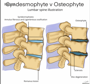
Three types of osteophytes related to the degenerative spine: (a) traction osteophytes (arrow), (b) claw osteophytes (arrow) and (c) wraparound bumper osteophytes (arrow)
Pathophysiology
Osteophytes are formed of fibrocartilage and bone, often forming at the peripheral margins of joints at the interface between cartilage and the periosteum. Osteophytes are thought to develop from chondrogenic differentiation of progenitor cells, commonly from within the periosteum. Osteophytes appear to be a cellular repair response to the altered growth factor environment following joint injury. In some cases osteophytes can contribute to the stability of the joints.[2]
Osteophytes are most commonly found in the following areas. Neck, shoulder, knee, lower back, fingers or big toe, foot or heel. Osteophytes cause problems for example if they: cause friction if rubbing against footwear; impinge nerves such as an existing spinal root; restrict movement as may occur in hip and knee OA.[3] Following are the pages of conditions where osteophytes are present:
Osteophytes when causing issues, for example with OA, can be managed along side management of OA, See link here Osteoarthritis. They are not managed specifically.
Removal of an osteophyte is not usual, unless it's irritating a nerve in the spine or restricting a joint's range of movement. If surgery is warranted, a surgeon will explain the procedure's risks and benefits.[3]
Differential Diagnosis
Syndesmophyte: Thin, vertically oriented ossification of annular ligament (Sharpey fibers) from ankylosing spondylitis[5]
Enthesophyte: located at an attachment of a ligament, fascia or tendon, not associated with a joint.[1]
It is difficult to tell the difference between an osteophyte and disc herniation solely by MRI. CT is a helpful adjunct; however, often central canal and foraminal narrowing are caused by a combination of a disc herniation and osteophyte.