Blood Flow Restriction Training: Difference between revisions
No edit summary |
No edit summary |
||
| (44 intermediate revisions by 7 users not shown) | |||
| Line 7: | Line 7: | ||
== Introduction == | == Introduction == | ||
Muscle weakness | [[Muscle]] weakness commonly occurs in a variety of conditions and pathologies. High load [[Strength Training|resistance training]] has been shown to be the most successful means in improving muscular strength and obtaining muscle hypertrophy. However, in certain populations that require muscle strengthening, eg individuals with [[Chronic Pain|chronic pain]] or post-operative patients, high load and high intensity exercises may not be clinically appropriate. Conditions that result in loss of muscle mass such as [[Oncology|cancer]], [[Human Immunodeficiency Virus (HIV)]], [[diabetes]] and [[COPD (Chronic Obstructive Pulmonary Disease)|COPD]] could potentially benefit from muscle strengthening and muscle hypertrophy but cannot tolerate high intensity/ loaded exercises.<ref>Hamilton, David & MacKenzie, Matthew & Baar, Keith. (2009). Molecular mechanisms of skeletal muscle hypertrophy Using molecular biology to understand muscle growth. Accessed from https://www.researchgate.net/publication/235702201_Using_molecular_biology_to_understand_muscle_growth/stats</ref><ref>Miller BC, Tirko AW, Shipe JM, Sumeriski OR, Moran K. [https://www.ncbi.nlm.nih.gov/pmc/articles/PMC8329318/ The systemic effects of blood flow restriction training: a systematic review]. Int J Sports Phys Ther. 2021 Aug 2;16(4):978-90. </ref><ref>Wooten SV, Fleming RYD, Wolf JS Jr, Stray-Gundersen S, Bartholomew JB, Mendoza D, et al. [https://www.sciencedirect.com/science/article/abs/pii/S0748798321005333 Prehabilitation program composed of blood flow restriction training and sports nutrition improves physical functions in abdominal cancer patients awaiting surgery]. Eur J Surg Oncol. 2021 Nov;47(11):2952-8. </ref><ref>Alves TC, Pugliesi Abdalla P, Bohn L, Da Silva LSL, Dos Santos AP, et al. [https://www.nature.com/articles/s41598-022-19857-3 Acute and chronic cardiometabolic responses induced by resistance training with blood flow restriction in HIV patients]. Sci Rep. 2022 Oct 10;12(1):16989.</ref><ref>Jones MT, Aguiar EJ, Winchester LJ. [https://www.mdpi.com/2673-4540/2/4/16/htm Proposed mechanisms of blood flow restriction exercise for the improvement of type 1 diabetes pathologies]. ''Diabetology''. 2021; 2(4):176-89.</ref><ref>Pitsillides A, Stasinopoulos D, Mamais I. [https://www.sciencedirect.com/science/article/abs/pii/S1360859221000899 Blood flow restriction training in patients with knee osteoarthritis: Systematic review of randomized controlled trials]. J Bodyw Mov Ther. 2021 Jul;27:477-86. </ref> | ||
'''Blood Flow Restriction (BFR) training''' is a technique that combines low intensity exercise with [[blood]] flow occlusion that produces similar results to high intensity training. It has been used in the gym setting for some time but it is gaining popularity in clinical settings.<ref name=":3">VanWye WR, Weatherholt AM, Mikesky AE. [https://www.ncbi.nlm.nih.gov/pmc/articles/PMC5609669/ Blood flow restriction training: Implementation into clinical practice.] International journal of exercise science. 2017;10(5):649.</ref><ref>Loenneke JP, Fahs CA, Rossow LM, Sherk VD, Thiebaud RS, Abe T, Bemben DA, Bemben MG. [https://www.ncbi.nlm.nih.gov/pmc/articles/PMC4133131/ Effects of cuff width on arterial occlusion: implications for blood flow restricted exercise]. European journal of applied physiology. 2012 Aug 1;112(8):2903-12.</ref> | |||
== BFR and | == Blood Flow Restriction (BFR) Training == | ||
BFR training was initially developed in the 1960s in Japan and known as '''KAATSU training'''.<ref name=":5">Accessed from<nowiki/>https://www.sportsmed.org/AOSSMIMIS/members/downloads/SMU/2017Spring.pdf</ref> It involves the application of a pneumatic cuff (tourniquet) proximally to the muscle that is being trained. It can be applied to either the upper or lower limb. The cuff is then inflated to a specific pressure with the aim of obtaining partial arterial and complete venous occlusion. The patient is then asked to perform [[Strength Training|resistance exercises]] at a low intensity of '''20-30% of 1 repetition max''' (1RM), with high repetitions per set (15-30) and short rest intervals between sets (30 seconds) <ref name=":4">Pope ZK, Willardson JM, Schoenfeld BJ. [https://journals.lww.com/nsca-jscr/fulltext/2013/10000/Exercise_and_Blood_Flow_Restriction.37.aspx Exercise and blood flow restriction.] The Journal of Strength & Conditioning Research. 2013 Oct 1;27(10):2914-26.</ref> | |||
=== | == BFR and Strength Training == | ||
=== Understanding the Physiology of Muscle Hypertrophy === | |||
Muscle hypertrophy is the increase in diameter of the muscle as well as an increase of the [[Muscle Proteins|protein]] content within the [[Muscle Fibre Types|fibres]]. An increase in cross-sectional area of the muscle directly correlates with an increase in [[Muscle Strength Testing|strength]]. <ref name=":1">Bonnieu A, Carnac G, Vernus B. [https://www.ncbi.nlm.nih.gov/pmc/articles/PMC2647158/ Myostatin in the pathophysiology of skeletal muscle]. Current genomics. 2007 Nov 1;8(7):415-22.</ref> | |||
''' | '''Muscle tension''' and '''metabolic stress''' are the two primary factors responsible for muscle hypertrophy. | ||
==== Mechanical Tension and Metabolic Stress ==== | |||
When a muscle is placed under mechanical stress, the concentration of anabolic [[Hormones|hormone]] levels increase. The activation of myogenic stem cells and the elevated anabolic hormones result in protein metabolism and as such muscle hypertrophy can occur. <ref name=":10">Luke O'Brien. [https://members.physio-pedia.com/learn/blood-flow-restriction-therapy/ Blood Flow Restriction Therapy Course]. Plus. 2019</ref><ref name=":2">Johnny Owens. Owens Recovery Science. Blood Flow Restriction Rehabilitation Accessed from https://www.owensrecoveryscience.com/?gclid=EAIaIQobChMIoda_9I6q8AIVxgorCh0YFgd2EAAYASAAEgK3APD_BwE</ref> | |||
Release of hormones, hypoxia and cell swelling occur when a muscle is under metabolic stress.<ref name=":0">de Freitas MC, Gerosa-Neto J, Zanchi NE, Lira FS, Rossi FE. [https://www.ncbi.nlm.nih.gov/pmc/articles/PMC5489423/#B11 Role of metabolic stress for enhancing muscle adaptations: practical applications.] World journal of methodology. 2017 Jun 26;7(2):46.</ref> These factors are all part of the anabolism of muscle tissue. | |||
===== Activation of myogenic stem cells ===== | |||
[[File:Myogenesis Schematic of satellite cell myogenesis and markers typical of each stage.jpg|thumb|Myogenesis]] | |||
Myogenic stem cells ([[Satellite Cell|satellite cells]]), are found between the basal lamina and plasma membrane of myofibres. They are normally inactive and become activated in response to [[Muscle Injuries|muscle injury]] or increased muscle tension. These cells are responsible for both repair of damaged muscle fibres and also the growth of the fibres themselves<ref name=":1" />. | |||
=== | ===== Release of hormones ===== | ||
[[File:HGH function.jpg|thumb|HGH function]] | |||
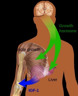
Any exercise, resistance or [[Aerobic Exercise|aerobic,]] brings about a significant increase in [[Growth Hormone|human growth hormone (HGH)]]. Insulin-like growth factor and growth hormone are responsible for increased [[collagen]] synthesis after exercise and aids muscle recovery. Growth hormone itself does not directly cause muscle hypertrophy but it aids muscle recovery and thereby potentially facilitates the muscle strengthening process.<ref>Wideman L, Weltman JY, Hartman ML, Veldhuis JD, Weltman A. [https://www.ncbi.nlm.nih.gov/pubmed/12457419 Growth hormone release during acute and chronic aerobic and resistance exercise.] Sports medicine. 2002 Dec 1;32(15):987-1004.</ref> The accumulation of lactate and hydrogen ions (eg in hypoxic training) further increases the release of growth hormone. <ref name=":2" /> | |||
High intensity training has been shown to down regulate myostatin and thereby provide an environment for muscle hypertrophy to occur.<ref name=":10" /> Myostatin controls and inhibits cell growth in muscle tissue. It needs to be essentially shut down for muscle hypertrophy to occur. | |||
===== Hypoxia ===== | |||
Resistance training results in the compression of blood vessels within the muscles being trained. This causes an hypoxic environment due to a reduction in oxygen delivery to the muscle. As a result of the hypoxia hypoxia-inducible factor (HIF-1α) is activated. This leads to an increase in '''anaerobic lactic metabolism''' and the '''production of lactate'''.<ref name=":0" /> | |||
===== Cell Swelling ===== | |||
When there is blood pooling and an accumulation of metabolites cell swelling occurs. This swelling within the cells causes an anabolic reaction and results in muscle hypertrophy.<ref name=":9">Wilson JM, Lowery RP, Joy JM, Loenneke JP, Naimo MA. [https://journals.lww.com/nsca-jscr/fulltext/2013/11000/Practical_Blood_Flow_Restriction_Training.20.aspx Practical blood flow restriction training increases acute determinants of hypertrophy without increasing indices of muscle damage.] The Journal of Strength & Conditioning Research. 2013 Nov 1;27(11):3068-75.</ref> The cell swelling may actually cause mechanical tension which will then activate the myogenic stem cells as discussed above. | |||
=== Effects of Blood Flow Restriction on Muscle Strength === | |||
The aim of BFR training is to '''mimic the effects of high intensity exercise''' by recreating a hypoxic environment using a cuff. The cuff is placed proximally to the muscle being exercise and low intensity exercises can then be performed. Because the outflow of blood is limited using the cuff capillary blood that has a low oxygen content collects and there is an increase in protons and lactic acid, the same physiological adaptations to the muscle (eg release of hormones, hypoxia and cell swelling) will take place during the BFR training and low intensity exercise as would occur with high intensity exercise.<ref name=":9" /> | |||
* '''Low intensity BFR training''' results in greater muscle circumference when compared with normal low intensity exercise. (1) | |||
* '''Low intensity BFR (LI-BFR)''' results in an increase in the water content of the muscle cells (cell swelling).<ref name=":9" /> It also speeds up the recruitment of fast-twitch muscle fibres.<ref name=":6">Spranger MD, Krishnan AC, Levy PD, O'Leary DS, Smith SA. [https://www.physiology.org/doi/full/10.1152/ajpheart.00208.2015?url_ver=Z39.88-2003&rfr_id=ori:rid:crossref.org&rfr_dat=cr_pub%3dpubmed Blood flow restriction training and the exercise pressor reflex: a call for concern]. American Journal of Physiology-Heart and Circulatory Physiology. 2015 Sep 4;309(9):H1440-52.</ref> It is also hypothesized that once the cuff is removed a hyperemia (excess of blood in the blood vessels) will form and this will cause further cell swelling.<ref name=":4" /> | |||
* '''Short duration, low intensity BFR training''' of around 4-6 weeks has been shown to cause a 10-20% increase in muscle strength. These increases were similar to gains obtained as a result of high-intensity exercise without BFR<ref name=":6" /> | |||
A study comparing (1) high intensity, (2) low intensity, (3) high and low intensity with BFR and (4) low intensity with BFR. While all 4 exercise regimes produced increases in torque, muscle activations and muscle endurance over a 6 week period - the high intensity (group 1) and BFR (groups 3 and 4) produced the greatest effect size and were comparable to each other. <ref>Sousa, Jbc et al. [https://www.ncbi.nlm.nih.gov/pmc/articles/PMC5377566/ “Effects of strength training with blood flow restriction on torque, muscle activation and local muscular endurance in healthy subjects.”] ''Biology of sport'' vol. 34,1 (2016): 83-90. doi:10.5114/biolsport.2017.63738</ref> | |||
== Equipment == | == Equipment == | ||
=== BFR Cuff === | |||
BFR requires a '''tourniquet''' to be placed on a limb. The cuff needs to be tightened to a specific pressure that occludes venous flow while still allowing arterial flow whilst exercises are being performed. | |||
Simple pieces of equipment such as '''surgical tubing or elastic straps''' have been used in gym settings to achieve this result.<ref name=":8">McEwen JA, Owens JG, Jeyasurya J. [https://link.springer.com/article/10.1007/s40846-018-0397-7#Sec8 Why is it Crucial to Use Personalized Occlusion Pressures in Blood Flow Restriction (BFR) Rehabilitation?.] Journal of Medical and Biological Engineering. 2019 Apr 2;39(2):173-7.</ref> These are not advisable as you are unable to monitor the amount of blood flow occlusion. A thin diameter may also cause too much local pressure and result in tissue damage. | |||
=== BFR Cuff Width === | |||
A wide cuff is preferred in the correct application of BFR. 10-12cm cuffs are generally used. A wide cuff of 15cm may be best to allow for even restriction. Modern cuffs are shaped to fit the natural contour of the arm or thigh with a proximal to distal narrowing. There are also specific upper and lower limb cuffs that allow for better fitment.<ref name=":7">Bond CW, Hackney KJ, Brown SL, Noonan BC. [https://www.jospt.org/doi/full/10.2519/jospt.2019.8375 Blood Flow Restriction Resistance Exercise as a Rehabilitation Modality Following Orthopaedic Surgery: A Review of Venous Thromboembolism Risk]. journal of orthopaedic & sports physical therapy. 2019 Jan;49(1):17-27.</ref> | |||
=== BFR Cuff Material === | |||
BFR cuffs can be made from either elastic or nylon. The narrower cuffs are normally elastic and the wider nylon. With elastic cuffs there is an initial pressure even before the cuff is inflated and this results in a different ability to restrict blood flow as compared with nylon cuffs.<ref>Loenneke JP, Fahs CA, Rossow LM, Thiebaud RS, Mattocks KT, Abe T, Bemben MG. [https://www.ncbi.nlm.nih.gov/pmc/articles/PMC3767914/#B8 Blood flow restriction pressure recommendations: a tale of two cuffs.] Frontiers in physiology. 2013 Sep 10;4:249</ref> | |||
Elastic cuffs have been shown to provide a significantly greater arterial occlusion pressure as opposed to nylon cuffs. <ref>Buckner SL, Dankel SJ, Counts BR, Jessee MB, Mouser JG, Mattocks KT, Laurentino GC, Abe T, Loenneke JP. [https://www.researchgate.net/publication/303378637_Influence_of_cuff_material_on_blood_flow_restriction_stimulus_in_the_upper_body Influence of cuff material on blood flow restriction stimulus in the upper body.] The Journal of Physiological Sciences. 2017 Jan 1;67(1):207-15.</ref> | |||
== | === BFR Cuff Pressure === | ||
Different blood flow restriction cuff pressure prescription methods:<ref name=":7" /> | |||
# a standard pressure (used for all patients) for e.g. 180 mmHg; | |||
# a pressure relative to the patient's systolic blood pressure, for e.g. 1.2- or 1.5-fold greater than systolic blood pressure; | |||
# a pressure relative to the patient's thigh circumference. | |||
It is the safest to use a pressure specific to each individual patient, because different pressures occlude the amount of blood flow for all individuals under the same conditions.<ref name=":7" /> | |||
A Doppler ultrasound or plethysmography can be used to determine the blood flow to the limb. The cuff is inflated to a specific pressure where the arterial blood flow is completely occluded. This known as limb occlusion pressure (LOP) or arterial occlusion pressure (AOP). The cuff pressure is then calculated as a percentage of the LOP, normally between 40%-80%. | |||
Using this method is preferable as it ensures patients are exercising at the correct pressure for them and the type of cuff being used. It is safer and makes sure that they are exercising at optimal pressures, not too high to cause tissue damage and also not too low to be ineffective.<ref name=":7" /> | |||
The pressure of the cuff depends upon the width of the cuff as well as the size of the limb on which the cuff is applied. | |||
The key to BFR is that the pressure needs to be high enough to occlude venous return and allow blood pooling but needs to be low enough to maintain the arterial inflow <ref>Loenneke JP, Kim D, Fahs CA, Thiebaud RS, Abe T, Larson RD, Bemben DA, Bemben MG. [https://www.ncbi.nlm.nih.gov/pubmed/25187395 Effects of exercise with and without different degrees of blood flow restriction on torque and muscle activation]. Muscle & nerve. 2015 May;51(5):713-21.</ref>Perceived wrap tightness, on a scale of 0-10 has also been used to conduct BFR training. Wilson et al (2013) found that a perceived wrap tightness of 7 out of 10 resulted in total venous occlusion but still allowed arterial inflow. <ref>Accessed from https://www.strengthandconditioningresearch.com/blood-flow-restriction-training-bfr/#5 on 16/04/19</ref><ref>Lowery RP, Joy JM, Loenneke JP, de Souza EO, Machado M, Dudeck JE, Wilson JM. [https://www.ncbi.nlm.nih.gov/pubmed/24188499 Practical blood flow restriction training increases muscle hypertrophy during a periodized resistance training programme]. Clinical physiology and functional imaging. 2014 Jul;34(4):317-21.</ref> | |||
The | |||
== | == Clinical Application == | ||
BFR has been used in athletes and recreational training to obtain muscle hypertrophy. It can also be used in clinical populations that cannot perform high intensity exercises because of the stage of their condition or pathology involved.<ref>PICÓN MM, CHULVI IM, CORTELL JM, Tortosa J, Alkhadar Y, Sanchís J, Laurentino G. [https://www.ncbi.nlm.nih.gov/pubmed/29795722 Acute cardiovascular responses after a single bout of blood flow restriction training.] International Journal of Exercise Science. 2018;11(2):20.</ref> | |||
Examples of BFR training in the clinic: | |||
===== | <div class="row"> | ||
<div class="col-md-4"> {{#ev:youtube|FbXSdCm8Q6U|250}} <div class="text-right"><ref>NovaCare SelectPT TV. The ACL Road to Recovery - Blood Flow Restriction. Available from: https://youtu.be/FbXSdCm8Q6U</ref></div></div> | |||
<div class="col-md-4">{{#ev:youtube|2fMUpxqJq48|250}} <div class="text-right"><ref>Performance Physical Therapy & Wellness - Blood Flow Restriction Therapy (BFR). Available from: https://youtu.be/2fMUpxqJq48</ref></div></div> | |||
<div class="col-md-4">{{#ev:youtube|pDiLSj6ixTo|250}} <div class="text-right"><ref>Kate Warren. Blood Flow Restriction Training and Physical Therapy | Breaking Athletic Barriers | EVOLVE PT. Available from: https://youtu.be/pDiLSj6ixTo</ref></div></div> | |||
</div> | |||
=== Procedure === | |||
'''Upper Limb:''' The tourniquet is placed on the upper arm. The cuff is inflated to restrict 50% of the arterial blood flow and 100% of the venous flow. | |||
'''Lower limb:''' The tourniquet is placed on the upper thigh. The cuff is inflated to restrict 80% of the arterial blood flow and 100% of the venous flow. With the cuff inflated to the correct pressure normal exercises are performed at about 20-30% of 1RM. | |||
=== Exercise Prescription === | |||
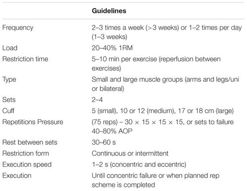
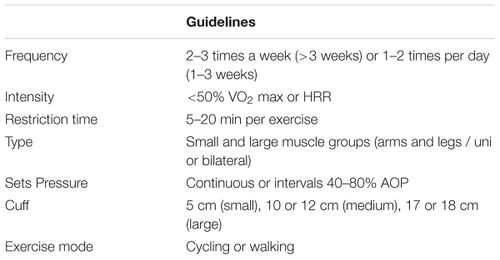
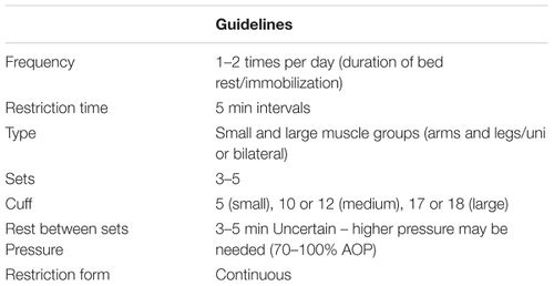
Exercise prescription for BFR varies, this is dependent on whether it is being applied during resistance training (BFR-RE), aerobic training (BFR-AE) or passively without exercise (P-BFR)<ref name=":12">Patterson SD, Hughes L, Warmington S, Burr J, Scott BR, Owens J, Abe T, Nielsen JL, Libardi CA, Laurentino G, Neto GR. Corrigendum: [https://pdfs.semanticscholar.org/3522/8993adf1ab323d3dd53bb6371ad9beaf43a0.pdf Blood Flow Restriction Exercise: Considerations of Methodology, Application, and Safety.] Frontiers in physiology. 2019;10.</ref>[[File:BFR1.jpg|thumb|Model of exercise prescription with BFR-RE<ref name=":12" />|500x500px]] | |||
==== BFR-RE (resistance training) ==== | |||
[[File:BFR2.jpg|thumb|Model of exercise prescription with BFR-AE<ref name=":12" />|500x500px]]For optimal results, resistance training should ideally be done 2-4 times per week. In theory, strength training with BFR can be done daily, however, this may not be the best long term strategy and training 1-2 times per day should only be done for shorter time periods of 1-3 weeks. BFR-RE is typically a single joint exercise modality for strength training.<ref name=":12" /> | |||
Muscle hypertrophy can be observed during BFR-RE within a 3 week period but most studies advocate for longer training durations of more than 3 weeks. | |||
A load of 20-40% 1RM has been shown to produce consistent muscle adaptations for BFR-RE. | |||
* The most commonly used training volume in literature is 75 repetitions across 4 sets (30, 15, 15, 15). | |||
* Rest periods between sets are normally about 30-60 seconds. | |||
* It is important to keep the cuff inflated during the rest periods to capture the metabolites. Intermittent pressure can be applied however this is not as effective as continuous.<ref name=":12" /> | |||
[[File:BFR3.jpg|thumb|Model of Exercise Prescription with P-BFR<ref name=":12" />|500x500px]]The amount of pressure needed to occlude blood flow in the limb depends on the limb size, underlying soft tissue, cuff width and device used. The arterial occlusion pressure applied is dependent on whether it is an upper or lower limb and should be between 40%-80%.<ref name=":12" /> | |||
==== | ==== BFR-AE (aerobic training) ==== | ||
BFR can be applied during aerobic exercise and in research has normally been applied during walking or cycling. It is somewhat more difficult to maintain cuff pressures and literature lacks standardization of cuff pressures during BFR-AE.<ref name=":12" />Also, there is limited evidence that BFR training improves aerobic capacity and performance in trained athletes.<ref>Castilla-López C, Molina-Mula J, Romero-Franco N. [https://www.sciencedirect.com/science/article/pii/S1728869X2200020X?via%3Dihub#sec2 Blood flow restriction during training for improving the aerobic capacity and sport performance of trained athletes: A systematic review and meta-analysis.] Journal of Exercise Science & Fitness. 2022 Mar 22.</ref> | |||
==== P-BFR (passively without exercise) ==== | |||
Passively applied BFR (i.e. BFR is applied and no exercise is performed) has not been widely researched. However, it has shown positive results in reducing muscle atrophy post ACL surgery. The studies conducted did not use standardised pressures and some pressures used were high enough to possibly completely occlude blood flow, which poses safety risks. P-BFR could potentially be beneficial in postoperative patients however more research is needed in this field.<ref name=":12" /> | |||
== Side Effects == | |||
Reported side effects while performing BFR exercises are fainting and dizziness, numbness, pain and discomfort, delayed onset muscle soreness<ref>Brandner, Christopher & May, Anthony & Clarkson, Matthew & Warmington, Stuart. (2018). [https://www.researchgate.net/publication/324233071_Reported_Side-effects_and_Safety_Considerations_for_the_Use_of_Blood_Flow_Restriction_During_Exercise_in_Practice_and_Research Reported Side-effects and Safety Considerations for the Use of Blood Flow Restriction During Exercise in Practice and Research.] Techniques in Orthopaedics. 33. 1. 10.1097/BTO.0000000000000259.</ref>. | |||
* | == Contraindications == | ||
All patients should be assessed for the risks and contraindications to tourniquet use before BFR application. | |||
* Patients possibly at risk of adverse reactions are those with poor [[Cardiovascular System|circulatory system]]<nowiki/>, [[obesity]], diabetes, arterial calcification, [[Sickle Cell Anemia|sickle cell]] trait, severe [[hypertension]], or renal compromise<ref>DePhillipo NN, Kennedy MI, Aman ZS, Bernhardson AS, O'Brien L, LaPrade RF. [https://www.ncbi.nlm.nih.gov/pmc/articles/PMC6203234/ Blood Flow Restriction Therapy After Knee Surgery: Indications, Safety Considerations, and Postoperative Protocol]. Arthroscopy techniques. 2018 Oct 1;7(10):e1037-43.</ref>. | |||
* Potential contraindications to consider are venous thromboembolism, peripheral vascular compromise, sickle cell anemia, extremity infection, lymphadenectomy, cancer or tumor, extremity with dialysis access, acidosis, open fracture, increased intracranial pressure vascular grafts, or medications known to increase clotting risk<ref name=":2" />. | |||
== Safety Implications == | |||
Safety implications around BFR are conflicting. Safety concerns are mainly around the formation of venous thromboembolism ([[Deep Vein Thrombosis|deep vein thrombosis]] and [[Pulmonary Embolism|pulmonary embolism]]) and muscle damage.<ref name=":7" /> Different safety concerns and implications are discussed below: | |||
== | |||
=== | ==== '''Blood hemostasis and BFR''' ==== | ||
Blood has the ability to clot through various systems of coagulation. Blood coagulation is kept in check in part by the fibrinolytic system. Fibrinolysis can help prevent the progression of a blood clot into a venous thromboembolism. | |||
A systematic review conducted by da Cunha Nascimento et al in 2019 examined the long and short term effects on blood hemostasis (the balance between fibrinolysis and coagulation). It concluded that more research needs to be conducted in the field before definitive guidelines can be given.<ref name=":11">da Cunha Nascimento D, Petriz B, da Cunha Oliveira S, Vieira DC, Funghetto SS, Silva AO, Prestes J. [https://www.dovepress.com/effects-of-blood-flow-restriction-exercise-on-hemostasis-a-systematic--peer-reviewed-article-IJGM Effects of blood flow restriction exercise on hemostasis: a systematic review of randomized and non-randomized trials.] International Journal of General Medicine. 2019;12:91.</ref> | |||
In this review, they raised concerns about the following<ref name=":11" /> | |||
* Adverse effects were not always reported | |||
* The level of prior training of subjects was not indicated which makes a significant difference in physiological response | |||
* Pressures applied in studies were extremely variable with different methods of occlusion as well as criteria of occlusion | |||
* Most studies were conducted on a short-term basis and long term responses were not measured | |||
* The studies focused on healthy subjects and not subjects with risk for thromboembolic disorders, impaired fibrinolysis, diabetes and obesity | |||
Their final conclusion on the safety of BFR was as such:<ref name=":11" /> | |||
'''“Thus practitioners must consider the evidence available and ask''' | |||
#'''If the client is sufficiently similar to the subjects in the studies you have examined''' | |||
#'''Does the treatment have a clinically relevant benefit that outweighs the potential risk?''' | |||
#'''Is another treatment available that would provide greater results?" <ref name=":11" />''' | |||
{{#ev:youtube|watch?v=OZjn6vAXJSE}}<ref>Resistance training and coagulation system - Video Abstract ID 194883 Dove Medical Press Available at https://www.youtube.com/watch?v=OZjn6vAXJSE </ref> | {{#ev:youtube|watch?v=OZjn6vAXJSE}}<ref>Resistance training and coagulation system - Video Abstract ID 194883 Dove Medical Press Available at https://www.youtube.com/watch?v=OZjn6vAXJSE </ref> | ||
==== '''Muscle Damage''' ==== | |||
In general, it is well established that unaccustomed exercise results in muscle damage and [[Delayed onset muscle soreness (DOMS)|delayed onset muscle soreness]] (DOMS), especially if the exercise involves a large number of eccentric actions. DOMS is normal after unaccustomed exercise, including after LL-BFR training, and should subside within 24–72 hours[[Blood Flow Restriction Training|[2]]]. | |||
High-load Resistance exercise in any form can result in muscle damage. Excessive breakdown of striated muscle is known as exertional [[rhabdomyolysis]] and can result in organ damage.<ref name=":12" /> The incidence of rhabdomyolysis from BFR-RE is very low at approximately 0,07%-0,2%. This seems to be similar to the occurrence of rhabdomyolysis during normal high load resistance training. There is concern that even with low-load BFR, the increased metabolic stress may trigger rhabdomyolysis but the incidence levels are so low the current evidence does not suggest there is increased risk of rhabdomyolysis during BFR-RE compared to other forms of resistance exercise. <ref name=":12" /> However, a recent systematic review analyzing the evidence about muscle damage after resistance training sessions with blood flow restriction suggests that the use of BFR at high loads of training until muscle failure leads to marked levels of muscle damage, and should be avoided. The findings emphasizes that the magnitude of the muscle damage seems to be attenuated after a first session of resistance training with BFR, demonstrating a protective load effect through this type of exercise. Therefore, professionals can use a principle of progressive overload in structuring resistance training with BFR programs in clinical contexts.<ref>de Queiros VS, Dos Santos ÍK, Almeida-Neto PF, Dantas M, de França IM, Vieira WH, Neto GR, Dantas PM, Cabral BG. [https://journals.plos.org/plosone/article?id=10.1371/journal.pone.0253521 Effect of resistance training with blood flow restriction on muscle damage markers in adults]: A systematic review. Plos one. 2021 Jun 18;16(6):e0253521.</ref> | |||
==== '''Use of Tourniquets''' ==== | |||
By using the 3rd generation system the risk of tourniquet complication is very low, ranging from 0.04% to 0.8%. However, there is an inherent risk to tourniquet use. Some of the common complications<ref name=":2" /> are: | By using the 3rd generation system the risk of tourniquet complication is very low, ranging from 0.04% to 0.8%. However, there is an inherent risk to tourniquet use. Some of the common complications<ref name=":2" /> are: | ||
* Nerve injury: Mechanical compression and neural ischemia play an important role.<ref>JP Sharma, R Salhotra - Indian journal of orthopaedics, 2012 [https://www.ncbi.nlm.nih.gov/pmc/articles/PMC3421924/ Tourniquets in orthopaedic surgery]. Indian J Orthop.Jul-Aug 2012, v.46(4). | * [[Nerve Injury Rehabilitation Physiotherapy|Nerve injury]]: Mechanical compression and neural ischemia play an important role.<ref>JP Sharma, R Salhotra - Indian journal of orthopaedics, 2012 [https://www.ncbi.nlm.nih.gov/pmc/articles/PMC3421924/ Tourniquets in orthopaedic surgery]. Indian J Orthop.Jul-Aug 2012, v.46(4). | ||
</ref> Nerve injury can range from mild transient loss of function to irreversible damage and paralysis. | </ref> Nerve injury can range from mild transient loss of function to irreversible damage and paralysis. | ||
* Skin injury | * [[Skin]] injury | ||
* Tourniquet pain | * Tourniquet pain | ||
* Chemical Burns | * Chemical Burns | ||
* Respiratory, Cardiovascular, Cerebral circulatory and | * Respiratory, Cardiovascular, Cerebral circulatory and hematological effects caused by prolonged ischemia | ||
* Temperature changes | * Temperature changes | ||
== Summary == | == Summary == | ||
BFR training can be viewed as an emerging clinical modality to achieve physiological adaptations for individuals who cannot safely tolerate high muscular tension exercise or those who cannot produce volitional muscle activity. However, continued research is needed to establish parameters for safe application prior to widespread clinical adoption<ref name=":3" />. | BFR training can be viewed as an emerging clinical modality to achieve physiological adaptations for individuals who cannot safely tolerate high muscular tension exercise or those who cannot produce volitional muscle activity. However, continued research is needed to establish parameters for safe application prior to widespread clinical adoption<ref name=":3" />. "Health care professionals must also make sure they have the proper training and are using the correct BFR equipment and pressure prescription techniques to ensure their patients' safety."<ref name=":7" /> | ||
{{#ev:youtube|watch?v=FZWhPx5u9K0}}<ref>Blood Flow Restriction Training American Physical Therapy Association Available at https://www.youtube.com/watch?v=FZWhPx5u9K0</ref> | == Additional Resources == | ||
<div class="row"> | |||
<div class="col-md-4"> {{#ev:youtube|watch?v=FZWhPx5u9K0|250}} <div class="text-right"><ref>Blood Flow Restriction Training American Physical Therapy Association Available at https://www.youtube.com/watch?v=FZWhPx5u9K0</ref></div></div> | |||
<div class="col-md-4"> {{#ev:youtube|WyQN8ct-TsU|250}} <div class="text-right"><ref>Sports Kongres. Symposium: Blood flow restricted exercise in rehabilitation. Available from: https://youtu.be/WyQN8ct-TsU</ref></div></div> | |||
<div class="col-md-4">{{#ev:youtube|0umm5WJBNZc|250}} <div class="text-right"><ref>TheIHMC. Jim Stray-Gundersen - Blood Flow Restriction Training: Anti-aging medicine for the busy baby boomer. Available from: https://youtu.be/0umm5WJBNZc</ref></div></div> | |||
== References == | == References == | ||
<references /> | <references /> | ||
[[Category:Course Pages]] | |||
[[Category:Plus Content]] | |||
[[Category:Rehabilitation]] | |||
[[Category:Sports Medicine]] | |||
[[Category:Musculoskeletal/Orthopaedics]] | |||
[[Category:Health Promotion]] | |||
[[Category:Procedures]] | |||
Revision as of 10:30, 8 November 2022
Original Editor - Vidya Acharya
Top Contributors - Vidya Acharya, Mandy Roscher, Kim Jackson, Tarina van der Stockt, Chelsea Mclene, Lucinda hampton, Jess Bell and Olajumoke Ogunleye
Introduction[edit | edit source]
Muscle weakness commonly occurs in a variety of conditions and pathologies. High load resistance training has been shown to be the most successful means in improving muscular strength and obtaining muscle hypertrophy. However, in certain populations that require muscle strengthening, eg individuals with chronic pain or post-operative patients, high load and high intensity exercises may not be clinically appropriate. Conditions that result in loss of muscle mass such as cancer, Human Immunodeficiency Virus (HIV), diabetes and COPD could potentially benefit from muscle strengthening and muscle hypertrophy but cannot tolerate high intensity/ loaded exercises.[1][2][3][4][5][6]
Blood Flow Restriction (BFR) training is a technique that combines low intensity exercise with blood flow occlusion that produces similar results to high intensity training. It has been used in the gym setting for some time but it is gaining popularity in clinical settings.[7][8]
Blood Flow Restriction (BFR) Training[edit | edit source]
BFR training was initially developed in the 1960s in Japan and known as KAATSU training.[9] It involves the application of a pneumatic cuff (tourniquet) proximally to the muscle that is being trained. It can be applied to either the upper or lower limb. The cuff is then inflated to a specific pressure with the aim of obtaining partial arterial and complete venous occlusion. The patient is then asked to perform resistance exercises at a low intensity of 20-30% of 1 repetition max (1RM), with high repetitions per set (15-30) and short rest intervals between sets (30 seconds) [10]
BFR and Strength Training[edit | edit source]
Understanding the Physiology of Muscle Hypertrophy[edit | edit source]
Muscle hypertrophy is the increase in diameter of the muscle as well as an increase of the protein content within the fibres. An increase in cross-sectional area of the muscle directly correlates with an increase in strength. [11]
Muscle tension and metabolic stress are the two primary factors responsible for muscle hypertrophy.
Mechanical Tension and Metabolic Stress[edit | edit source]
When a muscle is placed under mechanical stress, the concentration of anabolic hormone levels increase. The activation of myogenic stem cells and the elevated anabolic hormones result in protein metabolism and as such muscle hypertrophy can occur. [12][13]
Release of hormones, hypoxia and cell swelling occur when a muscle is under metabolic stress.[14] These factors are all part of the anabolism of muscle tissue.
Activation of myogenic stem cells[edit | edit source]
Myogenic stem cells (satellite cells), are found between the basal lamina and plasma membrane of myofibres. They are normally inactive and become activated in response to muscle injury or increased muscle tension. These cells are responsible for both repair of damaged muscle fibres and also the growth of the fibres themselves[11].
Release of hormones[edit | edit source]
Any exercise, resistance or aerobic, brings about a significant increase in human growth hormone (HGH). Insulin-like growth factor and growth hormone are responsible for increased collagen synthesis after exercise and aids muscle recovery. Growth hormone itself does not directly cause muscle hypertrophy but it aids muscle recovery and thereby potentially facilitates the muscle strengthening process.[15] The accumulation of lactate and hydrogen ions (eg in hypoxic training) further increases the release of growth hormone. [13]
High intensity training has been shown to down regulate myostatin and thereby provide an environment for muscle hypertrophy to occur.[12] Myostatin controls and inhibits cell growth in muscle tissue. It needs to be essentially shut down for muscle hypertrophy to occur.
Hypoxia[edit | edit source]
Resistance training results in the compression of blood vessels within the muscles being trained. This causes an hypoxic environment due to a reduction in oxygen delivery to the muscle. As a result of the hypoxia hypoxia-inducible factor (HIF-1α) is activated. This leads to an increase in anaerobic lactic metabolism and the production of lactate.[14]
Cell Swelling[edit | edit source]
When there is blood pooling and an accumulation of metabolites cell swelling occurs. This swelling within the cells causes an anabolic reaction and results in muscle hypertrophy.[16] The cell swelling may actually cause mechanical tension which will then activate the myogenic stem cells as discussed above.
Effects of Blood Flow Restriction on Muscle Strength[edit | edit source]
The aim of BFR training is to mimic the effects of high intensity exercise by recreating a hypoxic environment using a cuff. The cuff is placed proximally to the muscle being exercise and low intensity exercises can then be performed. Because the outflow of blood is limited using the cuff capillary blood that has a low oxygen content collects and there is an increase in protons and lactic acid, the same physiological adaptations to the muscle (eg release of hormones, hypoxia and cell swelling) will take place during the BFR training and low intensity exercise as would occur with high intensity exercise.[16]
- Low intensity BFR training results in greater muscle circumference when compared with normal low intensity exercise. (1)
- Low intensity BFR (LI-BFR) results in an increase in the water content of the muscle cells (cell swelling).[16] It also speeds up the recruitment of fast-twitch muscle fibres.[17] It is also hypothesized that once the cuff is removed a hyperemia (excess of blood in the blood vessels) will form and this will cause further cell swelling.[10]
- Short duration, low intensity BFR training of around 4-6 weeks has been shown to cause a 10-20% increase in muscle strength. These increases were similar to gains obtained as a result of high-intensity exercise without BFR[17]
A study comparing (1) high intensity, (2) low intensity, (3) high and low intensity with BFR and (4) low intensity with BFR. While all 4 exercise regimes produced increases in torque, muscle activations and muscle endurance over a 6 week period - the high intensity (group 1) and BFR (groups 3 and 4) produced the greatest effect size and were comparable to each other. [18]
Equipment[edit | edit source]
BFR Cuff[edit | edit source]
BFR requires a tourniquet to be placed on a limb. The cuff needs to be tightened to a specific pressure that occludes venous flow while still allowing arterial flow whilst exercises are being performed.
Simple pieces of equipment such as surgical tubing or elastic straps have been used in gym settings to achieve this result.[19] These are not advisable as you are unable to monitor the amount of blood flow occlusion. A thin diameter may also cause too much local pressure and result in tissue damage.
BFR Cuff Width[edit | edit source]
A wide cuff is preferred in the correct application of BFR. 10-12cm cuffs are generally used. A wide cuff of 15cm may be best to allow for even restriction. Modern cuffs are shaped to fit the natural contour of the arm or thigh with a proximal to distal narrowing. There are also specific upper and lower limb cuffs that allow for better fitment.[20]
BFR Cuff Material[edit | edit source]
BFR cuffs can be made from either elastic or nylon. The narrower cuffs are normally elastic and the wider nylon. With elastic cuffs there is an initial pressure even before the cuff is inflated and this results in a different ability to restrict blood flow as compared with nylon cuffs.[21]
Elastic cuffs have been shown to provide a significantly greater arterial occlusion pressure as opposed to nylon cuffs. [22]
BFR Cuff Pressure[edit | edit source]
Different blood flow restriction cuff pressure prescription methods:[20]
- a standard pressure (used for all patients) for e.g. 180 mmHg;
- a pressure relative to the patient's systolic blood pressure, for e.g. 1.2- or 1.5-fold greater than systolic blood pressure;
- a pressure relative to the patient's thigh circumference.
It is the safest to use a pressure specific to each individual patient, because different pressures occlude the amount of blood flow for all individuals under the same conditions.[20]
A Doppler ultrasound or plethysmography can be used to determine the blood flow to the limb. The cuff is inflated to a specific pressure where the arterial blood flow is completely occluded. This known as limb occlusion pressure (LOP) or arterial occlusion pressure (AOP). The cuff pressure is then calculated as a percentage of the LOP, normally between 40%-80%.
Using this method is preferable as it ensures patients are exercising at the correct pressure for them and the type of cuff being used. It is safer and makes sure that they are exercising at optimal pressures, not too high to cause tissue damage and also not too low to be ineffective.[20]
The pressure of the cuff depends upon the width of the cuff as well as the size of the limb on which the cuff is applied.
The key to BFR is that the pressure needs to be high enough to occlude venous return and allow blood pooling but needs to be low enough to maintain the arterial inflow [23]Perceived wrap tightness, on a scale of 0-10 has also been used to conduct BFR training. Wilson et al (2013) found that a perceived wrap tightness of 7 out of 10 resulted in total venous occlusion but still allowed arterial inflow. [24][25]
Clinical Application[edit | edit source]
BFR has been used in athletes and recreational training to obtain muscle hypertrophy. It can also be used in clinical populations that cannot perform high intensity exercises because of the stage of their condition or pathology involved.[26]
Examples of BFR training in the clinic:
Procedure[edit | edit source]
Upper Limb: The tourniquet is placed on the upper arm. The cuff is inflated to restrict 50% of the arterial blood flow and 100% of the venous flow.
Lower limb: The tourniquet is placed on the upper thigh. The cuff is inflated to restrict 80% of the arterial blood flow and 100% of the venous flow. With the cuff inflated to the correct pressure normal exercises are performed at about 20-30% of 1RM.
Exercise Prescription[edit | edit source]
Exercise prescription for BFR varies, this is dependent on whether it is being applied during resistance training (BFR-RE), aerobic training (BFR-AE) or passively without exercise (P-BFR)[30]

BFR-RE (resistance training)[edit | edit source]

For optimal results, resistance training should ideally be done 2-4 times per week. In theory, strength training with BFR can be done daily, however, this may not be the best long term strategy and training 1-2 times per day should only be done for shorter time periods of 1-3 weeks. BFR-RE is typically a single joint exercise modality for strength training.[30]
Muscle hypertrophy can be observed during BFR-RE within a 3 week period but most studies advocate for longer training durations of more than 3 weeks.
A load of 20-40% 1RM has been shown to produce consistent muscle adaptations for BFR-RE.
- The most commonly used training volume in literature is 75 repetitions across 4 sets (30, 15, 15, 15).
- Rest periods between sets are normally about 30-60 seconds.
- It is important to keep the cuff inflated during the rest periods to capture the metabolites. Intermittent pressure can be applied however this is not as effective as continuous.[30]

The amount of pressure needed to occlude blood flow in the limb depends on the limb size, underlying soft tissue, cuff width and device used. The arterial occlusion pressure applied is dependent on whether it is an upper or lower limb and should be between 40%-80%.[30]
BFR-AE (aerobic training)[edit | edit source]
BFR can be applied during aerobic exercise and in research has normally been applied during walking or cycling. It is somewhat more difficult to maintain cuff pressures and literature lacks standardization of cuff pressures during BFR-AE.[30]Also, there is limited evidence that BFR training improves aerobic capacity and performance in trained athletes.[31]
P-BFR (passively without exercise)[edit | edit source]
Passively applied BFR (i.e. BFR is applied and no exercise is performed) has not been widely researched. However, it has shown positive results in reducing muscle atrophy post ACL surgery. The studies conducted did not use standardised pressures and some pressures used were high enough to possibly completely occlude blood flow, which poses safety risks. P-BFR could potentially be beneficial in postoperative patients however more research is needed in this field.[30]
Side Effects[edit | edit source]
Reported side effects while performing BFR exercises are fainting and dizziness, numbness, pain and discomfort, delayed onset muscle soreness[32].
Contraindications[edit | edit source]
All patients should be assessed for the risks and contraindications to tourniquet use before BFR application.
- Patients possibly at risk of adverse reactions are those with poor circulatory system, obesity, diabetes, arterial calcification, sickle cell trait, severe hypertension, or renal compromise[33].
- Potential contraindications to consider are venous thromboembolism, peripheral vascular compromise, sickle cell anemia, extremity infection, lymphadenectomy, cancer or tumor, extremity with dialysis access, acidosis, open fracture, increased intracranial pressure vascular grafts, or medications known to increase clotting risk[13].
Safety Implications[edit | edit source]
Safety implications around BFR are conflicting. Safety concerns are mainly around the formation of venous thromboembolism (deep vein thrombosis and pulmonary embolism) and muscle damage.[20] Different safety concerns and implications are discussed below:
Blood hemostasis and BFR[edit | edit source]
Blood has the ability to clot through various systems of coagulation. Blood coagulation is kept in check in part by the fibrinolytic system. Fibrinolysis can help prevent the progression of a blood clot into a venous thromboembolism.
A systematic review conducted by da Cunha Nascimento et al in 2019 examined the long and short term effects on blood hemostasis (the balance between fibrinolysis and coagulation). It concluded that more research needs to be conducted in the field before definitive guidelines can be given.[34]
In this review, they raised concerns about the following[34]
- Adverse effects were not always reported
- The level of prior training of subjects was not indicated which makes a significant difference in physiological response
- Pressures applied in studies were extremely variable with different methods of occlusion as well as criteria of occlusion
- Most studies were conducted on a short-term basis and long term responses were not measured
- The studies focused on healthy subjects and not subjects with risk for thromboembolic disorders, impaired fibrinolysis, diabetes and obesity
Their final conclusion on the safety of BFR was as such:[34]
“Thus practitioners must consider the evidence available and ask
- If the client is sufficiently similar to the subjects in the studies you have examined
- Does the treatment have a clinically relevant benefit that outweighs the potential risk?
- Is another treatment available that would provide greater results?" [34]
Muscle Damage[edit | edit source]
In general, it is well established that unaccustomed exercise results in muscle damage and delayed onset muscle soreness (DOMS), especially if the exercise involves a large number of eccentric actions. DOMS is normal after unaccustomed exercise, including after LL-BFR training, and should subside within 24–72 hours[2].
High-load Resistance exercise in any form can result in muscle damage. Excessive breakdown of striated muscle is known as exertional rhabdomyolysis and can result in organ damage.[30] The incidence of rhabdomyolysis from BFR-RE is very low at approximately 0,07%-0,2%. This seems to be similar to the occurrence of rhabdomyolysis during normal high load resistance training. There is concern that even with low-load BFR, the increased metabolic stress may trigger rhabdomyolysis but the incidence levels are so low the current evidence does not suggest there is increased risk of rhabdomyolysis during BFR-RE compared to other forms of resistance exercise. [30] However, a recent systematic review analyzing the evidence about muscle damage after resistance training sessions with blood flow restriction suggests that the use of BFR at high loads of training until muscle failure leads to marked levels of muscle damage, and should be avoided. The findings emphasizes that the magnitude of the muscle damage seems to be attenuated after a first session of resistance training with BFR, demonstrating a protective load effect through this type of exercise. Therefore, professionals can use a principle of progressive overload in structuring resistance training with BFR programs in clinical contexts.[36]
Use of Tourniquets[edit | edit source]
By using the 3rd generation system the risk of tourniquet complication is very low, ranging from 0.04% to 0.8%. However, there is an inherent risk to tourniquet use. Some of the common complications[13] are:
- Nerve injury: Mechanical compression and neural ischemia play an important role.[37] Nerve injury can range from mild transient loss of function to irreversible damage and paralysis.
- Skin injury
- Tourniquet pain
- Chemical Burns
- Respiratory, Cardiovascular, Cerebral circulatory and hematological effects caused by prolonged ischemia
- Temperature changes
Summary[edit | edit source]
BFR training can be viewed as an emerging clinical modality to achieve physiological adaptations for individuals who cannot safely tolerate high muscular tension exercise or those who cannot produce volitional muscle activity. However, continued research is needed to establish parameters for safe application prior to widespread clinical adoption[7]. "Health care professionals must also make sure they have the proper training and are using the correct BFR equipment and pressure prescription techniques to ensure their patients' safety."[20]
Additional Resources[edit | edit source]
References[edit | edit source]
- ↑ Hamilton, David & MacKenzie, Matthew & Baar, Keith. (2009). Molecular mechanisms of skeletal muscle hypertrophy Using molecular biology to understand muscle growth. Accessed from https://www.researchgate.net/publication/235702201_Using_molecular_biology_to_understand_muscle_growth/stats
- ↑ Miller BC, Tirko AW, Shipe JM, Sumeriski OR, Moran K. The systemic effects of blood flow restriction training: a systematic review. Int J Sports Phys Ther. 2021 Aug 2;16(4):978-90.
- ↑ Wooten SV, Fleming RYD, Wolf JS Jr, Stray-Gundersen S, Bartholomew JB, Mendoza D, et al. Prehabilitation program composed of blood flow restriction training and sports nutrition improves physical functions in abdominal cancer patients awaiting surgery. Eur J Surg Oncol. 2021 Nov;47(11):2952-8.
- ↑ Alves TC, Pugliesi Abdalla P, Bohn L, Da Silva LSL, Dos Santos AP, et al. Acute and chronic cardiometabolic responses induced by resistance training with blood flow restriction in HIV patients. Sci Rep. 2022 Oct 10;12(1):16989.
- ↑ Jones MT, Aguiar EJ, Winchester LJ. Proposed mechanisms of blood flow restriction exercise for the improvement of type 1 diabetes pathologies. Diabetology. 2021; 2(4):176-89.
- ↑ Pitsillides A, Stasinopoulos D, Mamais I. Blood flow restriction training in patients with knee osteoarthritis: Systematic review of randomized controlled trials. J Bodyw Mov Ther. 2021 Jul;27:477-86.
- ↑ 7.0 7.1 VanWye WR, Weatherholt AM, Mikesky AE. Blood flow restriction training: Implementation into clinical practice. International journal of exercise science. 2017;10(5):649.
- ↑ Loenneke JP, Fahs CA, Rossow LM, Sherk VD, Thiebaud RS, Abe T, Bemben DA, Bemben MG. Effects of cuff width on arterial occlusion: implications for blood flow restricted exercise. European journal of applied physiology. 2012 Aug 1;112(8):2903-12.
- ↑ Accessed fromhttps://www.sportsmed.org/AOSSMIMIS/members/downloads/SMU/2017Spring.pdf
- ↑ 10.0 10.1 Pope ZK, Willardson JM, Schoenfeld BJ. Exercise and blood flow restriction. The Journal of Strength & Conditioning Research. 2013 Oct 1;27(10):2914-26.
- ↑ 11.0 11.1 Bonnieu A, Carnac G, Vernus B. Myostatin in the pathophysiology of skeletal muscle. Current genomics. 2007 Nov 1;8(7):415-22.
- ↑ 12.0 12.1 Luke O'Brien. Blood Flow Restriction Therapy Course. Plus. 2019
- ↑ 13.0 13.1 13.2 13.3 Johnny Owens. Owens Recovery Science. Blood Flow Restriction Rehabilitation Accessed from https://www.owensrecoveryscience.com/?gclid=EAIaIQobChMIoda_9I6q8AIVxgorCh0YFgd2EAAYASAAEgK3APD_BwE
- ↑ 14.0 14.1 de Freitas MC, Gerosa-Neto J, Zanchi NE, Lira FS, Rossi FE. Role of metabolic stress for enhancing muscle adaptations: practical applications. World journal of methodology. 2017 Jun 26;7(2):46.
- ↑ Wideman L, Weltman JY, Hartman ML, Veldhuis JD, Weltman A. Growth hormone release during acute and chronic aerobic and resistance exercise. Sports medicine. 2002 Dec 1;32(15):987-1004.
- ↑ 16.0 16.1 16.2 Wilson JM, Lowery RP, Joy JM, Loenneke JP, Naimo MA. Practical blood flow restriction training increases acute determinants of hypertrophy without increasing indices of muscle damage. The Journal of Strength & Conditioning Research. 2013 Nov 1;27(11):3068-75.
- ↑ 17.0 17.1 Spranger MD, Krishnan AC, Levy PD, O'Leary DS, Smith SA. Blood flow restriction training and the exercise pressor reflex: a call for concern. American Journal of Physiology-Heart and Circulatory Physiology. 2015 Sep 4;309(9):H1440-52.
- ↑ Sousa, Jbc et al. “Effects of strength training with blood flow restriction on torque, muscle activation and local muscular endurance in healthy subjects.” Biology of sport vol. 34,1 (2016): 83-90. doi:10.5114/biolsport.2017.63738
- ↑ McEwen JA, Owens JG, Jeyasurya J. Why is it Crucial to Use Personalized Occlusion Pressures in Blood Flow Restriction (BFR) Rehabilitation?. Journal of Medical and Biological Engineering. 2019 Apr 2;39(2):173-7.
- ↑ 20.0 20.1 20.2 20.3 20.4 20.5 Bond CW, Hackney KJ, Brown SL, Noonan BC. Blood Flow Restriction Resistance Exercise as a Rehabilitation Modality Following Orthopaedic Surgery: A Review of Venous Thromboembolism Risk. journal of orthopaedic & sports physical therapy. 2019 Jan;49(1):17-27.
- ↑ Loenneke JP, Fahs CA, Rossow LM, Thiebaud RS, Mattocks KT, Abe T, Bemben MG. Blood flow restriction pressure recommendations: a tale of two cuffs. Frontiers in physiology. 2013 Sep 10;4:249
- ↑ Buckner SL, Dankel SJ, Counts BR, Jessee MB, Mouser JG, Mattocks KT, Laurentino GC, Abe T, Loenneke JP. Influence of cuff material on blood flow restriction stimulus in the upper body. The Journal of Physiological Sciences. 2017 Jan 1;67(1):207-15.
- ↑ Loenneke JP, Kim D, Fahs CA, Thiebaud RS, Abe T, Larson RD, Bemben DA, Bemben MG. Effects of exercise with and without different degrees of blood flow restriction on torque and muscle activation. Muscle & nerve. 2015 May;51(5):713-21.
- ↑ Accessed from https://www.strengthandconditioningresearch.com/blood-flow-restriction-training-bfr/#5 on 16/04/19
- ↑ Lowery RP, Joy JM, Loenneke JP, de Souza EO, Machado M, Dudeck JE, Wilson JM. Practical blood flow restriction training increases muscle hypertrophy during a periodized resistance training programme. Clinical physiology and functional imaging. 2014 Jul;34(4):317-21.
- ↑ PICÓN MM, CHULVI IM, CORTELL JM, Tortosa J, Alkhadar Y, Sanchís J, Laurentino G. Acute cardiovascular responses after a single bout of blood flow restriction training. International Journal of Exercise Science. 2018;11(2):20.
- ↑ NovaCare SelectPT TV. The ACL Road to Recovery - Blood Flow Restriction. Available from: https://youtu.be/FbXSdCm8Q6U
- ↑ Performance Physical Therapy & Wellness - Blood Flow Restriction Therapy (BFR). Available from: https://youtu.be/2fMUpxqJq48
- ↑ Kate Warren. Blood Flow Restriction Training and Physical Therapy | Breaking Athletic Barriers | EVOLVE PT. Available from: https://youtu.be/pDiLSj6ixTo
- ↑ 30.00 30.01 30.02 30.03 30.04 30.05 30.06 30.07 30.08 30.09 30.10 Patterson SD, Hughes L, Warmington S, Burr J, Scott BR, Owens J, Abe T, Nielsen JL, Libardi CA, Laurentino G, Neto GR. Corrigendum: Blood Flow Restriction Exercise: Considerations of Methodology, Application, and Safety. Frontiers in physiology. 2019;10.
- ↑ Castilla-López C, Molina-Mula J, Romero-Franco N. Blood flow restriction during training for improving the aerobic capacity and sport performance of trained athletes: A systematic review and meta-analysis. Journal of Exercise Science & Fitness. 2022 Mar 22.
- ↑ Brandner, Christopher & May, Anthony & Clarkson, Matthew & Warmington, Stuart. (2018). Reported Side-effects and Safety Considerations for the Use of Blood Flow Restriction During Exercise in Practice and Research. Techniques in Orthopaedics. 33. 1. 10.1097/BTO.0000000000000259.
- ↑ DePhillipo NN, Kennedy MI, Aman ZS, Bernhardson AS, O'Brien L, LaPrade RF. Blood Flow Restriction Therapy After Knee Surgery: Indications, Safety Considerations, and Postoperative Protocol. Arthroscopy techniques. 2018 Oct 1;7(10):e1037-43.
- ↑ 34.0 34.1 34.2 34.3 da Cunha Nascimento D, Petriz B, da Cunha Oliveira S, Vieira DC, Funghetto SS, Silva AO, Prestes J. Effects of blood flow restriction exercise on hemostasis: a systematic review of randomized and non-randomized trials. International Journal of General Medicine. 2019;12:91.
- ↑ Resistance training and coagulation system - Video Abstract ID 194883 Dove Medical Press Available at https://www.youtube.com/watch?v=OZjn6vAXJSE
- ↑ de Queiros VS, Dos Santos ÍK, Almeida-Neto PF, Dantas M, de França IM, Vieira WH, Neto GR, Dantas PM, Cabral BG. Effect of resistance training with blood flow restriction on muscle damage markers in adults: A systematic review. Plos one. 2021 Jun 18;16(6):e0253521.
- ↑ JP Sharma, R Salhotra - Indian journal of orthopaedics, 2012 Tourniquets in orthopaedic surgery. Indian J Orthop.Jul-Aug 2012, v.46(4).
- ↑ Blood Flow Restriction Training American Physical Therapy Association Available at https://www.youtube.com/watch?v=FZWhPx5u9K0
- ↑ Sports Kongres. Symposium: Blood flow restricted exercise in rehabilitation. Available from: https://youtu.be/WyQN8ct-TsU
- ↑ TheIHMC. Jim Stray-Gundersen - Blood Flow Restriction Training: Anti-aging medicine for the busy baby boomer. Available from: https://youtu.be/0umm5WJBNZc


