When refering to evidence in academic writing, you should always try to reference the primary (original) source. That is usually the journal article where the information was first stated. In most cases Physiopedia articles are a secondary source and so should not be used as references. Physiopedia articles are best used to find the original sources of information (see the references list at the bottom of the article).
If you believe that this Physiopedia article is the primary source for the information you are refering to, you can use the button below to access a related citation statement.
The synovial membrane is soft and thin membrane yet has various significant roles that are very essentials for our body movements.
Synovium (also called the synovial membrane) is a specialized connective soft-tissue membrane that lines the inner surface of synovial joint capsules. Together with bone, articular cartilage, tendon, ligament, and fibrous capsule, it is an important component of the tissues that form an integrated joint. As such, it not only has its own specific functions but also interacts with other tissues in the joint both structurally and functionally[1].
Synovial fluid lubricates articular cartilage and provides nourishment through diffusion. It made from a ultrafiltrate of blood plasma, produced and regulated by the synovium.[2]
Anatomy
Synovial membrane: fibrous and the intimal layer.
The synovium is vascularized, unlike the avascular articular cartilage (the other inner joint cavity surface tissue). Therefore, it serves as an important communication channel for transport of nutrients, debris and waste removal, immune modulation, and inflammation in the joint. These specialized functions are achieved by major cell types in synovial membrane, both resident and infiltrated. The membrane consists of 2 layers:
Intimal layer:
Well innervated and vascularised containing different types of cells eg fibroblasts type cells, macrophages, adipocytes, nerve fibres, vascular endothelial cells, lymphocytes. Responsible for: production of synovial fluid components; absorption from the joint cavity; blood/synovial fluid exchanges.[3]
Features two types of synoviocytes:
Type A - macrophage-like cells are derived from hematopoietic monocyte lineage. They are located in the inner surface of the joint cavity, responsible for removing debris in synovial fluid through phagocytosis. Mitochondria are important for synovial macrophages to fulfill this energy-consuming task. Account for 25% of the residential cells in synovial membrane (resident cells live in matrix, permanently housed in the connective tissue, support, maintain, and repair extracellular matrix)[1].
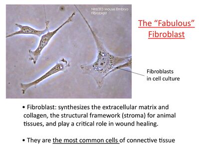
Type B cells - fibroblast-like, responsible for synthesizing and secreting major extracellular matrix proteins in synovial fluid including hyaluronic acid and lubricin. The endoplasmic reticulum of these cells is essential for synthesizing these extracellular matrix molecules in synovial fluid. Also some possess mesenchymal stem cell properties and can be used as source cells for cartilage repair[1].
Composed of fatty, fibrous or loose areolar tissue.[4]
Able to reshape itself to avoid any collision between the muscles and acting as the line-up between the bursae, tendon sheaths, and joints[5].
Synovial Fluid
This fluid is generated from an ultrafiltrate of blood plasma which is regulated by synovium. The purpose of this fluid is to lubricate the cartilage of the bone joint and provide nourishment through diffusion. It is made from a ultrafiltrate of blood plasma and is regulated by the synovium. eg healthy knee contains ~2mL of synovial fluid.
Consists of:
Hyaluronic acid: a viscous glycosaminoglycan serves as a structural element and lubricant.[6] A joint hydration supplement that uses hyaluronic acid
Lubricin: the most lubricating and anti-ahesive molecule in the human body. It is a surface-active mucinous glycoprotein secreted in the synovial joint and plays an important role in cartilage integrity. Lubricin molecules coat the cartilage surface, providing boundary lubrication and preventing cell and protein adhesion[7]
Proteinase: an enzyme that catalyzes proteolysis, the breakdown of proteins into smaller polypeptides or single amino acids.
Collagenases: enzymes that break the peptide bonds in collagen.
Prostaglandins: produced in nearly all cells and are part of the body's way of dealing with injury and illness.. They both sustain homeostatic functions and mediate pathogenic mechanisms, including the inflammatory response[8].
Synovial fluid exhibits non-Newtonian flow characteristics ie Viscosity coefficient is not a constant; Fluid is not linearly viscous; Viscosity increases as the shear rate decreases).[2]
In case of joint inflammation, swelling or redness, a synovial fluid analysis may be done. This sample is used to detect any traces of lactic dehydrogenase, uric acid, and protein which can be held responsible for inflammation[5].
Synovial fluid may also contain microparticles, which are endogenous crystals formed as a result of dysregulated metabolic processes. eg monosodium urate crystals, which are associated with gout; basic calcium phosphate (BCP) crystals, which are associated with osteoarthritis. These particulates contribute to synovial inflammation, cartilage destruction, and subchondral bone remodeling in these synovial related joint diseases.
Imaging
The synovium will be mostly imaged and assessed with MRI and ultrasound in the setting of inflammatory conditions such as arthritis, tenosynovitis and bursitis in which it becomes thickened and increased amounts of synovial fluid will be present.
Related pathology
Communication between synovium and structural tissues including cartilage, bone, capsule, and ligaments is important for maintaining normal joint physiology, but also contributes to several joint disease processes.
There are risk factors, conditions, and diseases that are evolved in situations when the membrane is irritated or thickened. Some conditions and disease are enlisted below.
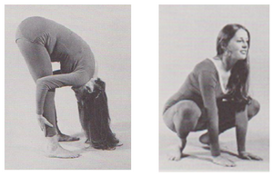
Importance of exercise: The synovial membrane produces synovial fluid, and production is increased when exercise increases circulation of fluid and nutrients to these membranes. Any physical activity can stimulate lubrication in your joints, but some exercises in particular may be most beneficial. These include:
Stretching
Strength training
Quadricep squats
Knee flexion
Heel raises
2. Education re diet : The foods that can help boost synovial fluid are the same foods that are good for your general health. Eating a healthy diet has a number of benefits, including improving joint health and reducing pain. High-fat diets have been shown to increase inflammation in the synovial fluid.
3. Therapies to Boost Synovial Fluid. For people who have severe loss of synovial fluid eg individuals with osteoarthritis—other therapies may be tried
Viscosupplementation injections: This therapy hasn’t been demonstrated to increase synovial fluid, but can help with pain or inflammation related to synovial fluid loss. Injections can be repeated a number of times over a period of several months.[9]
Platelet-rich plasma (PRP) treatment: This form of regenerative medicine aims to help your body heal and repair damaged tissues. This injection is then administered to the affected joint under ultrasound guidance. More large-scale research is needed to prove the benefits of this treatment, but small studies have shown that PRP is at least as effective as hyaluronic acid in treating osteoarthritis.[10]
4. Arthroplasty and the Synovium: The cells making up the re-formed synovial lining, as well as the lining of interface membranes following joint arthroplasty are similar to the typical Type A and B synoviocytes of normal joints. The synovial fluids around joint replacement devices are typically lower in viscosity than pre-arthroplasty fluids but the protein concentration and phospholipid concentrations are comparable, suggesting that the lining tissue function is preserved after arthroplasty. The widespread, long-term success of joint arthroplasty suggests that the lubricant formed from implanted joint synovium is adequate for good clinical performance in the majority of joints.[11]
↑Iwanaga T, Shikichi M, Kitamura H, Yanase H, Nozowa-inoue K. Morphology and functional roles of synoviocytes in the joint. Archives of histology and cytology. 2000;63(1):17-31.Available:https://pubmed.ncbi.nlm.nih.gov/10770586/ (accessed 19.6.2021)
↑The Synovial Lining and Synovial Fluid Properties after Joint Arthroplasty
Michael Shang Kung 1, John Markantonis 1, Scott D. Nelson 2 and Patricia Campbell 1. Lubricants 2015, 3, 394-412; doi:10.3390/lubricants3020394