
Cerebral Oedema
Original Editor - Abdelrahman Sameh Abdelkarim Attia
Top Contributors - Abdelrahman Sameh Abdelkarim Attia, Kirenga Bamurange Liliane and Kim Jackson
Introduction
Cerebral Oedema is a serious condition characterised by the abnormal accumulation of fluid in the brain tissue, leading to increased pressure within the skull and impaired brain function. It can occur as a result of various neurological and systemic conditions, such as traumatic brain injury, stroke, brain tumours, infections, and metabolic imbalances.
The brain swelling associated with cerebral oedema can compromise normal blood flow, resulting in secondary damage due to reduced blood supply to the brain tissue. In severe cases, it can lead to brain herniation, which can be fatal if not treated promptly.
The Monroe-Kellie doctrine provides a fundamental explanation of the pathophysiology of cerebral oedema. It states that the total volume of brain tissue ( approximately 1400 ml ), cerebrospinal fluid (CSF - approximately 150 ml ), and blood within the skull (approximately 150 ml ) must remain constant. An increase in one component, such as brain tissue swelling, requires a compensatory decrease in the others. When these compensatory mechanisms are overwhelmed, increased intracranial pressure can compress the brain structures, leading to widespread damage and potentially irreversible brain injury.
Cerebral oedema can present with a wide range of symptoms, from subtle cognitive changes to life-threatening conditions such as coma and dysfunction of the brainstem. Early recognition and timely intervention are crucial in preventing severe complications associated with this condition.
Clinically Relevant Anatomy
see Brain Anatomy for a comprehensive coverage of the Anatomy.
Classification of Cerebral Oedema
Cerebral oedema is classified into four distinct types based on the underlying pathophysiological mechanisms:
1. Vasogenic Oedema
- Pathophysiology: Vasogenic oedema occurs due to the disruption of the blood-brain barrier (BBB), leading to the leakage of fluid, plasma proteins, and other solutes from the intravascular compartment into the brain parenchyma, particularly the white matter. The compromised BBB allows fluid to accumulate in the extracellular space, particularly around tumours, areas of inflammation, or post-traumatic sites.[1]
- Aetiology: Common causes include brain tumours, abscesses, trauma, haemorrhages, and infections. Vascular endothelial growth factor (VEGF), produced locally in tumours, increases BBB permeability, contributing to peritumoral oedema.
- Clinical Relevance: Vasogenic oedema is the most common type of oedema associated with brain metastases and large tumours. It is also frequently seen in the acute phase following TBI or strokes, particularly haemorrhagic types.
2. Cytotoxic Oedema
- Pathophysiology: In contrast to vasogenic oedema, cytotoxic oedema is characterized by cellular swelling due to failure of the energy-dependent sodium-potassium (Na+/K+) ATPase pumps. This results in the accumulation of sodium inside the cells, which is followed by water influx, leading to intracellular oedema. Cytotoxic oedema primarily affects the gray matter and is a hallmark of ischaemic stroke and other conditions where energy depletion is the primary cause.[1]
- Aetiology: It is commonly seen in ischaemic stroke, hypoxic brain injury, and toxic/metabolic insults, such as in acute liver failure or severe hyponatraemia.
- Clinical Relevance: Cytotoxic oedema develops rapidly, often within minutes, following ischaemic events. In strokes, early cytotoxic oedema is a critical factor in patient prognosis.
3. Osmotic oedema
- Pathophysiology: Osmotic oedema arises when there is a breakdown in the osmolar balance between brain cells and the plasma. This usually occurs when plasma osmolality is reduced (e.g., in severe hyponatraemia) or increased intracellular osmolality draws water into the brain tissue, leading to cellular swelling.
- Aetiology: Conditions like diabetic ketoacidosis (DKA), severe hyponatraemia, or rapid hemodialysis can precipitate osmotic oedema.
- Clinical Relevance: Osmotic oedema is often seen in metabolic disorders where plasma and intracellular osmolarities are disrupted, leading to brain swelling and potential for rapid deterioration if not corrected.[2]
4. Interstitial Oedema
- Pathophysiology: Interstitial oedema results from the movement of cerebrospinal fluid (CSF) from the intraventricular space into the surrounding brain parenchyma, typically caused by elevated intraventricular pressure. This type of oedema primarily affects the periventricular white matter.[1]
- Aetiology: This form of oedema is most commonly associated with hydrocephalus and meningitis, where impaired CSF absorption or obstructed flow leads to its accumulation in the brain.
- Clinical Relevance: Hydrocephalus is the most typical clinical scenario where interstitial oedema is observed. The patient presents with ventricular enlargement on imaging and symptoms of raised ICP, including headache, vomiting, and papilledema.
Aetiology
Cerebral oedema can arise from a wide range of neurological and systemic conditions. These underlying causes can be broadly categorized into primary neurological and secondary systemic aetiologies, each leading to specific types of oedema (vasogenic, cytotoxic, osmotic, or interstitial). Understanding these causes is crucial for early diagnosis and targeted treatment.[2]
1. Neurological Causes
- Traumatic Brain Injury (TBI): One of the most common causes of cerebral oedema, trauma can lead to both vasogenic and cytotoxic oedema. Direct mechanical injury to the brain disrupts the blood-brain barrier (BBB), causing fluid to leak into brain tissue (vasogenic). Secondary energy failure following the trauma can lead to cellular swelling (cytotoxic oedema).
- Stroke (Ischaemic and Haemorrhagic):
- Ischaemic Stroke: Ischaemia leads to cytotoxic oedema due to failure of ATP-dependent sodium-potassium pumps, causing intracellular water accumulation. This type of oedema occurs rapidly after arterial occlusion.
- Haemorrhagic Stroke: In addition to cytotoxic oedema, blood leakage disrupts the BBB, resulting in vasogenic oedema.
- Brain Tumours: Tumours often cause vasogenic oedema, especially due to the secretion of factors such as vascular endothelial growth factor (VEGF), which increases BBB permeability. Peritumoral oedema is a hallmark of brain metastases and primary brain tumours.
- Infections:
- Meningitis and Encephalitis: These infections can lead to both cytotoxic and vasogenic oedema as a result of inflammation, BBB breakdown, and cellular damage. In cases of meningitis, interstitial oedema may develop due to impaired cerebrospinal fluid (CSF) absorption or flow.
- Intracranial Haemorrhage: The accumulation of blood in the brain can directly increase intracranial pressure, causing vasogenic oedema as fluid leaks from damaged blood vessels into surrounding brain tissue.
2. Systemic Causes
- Hyponatraemia: Severe hyponatraemia leads to osmotic oedema. The low sodium levels in the plasma cause water to shift into brain cells, leading to cellular swelling. This is commonly seen in patients with excessive fluid intake, syndrome of inappropriate antidiuretic hormone (SIADH), or after rapid correction of chronic hyponatraemia.
- Diabetic Ketoacidosis (DKA): In patients with DKA, osmotic imbalances can lead to osmotic oedema, where water shifts into brain cells, causing swelling and an increased risk of cerebral herniation, particularly in children.
- Reye Syndrome: This rare condition, often seen in children following viral infections treated with aspirin, can lead to cytotoxic oedema due to mitochondrial dysfunction and subsequent cellular energy failure.
- Hepatic Encephalopathy: In acute liver failure, the accumulation of toxic substances, particularly ammonia, results in cytotoxic oedema by disrupting astrocyte function and causing intracellular swelling.
- High-Altitude Cerebral Oedema (HACE): Occurs in individuals rapidly ascending to high altitudes. Vasogenic oedema develops due to hypoxia-induced disruption of the blood-brain barrier.
- Carbon Monoxide Poisoning: Hypoxic brain injury following carbon monoxide exposure leads to cytotoxic oedema by impairing oxygen delivery to brain cells and causing energy failure.
3. Rare Causes
- Pseudotumor Cerebri: Also known as idiopathic intracranial hypertension, this condition mimics the symptoms of a brain tumour, including elevated ICP and oedema. The exact aetiology is unclear, but it may involve impaired CSF absorption, leading to interstitial oedema.
Risk Factors
Several risk factors [1] predispose individuals to the development of cerebral oedema, including:
- Head Trauma: Individuals involved in contact sports, accidents, or falls are at increased risk of traumatic brain injury, a leading cause of cerebral oedema.
- Vascular Disorders: Patients with cardiovascular risk factors, including hypertension, diabetes mellitus, and atherosclerosis, are at increased risk of stroke, which can lead to cerebral oedema.
- Metabolic Disorders: Conditions such as diabetes, chronic kidney disease, and liver failure increase the risk of metabolic disturbances that can precipitate cerebral oedema (e.g., hyponatraemia, hepatic encephalopathy, diabetic ketoacidosis).
- Altitude Exposure: Rapid ascent to high altitudes, particularly above 2500 meters, increases the risk of high-altitude cerebral oedema (HACE).
- Cerebral Neoplasms: Individuals with brain tumours are predisposed to the development of vasogenic oedema due to increased vascular permeability around the tumour.
- Infections: Conditions such as bacterial meningitis, encephalitis, and sepsis can cause inflammation and oedema within the brain.
Pathophysiology
Cerebral oedema results from an imbalance between the production and clearance of fluid in the brain’s intracellular or extracellular compartments. The swelling causes an increase in intracranial pressure (ICP), which, according to the Monroe-Kellie doctrine, compromises cerebral perfusion and oxygen delivery, potentially leading to ischaemia and secondary neuronal injury. The development of cerebral oedema involves complex interactions between fluid dynamics, cellular homeostasis, vascular permeability, and metabolic disturbances.[2]
Understanding the pathophysiology of cerebral oedema is essential for guiding clinical management, including the use of therapeutic interventions such as osmotherapy, glucocorticoids, and surgical decompression, which aim to restore fluid balance, protect neuronal tissue, and reduce ICP. Subsequently, the correct understanding of pathophysiology, medical and surgical intervention is very critical to develop a rehabilitation protocol.[2]
The pathophysiological mechanisms behind the Cerebral oedema are four major types : vasogenic, cytotoxic, osmotic, and interstitial oedema.[1]
1. Vasogenic Oedema
Mechanism
Vasogenic oedema is the result of a breakdown in the blood-brain barrier (BBB), a critical structure that regulates the exchange of substances between the brain's vascular system and its surrounding tissue. Under normal circumstances, the BBB maintains brain homeostasis by preventing the free passage of proteins, ions, and water into the extracellular brain space. However, in certain pathological conditions, the BBB becomes compromised, allowing plasma proteins and other solutes to leak from the bloodstream into the brain’s interstitial spaces.
Steps of Development
- Disruption of the BBB: The structural integrity of the endothelial cells lining cerebral capillaries is lost, particularly due to the effects of inflammatory cytokines (e.g., TNF-α, IL-1β), growth factors (such as vascular endothelial growth factor (VEGF)), and mechanical insults (e.g., trauma).
- Increased Vascular Permeability: The loss of tight junctions between endothelial cells allows plasma constituents, especially proteins like albumin, to leak into the extracellular space.
- Osmotic Gradient Formation: Proteins in the extracellular space create an osmotic gradient, drawing water from the vasculature into the brain tissue, leading to swelling.
- White Matter Involvement: Vasogenic oedema primarily affects the white matter because this region offers less resistance to the flow of fluid compared to the denser gray matter.
Causes:
- Brain tumours (especially metastatic tumours and high-grade gliomas)
- Trauma
- Cerebral haemorrhage
- Abscesses
- Inflammatory conditions (e.g., meningitis, encephalitis)
Effects:
The increased fluid in the extracellular space compresses neurons and glial cells, disrupts neuronal signalling, and contributes to further damage by reducing blood flow and oxygenation to adjacent tissues. This process also leads to increased intracranial pressure, which compromises cerebral perfusion, resulting in a vicious cycle of injury.
2. Cytotoxic Oedema
Mechanism
Cytotoxic oedema refers to the swelling of brain cells, primarily astrocytes, neurons, and endothelial cells, due to energy failure and loss of cellular ionic balance. Unlike vasogenic oedema, where fluid accumulates in the extracellular space, cytotoxic oedema involves intracellular swelling caused by the dysfunction of ATP-dependent ion pumps, specifically the Na+/K+ ATPase pump.
Steps of Development
- Energy Depletion: In conditions such as ischaemic stroke or hypoxia, cells experience a depletion of ATP due to a lack of oxygen and glucose. Without ATP, energy-dependent ion pumps cease functioning.
- Ionic Imbalance: The failure of the Na+/K+ ATPase pump causes an intracellular accumulation of sodium ions, which are followed by chloride ions (Cl⁻) to maintain electrochemical neutrality. This ionic influx creates an osmotic gradient.
- Water Influx: Water moves into the cells to balance the osmotic gradient, leading to cellular swelling.
- Gray Matter Involvement: Unlike vasogenic oedema, cytotoxic oedema predominantly affects the gray matter due to its high metabolic demand and greater density of neurons and glial cells.
Causes
- Ischaemic stroke: Ischaemia leads to rapid depletion of oxygen and glucose, causing energy failure in affected brain regions.
- Traumatic brain injury (TBI): Direct cellular injury from trauma triggers energy depletion.
- Toxic/metabolic conditions: Conditions such as hepatic encephalopathy, diabetic ketoacidosis (DKA), and hypoglycaemia can also result in cytotoxic oedema.
Effects
Cytotoxic oedema develops within minutes to hours of the initial insult and contributes to cell death by disrupting normal cellular function, leading to cytotoxicity, neuronal apoptosis, and necrosis. This type of oedema can result in widespread neuronal damage, particularly in stroke patients, where rapid intervention is critical to minimize long-term deficits.
3. Osmotic Oedema
Mechanism
Osmotic oedema occurs when there is an imbalance in osmolarity between the brain cells and the extracellular fluid. This typically happens when systemic osmolarity is reduced (e.g., in hyponatraemia) or when cellular osmolarity increases (e.g., in diabetic ketoacidosis), causing water to move into the brain cells. This influx of water leads to cellular swelling across the brain.
Steps of Development
- Systemic Osmolar Imbalance: In metabolic conditions such as hyponatraemia or hyperglycaemia, plasma osmolarity decreases or intracellular osmolarity increases.
- Osmotic Gradient: A gradient develops between the plasma and the intracellular compartment of brain cells.
- Water Shift: To balance the osmotic gradient, water moves into brain cells, resulting in generalized swelling across both gray and white matter.
Causes
- Severe hyponatraemia: A reduction in plasma sodium levels results in water moving into brain cells.
- Diabetic ketoacidosis (DKA): In hyperglycaemia, the osmotic load inside the brain cells increases, drawing water in.
- Rapid dialysis: A sudden change in plasma osmolality during dialysis can lead to osmotic shifts in the brain.
Effects
Osmotic oedema can lead to rapid and diffuse brain swelling, which increases intracranial pressure. It is especially dangerous in conditions like DKA, where cerebral oedema can lead to coma or death if not promptly treated.
4. Interstitial Oedema
Mechanism
Interstitial oedema develops when cerebrospinal fluid (CSF) leaks from the ventricles into the surrounding brain tissue, particularly in cases of hydrocephalus or meningitis. This results in an accumulation of fluid in the brain's extracellular space, particularly in the periventricular white matter.
Steps of Development
- Increased Intraventricular Pressure: Obstruction of CSF flow, such as in non-communicating hydrocephalus, leads to an elevation in intraventricular pressure.
- CSF Leakage: The elevated pressure forces CSF to pass through the ependymal lining of the ventricles into the brain’s interstitial spaces.
- White Matter Involvement: Fluid accumulates predominantly in the white matter, leading to interstitial oedema.
Causes:
- Hydrocephalus: An obstruction of CSF flow (e.g., due to a tumour, congenital malformation, or aqueductal stenosis) leads to a build-up of fluid in the ventricles.
- Meningitis: Inflammation of the meninges can impair CSF absorption, resulting in increased intraventricular pressure and subsequent interstitial oedema.
Effects:
Interstitial oedema contributes to ventricular enlargement and white matter compression, potentially leading to neurological deficits such as motor dysfunction, gait instability, and cognitive impairment. If untreated, increased pressure may cause brain herniation.
Summary of Pathophysiological Effects
Across all types of cerebral oedema, the consequences of increased brain volume are largely the same—rising intracranial pressure, reduced cerebral perfusion pressure (CPP), and compromised oxygen and nutrient delivery to brain tissue. Left untreated, cerebral oedema progresses to herniation syndromes, where parts of the brain are displaced across rigid structures (e.g., the tentorium or foramen magnum), leading to brainstem compression, respiratory arrest, and death. The type of oedema and the underlying cause determine the rate of progression and the potential reversibility of brain damage.[1]
Clinical Presentation of Cerebral Oedema
The clinical presentation of cerebral oedema is highly variable, depending on the underlying cause, location, extent of the swelling, and the rapidity with which the oedema develops. Symptoms arise as increased intracranial pressure (ICP) affects brain function, causing focal neurological deficits, global cognitive impairment, or signs of autonomic dysregulation. In severe cases, cerebral oedema can progress to coma, herniation, and death if not treated promptly.[3] [4]
General Symptoms of Increased Intracranial Pressure (ICP):
Regardless of the underlying cause, increased ICP is a common feature of cerebral oedema, manifesting as a constellation of signs and symptoms. Key symptoms include:[5]
- Headache:
- A common early symptom, usually generalized, worsening with coughing, sneezing, or lying down (due to the increase in ICP). It may be throbbing and constant, and often more severe in the mornings.
- Nausea and Vomiting:
- Associated with raised ICP, vomiting is often described as projectile and can occur without preceding nausea. This symptom is more common when pressure is rapidly rising, as seen in acute conditions like trauma or stroke.
- Altered Mental Status:
- As ICP increases, patients may experience cognitive impairment, confusion, drowsiness, or even agitation. In more severe cases, patients may develop lethargy, obtundation, and eventually coma. Cognitive slowing and difficulty concentrating are often early signs in slower-developing conditions, such as tumors or hydrocephalus.
- Seizures:
- Seizures can occur in patients with significant cerebral edema, particularly if the underlying cause is trauma, hemorrhage, or tumor. Seizures may be focal or generalized, and status epilepticus can develop in some cases.
- Visual Disturbances:
- Patients may experience blurred vision, diplopia (double vision), or visual field deficits due to compression of the optic nerves or cranial nerves involved in eye movement (particularly cranial nerve VI). Papilledema, or swelling of the optic disc, is a hallmark sign of increased ICP and may be identified on fundoscopic examination.
- Cushing's Triad (Late Sign of Increased ICP):
- This is a sign of impending brain herniation and includes:
- Hypertension: Widened pulse pressure (increased systolic pressure with normal or reduced diastolic pressure).
- Bradycardia: A slowing heart rate due to increased pressure on the brainstem.
- Irregular Respirations: Abnormal respiratory patterns, such as Cheyne-Stokes respiration, indicating brainstem dysfunction.
- This is a sign of impending brain herniation and includes:
Symptoms by Type of Edema and Underlying Cause
1. Vasogenic Edema
- Focal Neurological Deficits:
- Weakness, numbness, or hemiparesis may occur due to compression of adjacent brain tissue by the expanding edema. These deficits are typically associated with the location of the swelling, such as hemiplegia or aphasia when edema involves the motor or speech areas.
- Visual disturbances, including visual field cuts or hemianopia, may develop if the edema affects the occipital lobe or optic pathways.
- Cognitive Impairment:
- Progressive decline in cognitive function is often seen in conditions like brain tumors or abscesses, where edema develops more gradually. These patients may exhibit memory problems, personality changes, or difficulty with executive functions.
- Symptoms in Brain Tumors:
- Peritumoral edema, which often involves vasogenic edema, leads to mass effect, causing symptoms based on the tumor location. For instance, patients may present with motor weakness in tumors near the motor cortex or aphasia if the tumor is located in the left hemisphere's speech areas.
2. Cytotoxic Edema
- Rapid Deterioration:
- Cytotoxic edema develops quickly, especially in cases of ischemic stroke or hypoxia. Patients may rapidly progress from mild symptoms (e.g., weakness or confusion) to more severe neurological deficits or coma.
- Focal Neurological Signs:
- In ischemic stroke, cytotoxic edema leads to hemiplegia, facial droop, speech difficulties, and sensory loss, depending on the area of brain affected by the infarct.
- Traumatic brain injury (TBI): Patients may experience initial confusion, followed by the rapid development of focal deficits, loss of consciousness, and post-traumatic seizures due to cytotoxic edema affecting the injured brain tissue.
3. Osmotic Edema
- Hyponatremia-related Symptoms:
- In severe cases of hyponatremia, cerebral edema causes confusion, altered mental status, and seizures. Rapid onset of hyponatremia may lead to severe brain swelling, coma, and even brainstem herniation if untreated.
- Diabetic Ketoacidosis (DKA):
- In pediatric patients with DKA, osmotic edema manifests as altered consciousness, lethargy, or irritability. Severe cases can progress to coma and respiratory failure.
4. Interstitial Edema
- Hydrocephalus:
- In cases of obstructive hydrocephalus, interstitial edema leads to ventricular enlargement and increased ICP. Common symptoms include gait disturbances, urinary incontinence, and cognitive decline (often described as the "classic triad" of normal pressure hydrocephalus in chronic cases).
- Acute hydrocephalus may present with headache, nausea, and rapid decline in consciousness, requiring emergency intervention to relieve the pressure.
- Meningitis:
- Infectious causes of interstitial edema, such as meningitis, often present with fever, neck stiffness, photophobia, and severe headache. As the infection progresses, patients may experience seizures, altered mental status, and signs of increased ICP, such as papilledema or cranial nerve palsies.
Progression to Herniation Syndromes
If cerebral edema is left untreated, it can lead to herniation syndromes, where portions of the brain are displaced across fixed structures in the skull, resulting in life-threatening complications. Key herniation syndromes include:
- Uncal Herniation: The uncus of the temporal lobe is displaced downward, compressing the midbrain.
- Symptoms include ipsilateral pupillary dilation, contralateral motor weakness, and a decreased level of consciousness.
- Tonsillar Herniation: The cerebellar tonsils are pushed downward through the foramen magnum, compressing the brainstem.
- Symptoms include respiratory arrest, bradycardia, hypertension, and coma, often leading to death if untreated.
- Subfalcine Herniation: The cingulate gyrus herniates under the falx cerebri, leading to compression of the anterior cerebral artery, which can cause leg weakness.
Diagnosis & Evaluation
The diagnosis of cerebral edema requires a comprehensive approach, combining clinical assessment with imaging studies and sometimes invasive monitoring. Early detection is crucial to prevent the devastating consequences of increased intracranial pressure (ICP) and brain herniation. Clinicians must differentiate cerebral edema from other causes of altered mental status and neurological deficits to guide timely and appropriate management.
1. Clinical Assessment
The initial step in evaluating cerebral edema is a thorough neurological examination and assessment of intracranial pressure. Key aspects of the clinical assessment include:[6]
- History: Identifying the underlying cause, such as trauma, stroke, metabolic derangements (e.g., hyponatremia), infections, or tumors, can provide clues. Acute onset of symptoms (e.g., following trauma or stroke) may suggest rapid progression of edema.
- Neurological Examination: This may reveal focal neurological deficits (e.g., hemiparesis, aphasia), cranial nerve involvement (e.g., diplopia, pupillary changes), and signs of raised ICP (e.g., papilledema, altered consciousness). Progressive symptoms like confusion, lethargy, and seizures often warrant immediate further evaluation.
2. Imaging Studies
Imaging is critical in diagnosing cerebral edema and identifying its underlying cause. The most commonly used imaging modalities include:
Computed Tomography (CT) Scan
- First-line imaging modality due to its rapid availability in emergency settings.[7]
- Findings:
- Areas of hypoattenuation (low density) indicating the presence of edema.
- Loss of gray-white matter differentiation, particularly in cytotoxic edema (common in stroke and trauma).
- Effacement of sulci and cisterns due to swelling, indicating increased ICP.
- Compression of the ventricles and displacement of midline structures, suggestive of mass effect from edema.
- Signs of herniation, such as downward displacement of the brainstem or lateral shift of midline structures.
- Vasogenic edema is often seen surrounding tumors and areas of inflammation (e.g., abscesses), appearing as localized hypoattenuated areas.
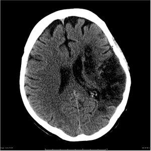
Cerebral Oedema as a result of stroke-evolution 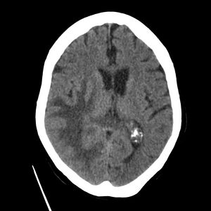
CT for Brain metastases from breast cancer


Magnetic Resonance Imaging (MRI)
- More sensitive than CT for detecting early or subtle cerebral edema, particularly in conditions like ischemic stroke and encephalitis.[7]
- Findings:
- T2-weighted and FLAIR sequences show areas of hyperintensity in both vasogenic and cytotoxic edema.
- Diffusion-weighted imaging (DWI) is particularly useful in identifying cytotoxic edema early in ischemic stroke, where restricted diffusion is seen in affected areas.
- Peritumoral edema can be visualized in more detail compared to CT, particularly in the white matter surrounding tumors.
- Hydrocephalus and ventricular enlargement can be better characterized using MRI, making it useful for evaluating interstitial edema.
3. Intracranial Pressure Monitoring
For patients with severe cerebral edema or those at risk of impending herniation, direct monitoring of intracranial pressure may be necessary, especially in the intensive care unit (ICU). This is typically indicated in cases of traumatic brain injury (TBI), massive stroke, or in patients who have deteriorating mental status without a clear cause.[6]
- Methods of Monitoring:
- Intraventricular Catheter (Ventriculostomy): A catheter placed in the lateral ventricle provides continuous ICP monitoring and allows for drainage of cerebrospinal fluid (CSF) to relieve pressure.
- Subdural or Epidural Bolts: Less invasive than ventriculostomy, these provide accurate ICP measurements but do not allow for CSF drainage.
- Indications:
- ICP monitoring is typically indicated in patients with a Glasgow Coma Scale (GCS) score of ≤ 8, severe TBI, or extensive stroke with signs of herniation.
- Normal ICP: Normal ICP is considered between 7-15 mm Hg in adults. Values exceeding 20-25 mm Hg require urgent intervention.
4. Laboratory Evaluation
Laboratory tests are essential to identify metabolic derangements or systemic causes contributing to cerebral edema. Common tests include:[6]
- Serum Sodium: To evaluate for hyponatremia, a common cause of osmotic edema.
- Blood Glucose Levels: Hyperglycemia or diabetic ketoacidosis (DKA) can precipitate osmotic edema, particularly in children.
- Liver Function Tests: Elevated ammonia levels in patients with hepatic encephalopathy can contribute to cytotoxic edema.
- Infectious Workup: If an infection (e.g., meningitis or encephalitis) is suspected, CSF analysis and blood cultures are indicated to identify the pathogen.
Differential Diagnosis
When assessing cerebral edema, clinicians must distinguish it from other causes of neurological decline, as many conditions can present with similar symptoms of increased intracranial pressure, altered mental status, and neurological deficits. The following are key conditions to consider in the differential diagnosis:[6]
1. Stroke (Ischemic and Hemorrhagic)
- Ischemic stroke can mimic cerebral edema, as infarcted brain tissue rapidly swells due to cytotoxic edema. Hemorrhagic stroke, on the other hand, results in mass effect from both the bleeding and subsequent edema.
- Distinguishing feature: Stroke often presents with acute focal neurological deficits and characteristic findings on CT or MRI (e.g., hypodense area in ischemic stroke, hyperdense in hemorrhagic stroke).
2. Tumors (Primary or Metastatic)
- Brain tumors are often surrounded by vasogenic oedema, which can cause significant mass effect and neurological symptoms.
- Distinguishing feature: Tumors tend to present with progressive neurological deficits and focal symptoms related to the tumor's location (e.g., seizures, cognitive decline). Imaging typically shows a mass with surrounding edema.
3. Meningitis and Encephalitis
- Both infections can lead to cytotoxic and vasogenic edema. Meningitis often presents with fever, neck stiffness, and photophobia, while encephalitis may present with confusion, seizures, and personality changes.
- Distinguishing feature: CSF analysis in meningitis reveals elevated white blood cells and protein levels. Imaging may show meningeal enhancement or diffuse brain swelling.
4. Subdural or Epidural Hematoma
- A subdural hematoma is the accumulation of blood between the dura and arachnoid mater, while an epidural hematoma occurs between the dura and the skull. Both can mimic the signs of cerebral edema by causing mass effect and raised ICP.
- Distinguishing feature: Imaging shows a crescent-shaped collection (subdural) or a biconvex lens-shaped collection (epidural) of blood. The onset may be delayed following trauma.
5. Hydrocephalus
- Obstructive hydrocephalus can cause interstitial edema due to impaired CSF circulation. Symptoms include gait disturbances, urinary incontinence, and cognitive decline.
- Distinguishing feature: Enlarged ventricles and periventricular edema are seen on imaging. The classic triad of gait instability, dementia, and urinary incontinence is often seen in normal pressure hydrocephalus (NPH).
6. Hypertensive Encephalopathy
- Severe acute hypertension (often >180/120 mm Hg) can lead to cerebral vasogenic edema through hyperperfusion and BBB breakdown. Patients may present with headache, confusion, visual disturbances, and seizures.
- Distinguishing feature: Patients with hypertensive encephalopathy typically have markedly elevated blood pressure and show posterior reversible encephalopathy syndrome (PRES) on imaging, with edema predominantly in the occipital and parietal lobes.
7. Metabolic Encephalopathies
- Hypoglycemia, hyperglycemia, and hepatic encephalopathy can cause neurological deterioration and may be associated with cytotoxic or osmotic edema.
- Distinguishing feature: Blood glucose or ammonia levels are often abnormal. Imaging may show diffuse brain swelling or localized changes in the setting of liver failure.
8. Toxic Ingestion or Poisoning
- Substances like carbon monoxide (CO) or drugs that cause CNS depression (e.g., opioids, sedatives) can present with altered mental status and mimic cerebral edema.
- Distinguishing feature: A detailed history and toxicology screen can help identify the offending agent. Imaging may show hypoxic brain injury in CO poisoning.
Treatment
The management of cerebral oedema involves two primary goals: reducing intracranial pressure (ICP) and treating the underlying cause of the oedema. The treatment strategies are tailored to prevent further brain injury, reverse or halt the oedema, and prevent life-threatening complications such as brain herniation. The choice of treatment depends on the type of oedema (vasogenic, cytotoxic, osmotic, or interstitial), the cause, and the severity of the patient's condition.[1]
1. Medical Management
Osmotherapy
Osmotherapy is one of the mainstays of treatment for cerebral oedema, aimed at reducing brain swelling by drawing fluid out of the brain tissue into the bloodstream.
- Mannitol:
- Mannitol is a hyperosmolar agent that increases plasma osmolality, drawing water from the brain parenchyma across the blood-brain barrier. It also reduces blood viscosity, improving cerebral blood flow.
- Dosage: Administered at 0.25–1 g/kg intravenously every 4–6 hours, mannitol can reduce ICP within minutes.
- Side Effects: Mannitol can lead to dehydration, electrolyte imbalances, and renal failure if serum osmolality exceeds 320 mOsm/L.
- Hypertonic Saline:
- Hypertonic saline is another osmotic agent that works by drawing water out of swollen brain cells. It is often used in cases of refractory ICP that do not respond to mannitol.
- Administration: Can be given as a continuous infusion or bolus, with close monitoring of serum sodium levels. Hypertonic saline is considered safe as long as serum sodium does not exceed 160 mmol/L.
- Benefits: Hypertonic saline is preferred in some cases because it maintains blood pressure and cerebral perfusion pressure (CPP) while reducing ICP.
Corticosteroids
Corticosteroids, particularly dexamethasone, are effective in treating vasogenic oedema, especially when caused by brain tumours or abscesses. They stabilize the blood-brain barrier (BBB) by reducing inflammation and limiting fluid leakage into the extracellular space.
- Dexamethasone: Typically administered in doses of 4–10 mg IV every 6 hours.
- Indications: Primarily used in vasogenic oedema associated with brain tumours, metastases, or abscesses.
- Contraindications: Corticosteroids are generally not recommended for cytotoxic oedema, such as that seen in ischaemic stroke or trauma, where they have not shown significant benefits.
Sedation and Analgesia
Sedation reduces cerebral metabolic demand, thereby decreasing ICP. Barbiturate coma can be induced in cases of refractory ICP, although it is typically reserved for severe cases.
- Propofol and benzodiazepines are often used for sedation in the ICU, especially in patients with traumatic brain injury or stroke.
- Barbiturates: These are used in cases where ICP is refractory to conventional treatments. Thiopental or pentobarbital are administered to induce a state of cerebral metabolic suppression.
Ventilation and Hyperventilation
- Hyperventilation: Controlled hyperventilation reduces PaCO₂, which causes cerebral vasoconstriction, temporarily lowering ICP. However, prolonged hyperventilation may reduce cerebral blood flow and can lead to ischaemia, so it should only be used as a short-term measure.
- Target PaCO₂: Typically between 30–35 mm Hg for brief periods.
Temperature Management
- Therapeutic Hypothermia: Induced hypothermia can reduce cerebral metabolism and inflammation, which lowers ICP. This strategy is often employed after cardiac arrest or severe traumatic brain injury.
- Risks: Prolonged hypothermia increases the risk of infections, coagulopathy, and electrolyte disturbances.
Anticonvulsants
Seizures are common in patients with cerebral oedema, especially those with tumours, trauma, or haemorrhages. Prophylactic anticonvulsants such as phenytoin or levetiracetam may be used in patients at risk of seizures.
2. Surgical Management
Decompressive Craniectomy
Decompressive craniectomy is a life-saving procedure used in patients with severe or refractory cerebral oedema. In this procedure, a portion of the skull is removed to allow the swollen brain tissue to expand without causing compression of critical structures.
- Indications: Typically performed in patients with:
- Severe traumatic brain injury with intractable ICP.
- Malignant middle cerebral artery (MCA) infarction with significant mass effect.
- Severe hydrocephalus or brain tumours causing uncontrollable oedema.
- Timing: Early intervention, particularly in patients with malignant MCA infarction, improves outcomes by preventing progression to brain herniation.
Ventriculostomy (External Ventricular Drainage)
In cases of hydrocephalus or interstitial oedema, drainage of cerebrospinal fluid (CSF) via a ventriculostomy can rapidly reduce ICP and relieve symptoms of cerebral oedema.
- Procedure: A catheter is inserted into the lateral ventricle to monitor ICP and drain excess CSF.
- Common in patients with hydrocephalus, subarachnoid haemorrhage, or intraventricular haemorrhage.
Tumour Resection
For patients with vasogenic oedema due to brain tumours, surgical resection of the tumour is necessary to remove the source of the oedema.
- Combined Approach: Resection is often combined with corticosteroid therapy to minimize postoperative oedema and manage peritumoral swelling.
3. Supportive Measures
Other supportive measures are essential for managing patients with cerebral oedema and preventing secondary complications:
- Elevation of the Head: Raising the head of the bed to 30 degrees promotes venous outflow and reduces ICP.
- Maintenance of Normovolemia: Avoiding hypotonic fluids (which can exacerbate oedema) and using isotonic fluids to maintain adequate intravascular volume.
- Avoidance of Straining and Coughing: Excessive strain, such as from coughing or physical activity, can increase ICP. Neuromuscular blocking agents may be used in intubated patients to prevent this.
Role of Physiotherapy
The management of cerebral oedema often requires a multidisciplinary approach, with physiotherapy playing a crucial role in the rehabilitation process.
Objectives of Physiotherapy in Cerebral Oedema
- Enhancing Mobility: Improve functional independence and mobility in patients.
- Reducing Complications: Prevent complications such as muscle atrophy, contractures, and pressure sores.
- Improving Neuromuscular Function: Facilitate neuromuscular re-education for better coordination, strength, and balance.
- Promoting Circulation: Use positioning and gentle mobilization to enhance circulation.
- Supporting Respiratory Function: Optimize respiratory mechanics and function.
Assessment and Individualized Treatment Planning
Initial Assessment
A comprehensive assessment is critical in developing an effective physiotherapy plan. Key components include:
- Medical History: Understanding the underlying cause and severity of cerebral oedema.
- Neurological Assessment: Evaluating muscle strength, tone, reflexes, and sensory function.
- Functional Mobility Assessment: Assessing the patient's ability to move independently and perform activities of daily living (ADLs).
- Posture and Alignment Evaluation: Identifying any postural abnormalities that may need to be addressed.
Goal Setting
Treatment goals should be individualized based on the patient's condition, level of function, and personal aspirations. Common goals may include:
- Restoring range of motion
- Increasing strength in affected muscle groups
- Enhancing balance and coordination
- Improving endurance for daily activities
Physiotherapy Interventions
1. Therapeutic Exercises
- Range of Motion (ROM) Exercises: Both passive and active ROM exercises can help maintain joint mobility and prevent contractures.[8]
- Strength Training: Focused on major muscle groups to improve overall strength and facilitate functional mobility.
- Balance and Coordination Activities: Exercises that challenge the patient’s stability to enhance balance and reduce fall risk.
2. Gait Training
For patients with mobility impairments, gait training may involve:
- Use of Assistive Devices: Teaching the use of walkers, canes, or crutches for safe ambulation.
- Task-specific Training: Practising walking on different surfaces and during various activities to enhance confidence and ability.
3. Vestibular Rehabilitation
For patients experiencing balance disturbances due to vestibular involvement:[9]
- Vestibular Exercises: Techniques aimed at habituation and compensation for vestibular deficits, improving balance and reducing dizziness.
- Balance Training: Incorporating specific exercises to challenge and enhance the vestibular system’s function.
4. Motor Retraining
This involves:
- Neuromuscular Re-education: Focused exercises to retrain movement patterns and improve motor control, especially for affected limbs.
- Functional Task Practice: Engaging in meaningful activities that promote the relearning of functional movements.
5. Early Mobilization
Initiating mobilization as early as possible is crucial:[10]
- Progressive Sitting and Standing: Encouraging patients to progress from lying to sitting, and then to standing and walking as tolerated.
- Bed Mobility Training: Teaching patients to perform safe movements in bed, promoting independence and preventing complications associated with immobility.
6. Positioning Techniques
Proper positioning is essential to minimize the risk of pressure sores and enhance comfort:
- Supine, Prone, and Side-lying Positions: Regularly changing positions can prevent skin breakdown and promote optimal alignment.
- Head Elevation: Maintaining head elevation may help reduce intracranial pressure.
7. Respiratory Physiotherapy
To address potential respiratory complications[11]:
- Breathing Exercises: Techniques such as diaphragmatic breathing and incentive spirometry can promote lung expansion and improve oxygenation.
- Airway Clearance Techniques: Necessary if patients present with secretions or respiratory complications.
8. Education and Family Involvement
Educating patients and their families about the condition, management strategies, and the importance of rehabilitation is vital for optimal recovery. Involving family members in therapy sessions can enhance support and motivation.
Monitoring Progress
Regular reassessment is essential to evaluate progress and adjust treatment plans accordingly. Physiotherapists should continuously monitor the patient’s response to interventions, looking for improvements in mobility, strength, and overall functional ability.
References
- ↑ 1.0 1.1 1.2 1.3 1.4 1.5 1.6 Jha SK. Cerebral edema and its management. Medical Journal Armed Forces India. 2003 Oct 1;59(4):326-31.
- ↑ 2.0 2.1 2.2 2.3 Yang Y, Rosenberg GA. Blood–brain barrier breakdown in acute and chronic cerebrovascular disease. Stroke. 2011 Nov;42(11):3323-8.
- ↑ Jha SK. Cerebral edema and its management. Medical Journal Armed Forces India. 2003 Oct 1;59(4):326-31. BibTeXEndNoteRefManRefWorks
- ↑ Yang Y, Rosenberg GA. Blood-brain barrier breakdown in acute and chronic cerebrovascular disease. Stroke. 2011;42(11):3323-8.
- ↑ Lane PL, Skoretz TG, Doig G, Girotti MJ. Intracranial pressure monitoring and outcomes after traumatic brain injury. Canadian Journal of Surgery. 2000 Dec;43(6):442.
- ↑ 6.0 6.1 6.2 6.3 Nehring SM, Tadi P, Tenny S, editors. Cerebral Edema. StatPearls [Internet]. 2023 Jul 3 [cited 2024 Nov 11]. Available from: https://www.statpearls.com/point-of-care/19182#History%20and%20Physical
- ↑ 7.0 7.1 Radiopaedia Cerebral edema Last revised by Ryan Thibodeau on 10 Jul 2024
- ↑ Huang, M. C., & Wong, H. H. (2019). Early mobilization and its effects on brain edema in stroke patients: A systematic review. Journal of Stroke and Cerebrovascular Diseases, 28(3), 654-661.
- ↑ Hall CD, Herdman SJ, Whitney SL, Anson ER, Carender WJ, Hoppes CW, Cass SP, Christy JB, Cohen HS, Fife TD, Furman JM. Vestibular rehabilitation for peripheral vestibular hypofunction: an updated clinical practice guideline from the academy of neurologic physical therapy of the American Physical Therapy Association. Journal of neurologic physical therapy. 2022 Apr 1;46(2):118-77. BibTeXEndNoteRefManRefWorks
- ↑ Rocca A, Pignat JM, Berney L, Jöhr J, Van de Ville D, Daniel RT, Levivier M, Hirt L, Luft AR, Grouzmann E, Diserens K. Sympathetic activity and early mobilization in patients in intensive and intermediate care with severe brain injuries: a preliminary prospective randomized study. BMC neurology. 2016 Dec;16:1-9. BibTeXEndNoteRefManRefWorks
- ↑ Selsby DS. Chest physiotherapy. BMJ: British Medical Journal. 1989 Mar 3;298(6673):541. BibTeXEndNoteRefManRefWorks