When refering to evidence in academic writing, you should always try to reference the primary (original) source. That is usually the journal article where the information was first stated. In most cases Physiopedia articles are a secondary source and so should not be used as references. Physiopedia articles are best used to find the original sources of information (see the references list at the bottom of the article).
If you believe that this Physiopedia article is the primary source for the information you are refering to, you can use the button below to access a related citation statement.
Brain tumours refers to a diverse group of neoplasms originating from intracranial tissues and the meninges with malignancy levels ranging from benign to aggressive.
Each tumour type has its own biology, treatment, and prognosis and risk factors.[1]
More than 50 per cent are malignant with poor prognosis despite surgery, radiotherapy, and chemotherapy[2]. Even benign or low -grade brain tumors can cause significant disability[3].
Epidemiology
Brain tumours increase in frequency with age, with a few notable exceptions and some uncommon tumours found in infancy.
Apart from genetic tendency, the largest environmental risk factor for brain tumours is exposure to high doses of ionising radiation.[1]
Classification
Brain tumour is an umbrella term for numerous individual tumours. Note figures shown vary significantly dependent on study. They are mainly classified into the below divisions.[4]
Gliomastumours: start in the glial (neuroglia) cells of the brain, also known as neuro-epithelial, most common (50%). They include: astrocytoma, most common tumours arising from glial cells (44%); ependymoma (3%); medulloblastoma (3%); oligodendroglioma (2%).
There are three common types of gliomas, based on the glial cell type:
Astrocytomas: Originated from astrocytes and can be encapsulated. Low grades are common in children while high grades are common in young adults and older patients. Glioblastomas, a type of high-grade astrocytoma, make up > 50% of all gliomas, common in both adults and children.
Oligodendrogliomas: Originated from oligodendrocyte cells. Less infiltrating than astrocytomas, common in adults.
Ependymomas: Originated from ependymal cells (cells lining the ventricular cavities and the central canal of the spinal cord). Common in the pediatric patient population.
Headaches are the most common first symptom of patients with glioma, thought to be a result of tumour growth that causing a mass effect on surrounding tissue. Gliomas generally, notably glioblastomas, are hard difficult to treat. Despite progress in understanding the pathophysiology of gliomas, no significant impact has been made toward preventing the lethality of high-grade gliomas.[5]
Gliomas can spread to the spinal axis via the cerebrospinal fluid (CSF).Spinal seeding is common in patients with glioblastoma (up to 20% of cases have positive CSF) or anaplastic astrocytoma, but it is uncommon in patients with oligodendroglioma, both low grade and anaplastic.[6]
Non-Glioma Tumour
Meningioma MRI Segmentation
Include:
Meningioma, most common tumour of the meninges (15%).Originate from the meningeal layer of either the brain or the spinal cord (non-glial neoplasm). Most meningiomas are benign (grade 1), roughly 1 to 3% of meningiomas can change into malignant tumours with a poor prognosis (5-year survival rate of 32 to 64%). Risk factors for recurrence include genetic disorders such as NF2-Related Schwannomatosis, exposure to radiation, hormonal therapy, and family history. Manifestation are dependent on size and location with wide differences in symptoms eg asymptomatic to severe experience neurological deficits.[7]
Pituitary tumours (8%). Starts in the pituitary gland, usually benign[8]
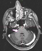
Acoustic Neuroma (pink)Schwannoma (8%). Starts in Schwann cells which surround nerves in the brain, usually benign, includes Facial Schwannoma,Acoustic Neuroma.[8]
This uncommon, usually benign childhood tumour grows slowly and can increase CSF production and block CSF flow, resulting in headaches and increased intracranial pressure. A less common cancerous form can spread through the cerebrospinal fluid.[9]
Germ Cell Tumors
These extremely rare childhood tumours may develop from cells that do not leave the CNS during development. Germ cell tumours typically develop in the centre of the brain and can spread to other parts of the brain and spinal cord. Tumours are named after the type of germ cell they contain. [9]
Treatment
Treatment aims vary, being to remove the tumour completely, slow its growth, or relieve symptoms by reducing the tumour size and decrease swelling.
Treatment is personalised and is dependent on many factors including:
Type, size, grade, location and genetic make-up of the tumour
Clients age, medical history and health status
Symptoms.
A benign tumour may only need surgery to removed, but with a malignant brain treatment options include surgery, radiation therapy and chemotherapy (alone or together). Medicines aim to reduce symptoms or through a clinical trial new or modified treatments may.
A multidisciplinary team will be involved which may include a medical oncologist, neuropathologist, rehabilitation specialist and Physiotherapist.[8]
Laser used to pinpoint tumour in a CT scanner to obtain images of tumour before radiation therapy
↑de Robles P, Fiest KM, Frolkis AD, Pringsheim T, Atta C, St. Germaine-Smith C, Day L, Lam D, Jette N. The worldwide incidence and prevalence of primary brain tumors: a systematic review and meta-analysis. Neuro-oncology. 2015 Jun 1;17(6):776-83.
↑Vargo M. Brain tumor rehabilitation. American journal of physical medicine & rehabilitation. 2011 May 1;90(5):S50-62.