Sciatica: Difference between revisions
Rosie Swift (talk | contribs) No edit summary |
Rosie Swift (talk | contribs) (Updated evidence - aded 3 references. Updated management sections and added link to caudal equine) |
||
| Line 5: | Line 5: | ||
Sciatica is a debilitating condition in which the patient experiences pain and/or paresthesias and/or weakness in the distribution of the [[Sciatic Nerve|sciatic nerve]] or an associated [[Sacral Plexus|lumbosacral nerve root]]. | Sciatica is a debilitating condition in which the patient experiences pain and/or paresthesias and/or weakness in the distribution of the [[Sciatic Nerve|sciatic nerve]] or an associated [[Sacral Plexus|lumbosacral nerve root]]. | ||
* A common mistake is referring to any [[Low Back Pain|low back pain]] or [[Radiculopathy|radicular]] leg pain as sciatica. | * A common mistake is referring to any [[Low Back Pain|low back pain]] or [[Radiculopathy|radicular]] leg pain as sciatica. | ||
* Sciatica is | * Sciatica is a clinical diagnosis based the presence of radiating pain in one leg, with or without the associated neurological deficits of parasthesia and muscle weakness<ref name=":3">Jensen RK, Kongsted A, Kjaer P, Koes B. Diagnosis and treatment of Sciatica. BMJ 2019;367:l6273. Available from: https://www.bmj.com/content/367/bmj.l6273.full [accessed 13 Nov 2021]</ref>, which are the direct result of sciatic nerve or sciatic nerve root pathology. | ||
*The sciatic nerve is made up of the L4 through S2 nerve roots which coalesce at the [[pelvis]] to form the sciatic nerve. At up to 2 cm in diameter, the sciatic nerve is easily the largest nerve in the body. | *The sciatic nerve is made up of the L4 through S2 nerve roots which coalesce at the [[pelvis]] to form the sciatic nerve. At up to 2 cm in diameter, the sciatic nerve is easily the largest nerve in the body. | ||
* Sciatica pain often is worsened with flexion of the [[Lumbar Anatomy|lumbar spine]], twisting, bending, or coughing. | * Sciatica pain often is worsened with flexion of the [[Lumbar Anatomy|lumbar spine]], twisting, bending, or coughing. | ||
| Line 51: | Line 51: | ||
== Epidemiology == | == Epidemiology == | ||
* Annual incidence of 1% to 5% | |||
* Lifetime incidence reported between 10% to 40% | |||
* No gender predominance | * No gender predominance | ||
* Peak incidence occurs in patients in their fourth decade | * Peak incidence occurs in patients in their fourth decade | ||
* Rarely occurs before age 20 (unless traumatic) | * Rarely occurs before age 20 (unless traumatic) | ||
* No association with body height has been established except in the age 50 to 60 group | |||
* Increased incidence in those with poor general health (including presence of co-morbidities and smoking) and the presence of psychological factors such as depression<ref name=":4">Parreira P, Maher C, Steffens D, Hancock M, Ferreira M. Risk factors for low back pain and sciatica: an umbrella review. The Spine Journal 2018; 18,9:1715-1721. Available from:https://www.sciencedirect.com/science/article/abs/pii/S1529943018302432 [Accessed 13 Nov 2021]</ref> | |||
* [[Physical Activity|Physical activity]] increases incidence in those with prior sciatic symptoms and decreased in those with no prior symptoms. | * [[Physical Activity|Physical activity]] increases incidence in those with prior sciatic symptoms and decreased in those with no prior symptoms. | ||
* Occupational predisposition has been shown in machine operators, truck drivers, and jobs where workers are subject to physically awkward positions<ref name=":0" /> | * Occupational predisposition has been shown in machine operators, truck drivers, and jobs where workers are subject to physically awkward positions<ref name=":0" /> or physical stress on the spine such as vibration<ref name=":4" /> | ||
== Clinical Presentation == | == Clinical Presentation == | ||
[[File:Lower-dermatomes.jpg|right|frameless]] | [[File:Lower-dermatomes.jpg|right|frameless]] | ||
<span style="line-height: 1.5em;">Patients with sciatica can present with neurological symptoms such as: </span> | <span style="line-height: 1.5em;">Patients with sciatica can present with neurological symptoms such as: </span> | ||
*<span style="line-height: 1.5em;"> | *<span style="line-height: 1.5em;">Radicular pain in the distribution of the lumbosacral nerve root</span> | ||
*<span style="line-height: 1.5em;"> | *Numbness and altered sensation | ||
*<span style="line-height: 1.5em;">Sensory impairment/<span style="line-height: 1.5em;">disturbance, such as hot and cold or tingling/ burning sensations in the legs</span></span> | |||
*<span style="line-height: 1.5em;">Muscular weakness</span> | *<span style="line-height: 1.5em;">Muscular weakness</span> | ||
*<span style="line-height: 1.5em;">[[Reflexes|Reflex]] impairment</span> | |||
*<span style="line-height: 1.5em;">[[Gait and Lower Limb Observation of Paediatrics - (GALLOP)|Gait]] dysfunction</span> | *<span style="line-height: 1.5em;">[[Gait and Lower Limb Observation of Paediatrics - (GALLOP)|Gait]] dysfunction</span> | ||
Sciatica symptoms can | |||
Sciatica symptoms of p<span style="line-height: 1.5em;">aresthesias or dysesthesias and oedema in the lower extremity can differ, depending on which nerve is affected<ref name="Koes" /><ref name="Kika" /> | |||
*L4: When the L4 nerve is compressed or irritated, the patient feels pain, tingling and numbness in the thigh. The patient also feels weak when straightening the leg and may have a diminished knee jerk reflex. | *L4: When the L4 nerve is compressed or irritated, the patient feels pain, tingling and numbness in the thigh. The patient also feels weak when straightening the leg and may have a diminished knee jerk reflex. | ||
*L5: When the L5 nerve is compressed or irritated, the pain, tingling and numbness may extend to the foot and big toes. | *L5: When the L5 nerve is compressed or irritated, the pain, tingling and numbness may extend to the foot and big toes. | ||
| Line 82: | Line 81: | ||
A thorough differential list is important in considering a diagnosis of sciatica and should include. | A thorough differential list is important in considering a diagnosis of sciatica and should include. | ||
* Herniated lumbosacral [[Disc Herniation|disc]] | * Herniated lumbosacral [[Disc Herniation|disc]] | ||
* [[Cauda Equina Syndrome|Cauda Equina]] | |||
* [[Muscle Injuries|Muscle]] spasm | * [[Muscle Injuries|Muscle]] spasm | ||
* Nerve root impingement | * Nerve root impingement | ||
| Line 103: | Line 103: | ||
2. Imaging | 2. Imaging may be used if pain persists for more than 12 weeks or the patient develops progressive neurological deficits<ref name=":3" /> | ||
* [[X-Rays|Plain films]] of the lumbosacral spine may evaluate for fracture or spondylolisthesis. | * [[X-Rays|Plain films]] of the lumbosacral spine may evaluate for fracture or spondylolisthesis. | ||
* Noncontrast [[CT Scans|CT scan]] may be performed to evaluate fracture if plain films are negative. Pain that has been persistent for 6 to 8 weeks and not responding to conservative management should be imaged. | * Noncontrast [[CT Scans|CT scan]] may be performed to evaluate fracture if plain films are negative. Pain that has been persistent for 6 to 8 weeks and not responding to conservative management should be imaged. | ||
| Line 123: | Line 123: | ||
== Examination == | == Examination == | ||
Clinicians should always look for and inquire about [[The Flag System|red flags]] when evaluating sciatica or in patients who present with any low back pain. | Clinicians should always look for and inquire about [[The Flag System|red flags]] when evaluating sciatica or in patients who present with any low back pain. Patients with signs of urinary retention or decreased anal sphincter tone should be urgently referred for investigation as this suggests [[Cauda Equina Syndrome|cauda equina syndrome]]. | ||
*See [[Lumbar Assessment]] | *See [[Lumbar Assessment]] | ||
| Line 129: | Line 129: | ||
== Medical Management == | == Medical Management == | ||
[[File:Massage image.jpg|right|frameless]] | [[File:Massage image.jpg|right|frameless]] | ||
Most patients improve over time with conservative treatment including exercise, manual therapy, and pain management<ref name=":3" /> | |||
* | * Pharmacology: a short course of oral [[NSAIDs in the Management of Rheumatoid Arthritis|NSAIDs]]; [[Pain Medications|Opioid and non-opioid analgesics]]; muscle relaxants; anticonvulsants for neurogenic pain; localized corticosteroid injections. | ||
* Surgical evaluation: to address structural abnormalities such as: disc herniation, epidural hematoma, epidural abscess or tumour may be considered if no improvement following 6-8 weeks of conservative treatment<ref name=":3" />. One study found that although it may speed up recovery, the effect is similar to conservative care at one year<ref name=":3" /> | |||
* Physical therapy management (see below) | |||
* Surgical evaluation | |||
| Line 144: | Line 139: | ||
{{#ev:youtube|4t8L4zHQ2nQ|412}} | {{#ev:youtube|4t8L4zHQ2nQ|412}} | ||
In most cases of sciatica, conservative treatment is favoured. The evidence does not show that one treatment is superior to the other<ref name="p2">Effectiveness of conservative treatments for the lumbosacral radicular syndrome: a systematic review. Pim A. J. Luijsterburg, Arianne P. Verhagen, Raymond W. J. G. Ostelo, Ton A. G. van Os, Wilco C. Peul, Bart W. Koes. European Spine Journal July 2007, Volume 16, Issue 7, pp 881-899 (1A)</ref> | In most cases of sciatica, conservative treatment is favoured. The evidence does not show that one treatment is superior to the other<ref name="p2">Effectiveness of conservative treatments for the lumbosacral radicular syndrome: a systematic review. Pim A. J. Luijsterburg, Arianne P. Verhagen, Raymond W. J. G. Ostelo, Ton A. G. van Os, Wilco C. Peul, Bart W. Koes. European Spine Journal July 2007, Volume 16, Issue 7, pp 881-899 (1A)</ref> | ||
* Patient Education: to include information on the nature of low back back, advice on self-management techniques and encouragement to continue normal activities<ref name=":5">National Institute for Health and Care Excellence (NICE). Low back pain and sciatica in over 16s: assessment and management: NICE Guideline [NG59] 2016. Available from <nowiki>https://www.nice.org.uk/guidance/ng59</nowiki> [Accessed 13 Nov 2021]</ref> | |||
* Promote self management techniques such as: use of [[Thermotherapy|hot]] or [[Cryotherapy|cold packs]] for comfort and to decreased inflammation; avoidance of inciting activities or prolonged sitting/standing, Regularly changing position i.e. from sitting to standing, practicing good, erect [[posture]], use of proper [[Lifting|lifting techniques]] | |||
* | * Exercise: exercises to increase [[Core Strengthening|core strength]], gentle [[stretching]] of the lumbar spine and hamstrings, regular light exercise such as walking, swimming, or [[aquatherapy]] | ||
* Manual therapy: spinal manipulation, mobilisation or soft tissue techniques such as massage - used alongside exercise and patient education<ref name=":5" /> | |||
* | |||
* | |||
Revision as of 06:41, 13 November 2021
Description[edit | edit source]

Sciatica is a debilitating condition in which the patient experiences pain and/or paresthesias and/or weakness in the distribution of the sciatic nerve or an associated lumbosacral nerve root.
- A common mistake is referring to any low back pain or radicular leg pain as sciatica.
- Sciatica is a clinical diagnosis based the presence of radiating pain in one leg, with or without the associated neurological deficits of parasthesia and muscle weakness[1], which are the direct result of sciatic nerve or sciatic nerve root pathology.
- The sciatic nerve is made up of the L4 through S2 nerve roots which coalesce at the pelvis to form the sciatic nerve. At up to 2 cm in diameter, the sciatic nerve is easily the largest nerve in the body.
- Sciatica pain often is worsened with flexion of the lumbar spine, twisting, bending, or coughing.
- The sciatic nerve provides direct motor function to the hamstrings, lower extremity adductors, and indirect motor function to the calf muscles, anterior lower leg muscles, and some intrinsic foot muscles.
- Indirectly through its terminal branches, the sciatic nerve provides sensation to the posterior and lateral lower leg as well as the plantar foot.
The following video gives a summary of sciatica.
Video:
Etiology[edit | edit source]
Any condition that may structurally impact or compress the sciatic nerve may cause sciatica symptoms.
Sciatic nerve injury can also result in sciatica symptoms (such as pain, muscle weakness and parasthesia) and is usually caused by a traumatic injury (pressure, stretching or cutting), rather than compression or irritation of the nerve. Please read our sciatic nerve injury page for more information.
The causes of sciatica can be categorised into spinal or non-spinal causes or iatrogenic[2]:
Spinal causes:
- Spinal stenosis (due to degenerative bone disorders, trauma, inflammatory disease)
- Spondylolisthesis
- Herniated or bulging lumbar intervertebral disc
- Spinal or paraspinal mass (malignancy, epidural hematoma or abscess)[3]
Non Spinal causes:
- Piriformis syndrome
- Pregnancy
- Lumbar Radiculopathy
- Pelvic tumours
- Trauma to leg
Iatrogenic causes:
- Direct surgical trauma
- Faulty positioning during anaesthesia
- Injection of neurotoxic substances
- Tourniquets
- Dressings, casts or faulty fitting orthotics
- Radiation
Epidemiology[edit | edit source]
- Annual incidence of 1% to 5%
- Lifetime incidence reported between 10% to 40%
- No gender predominance
- Peak incidence occurs in patients in their fourth decade
- Rarely occurs before age 20 (unless traumatic)
- No association with body height has been established except in the age 50 to 60 group
- Increased incidence in those with poor general health (including presence of co-morbidities and smoking) and the presence of psychological factors such as depression[4]
- Physical activity increases incidence in those with prior sciatic symptoms and decreased in those with no prior symptoms.
- Occupational predisposition has been shown in machine operators, truck drivers, and jobs where workers are subject to physically awkward positions[3] or physical stress on the spine such as vibration[4]
Clinical Presentation[edit | edit source]

Patients with sciatica can present with neurological symptoms such as:
- Radicular pain in the distribution of the lumbosacral nerve root
- Numbness and altered sensation
- Sensory impairment/disturbance, such as hot and cold or tingling/ burning sensations in the legs
- Muscular weakness
- Reflex impairment
- Gait dysfunction
Sciatica symptoms of paresthesias or dysesthesias and oedema in the lower extremity can differ, depending on which nerve is affected[5][6]
- L4: When the L4 nerve is compressed or irritated, the patient feels pain, tingling and numbness in the thigh. The patient also feels weak when straightening the leg and may have a diminished knee jerk reflex.
- L5: When the L5 nerve is compressed or irritated, the pain, tingling and numbness may extend to the foot and big toes.
- S1: When the S1 nerve is compressed or irritated, the patient feels pain, tingling and numbness on the outer part of the foot. The patient also experiences weakness when elevating the heel off the ground and standing on tiptoes. The ankle jerk reflex may be diminished.
Differential Diagnosis[edit | edit source]
A thorough differential list is important in considering a diagnosis of sciatica and should include.
- Herniated lumbosacral disc
- Cauda Equina
- Muscle spasm
- Nerve root impingement
- Epidural abscess
- Epidural hematoma
- Tumor
- Potts Disease, also known as spinal tuberculosis
- Piriformis syndrome[3]
Evaluation[edit | edit source]
Sciatica is most commonly diagnosed by:
1. History
- Complaints of radiating pain in the leg, which follows a dermatomal pattern[5].
- Pain generally radiates below the knee, into the foot[6].
- Dermatome maps used to locate the distribution of the pain[5].
- Patients complain about low back pain, which is usually less severe than the leg pain[5].
- Patients may also report sensory symptoms.
2. Imaging may be used if pain persists for more than 12 weeks or the patient develops progressive neurological deficits[1]
- Plain films of the lumbosacral spine may evaluate for fracture or spondylolisthesis.
- Noncontrast CT scan may be performed to evaluate fracture if plain films are negative. Pain that has been persistent for 6 to 8 weeks and not responding to conservative management should be imaged.
- In cases where the neurologic deficit is the present or mass effect is suspected, immediate MRI is the standard of care in establishing the cause of the pain and ruling out pressing surgical pathology[3]
Outcome Measures[edit | edit source]
Many to choose from, below are but a few, all dependant on cause and assessment
- Short Form-36 bodily pain (SF-36 BP)
- Oswestry disability index
- Roland-Morris disability index
- VAS-score: one of leg pain and one of back pain[7]
- McGill pain Questionnaire[7]: this questionnaire looks at the location, intensity, quality and pattern of the pain as well as alleviating and aggrevating factors[8].[8][8][8][12][11][11]
- VAS
- TUG
- Tampa Scale for Kinesiophobia[9]
- Pain Catastrophising Scale[9]
Examination[edit | edit source]
Clinicians should always look for and inquire about red flags when evaluating sciatica or in patients who present with any low back pain. Patients with signs of urinary retention or decreased anal sphincter tone should be urgently referred for investigation as this suggests cauda equina syndrome.
Medical Management[edit | edit source]

Most patients improve over time with conservative treatment including exercise, manual therapy, and pain management[1]
- Pharmacology: a short course of oral NSAIDs; Opioid and non-opioid analgesics; muscle relaxants; anticonvulsants for neurogenic pain; localized corticosteroid injections.
- Surgical evaluation: to address structural abnormalities such as: disc herniation, epidural hematoma, epidural abscess or tumour may be considered if no improvement following 6-8 weeks of conservative treatment[1]. One study found that although it may speed up recovery, the effect is similar to conservative care at one year[1]
- Physical therapy management (see below)
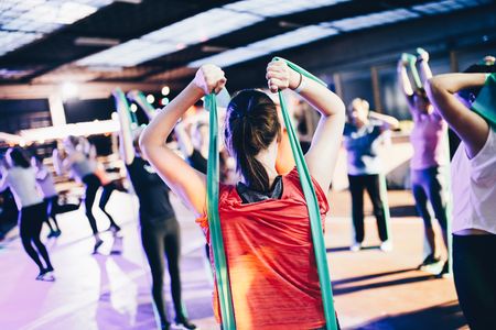
Physical Therapy Management[edit | edit source]
In most cases of sciatica, conservative treatment is favoured. The evidence does not show that one treatment is superior to the other[10]
- Patient Education: to include information on the nature of low back back, advice on self-management techniques and encouragement to continue normal activities[11]
- Promote self management techniques such as: use of hot or cold packs for comfort and to decreased inflammation; avoidance of inciting activities or prolonged sitting/standing, Regularly changing position i.e. from sitting to standing, practicing good, erect posture, use of proper lifting techniques
- Exercise: exercises to increase core strength, gentle stretching of the lumbar spine and hamstrings, regular light exercise such as walking, swimming, or aquatherapy
- Manual therapy: spinal manipulation, mobilisation or soft tissue techniques such as massage - used alongside exercise and patient education[11]
For comprehensive treatment see Links below
- Disc Herniation
- Lumbar Discogenic Pain
- Spinal Stenosis
- Degenerative Disc Disease
- Spondylolisthesis
- Piriformis Syndrome
- Sacroiliac Joint Dysfunction
- Sciatic Nerve Injury
- Corticosteroid injections
- Acupuncture
- Massage therapy has proven to be useful with the treatment of back pain. It promotes blood circulation, muscle relaxation and the release of endorphins[12],[13],[14].
Concluding Remarks[edit | edit source]
- The key to sciatica is patient education.
- There are many causes of sciatica and the disorder is best managed with a team of healthcare professionals that includes an orthopedic surgeon, physical therapist, neurologist, rehabilitation nurse, and a pain specialist.
- Unless there is an acute compression of the spinal nerves, the majority of cases of sciatica are best managed conservatively.
- Patients should be encouraged by the clinician and nurse to lose weight, stop smoking and enroll in a physical therapy program.
- Bed rest should be limited.
- The pharmacist should caution the patient against the use of prescription-strength medications to avoid dependence and other adverse effects.
- Surgery should only be undertaken when conservative methods have failed.
- Regular exercise is essential[3]

References[edit | edit source]
- ↑ 1.0 1.1 1.2 1.3 1.4 Jensen RK, Kongsted A, Kjaer P, Koes B. Diagnosis and treatment of Sciatica. BMJ 2019;367:l6273. Available from: https://www.bmj.com/content/367/bmj.l6273.full [accessed 13 Nov 2021]
- ↑ 2.0 2.1 Osmosis. Sciatica. Available from: https://www.youtube.com/watch?v=VYj-JfX0wT0 (last accessed 15.3.2019)
- ↑ 3.0 3.1 3.2 3.3 3.4 Davis DH, Wilkinson JT, Teaford AK, Smigiel MR. Sciatica produced by a sacral perineurial cyst. Texas Medicine. 1987 Mar 1;83(3):55-6.Available from:https://www.statpearls.com/kb/viewarticle/28772/ (last accessed 12.9.2020)
- ↑ 4.0 4.1 Parreira P, Maher C, Steffens D, Hancock M, Ferreira M. Risk factors for low back pain and sciatica: an umbrella review. The Spine Journal 2018; 18,9:1715-1721. Available from:https://www.sciencedirect.com/science/article/abs/pii/S1529943018302432 [Accessed 13 Nov 2021]
- ↑ 5.0 5.1 5.2 5.3 B.W Koes, M.W Van Tulder, W.C Peul. Diagnosis and treatment of sciatica. BMJ, 23 JUNE 2007, VOLUME 334, p.1313-1314 (1A)
- ↑ 6.0 6.1 Kika Konstantinou, Martyn Lewis, Kate M. Dunn. Agreement of self-reported items and clinically assessed nerve root involvement (or sciatica) in a primary care setting. Eur Spine J (2012) 21:2306–2315. (1B)
- ↑ 7.0 7.1 Brouwer, Patrick A., et al. "Effectiveness of percutaneous laser disc decompression versus conventional open discectomy in the treatment of lumbar disc herniation; design of a prospective randomized controlled trial." BMC musculoskeletal disorders 10.1 (2009)
- ↑ Ngamkham, Srisuda, et al. "The McGill Pain Questionnaire as a multidimensional measure in people with cancer: an integrative review." Pain Management Nursing 13.1 (2012)
- ↑ 9.0 9.1 Monticone M. et al., Management of catastrophising and kinesiophobia improves rehabilitation after fusion for lumbar spondylolisthesis and stenosis. A randomised controlled trial. Eur Spine J. (2014) Jan 23(1):87-95
- ↑ Effectiveness of conservative treatments for the lumbosacral radicular syndrome: a systematic review. Pim A. J. Luijsterburg, Arianne P. Verhagen, Raymond W. J. G. Ostelo, Ton A. G. van Os, Wilco C. Peul, Bart W. Koes. European Spine Journal July 2007, Volume 16, Issue 7, pp 881-899 (1A)
- ↑ 11.0 11.1 National Institute for Health and Care Excellence (NICE). Low back pain and sciatica in over 16s: assessment and management: NICE Guideline [NG59] 2016. Available from https://www.nice.org.uk/guidance/ng59 [Accessed 13 Nov 2021]
- ↑ Abdelilah el Barzouhi, M.D., Carmen L.A.M. Vleggeert-Lankamp, M.D., Ph.D., Geert J. Lycklama, Nijeholt, M.D., Ph.D., Bas F. Van der Kallen, M.D.,Wilbert B. van den Hout, Ph.D., Wilco C.H. Jacobs, Ph.D.,Bart W. Koes, Ph.D., and Wilco C. Peul, M.D., Ph.D. Magnetic Resonance Imaging in Follow-up Assessment of Sciatica. New England Journal of Medicine, 368;11 nejm.org march 14, 2013, P.1000 (1B)
- ↑ Dionne CE. A consensus approach toward the standardization of back pain definitions for use in prevalence studies. 2008
- ↑ John Barrett,Douglas Noel Golding. The practical treatment of backache and sciatica. Redwood Burn Limited. 1984.p97-103.

