Ankle Joint: Difference between revisions
Evan Thomas (talk | contribs) mNo edit summary |
Rachael Lowe (talk | contribs) mNo edit summary |
||
| Line 151: | Line 151: | ||
| align="center" valign="middle" | Sustentaculum Tali<span class="Apple-tab-span" style="white-space:pre"> </span> | | align="center" valign="middle" | Sustentaculum Tali<span class="Apple-tab-span" style="white-space:pre"> </span> | ||
| Reinforces Ankle Joint | | Reinforces Ankle Joint | ||
|} | |} | ||
=== Muscles === | === Muscles === | ||
| Line 335: | Line 333: | ||
Medial Cuniform | Medial Cuniform | ||
|} | |} | ||
==== Dorsiflexion ==== | ==== Dorsiflexion ==== | ||
| Line 470: | Line 466: | ||
*Dorsiflexion 0° - 20° | *Dorsiflexion 0° - 20° | ||
*Plantarflexion 0° - 50° | *Plantarflexion 0° - 50° | ||
=== Closed Packed Position === | === Closed Packed Position === | ||
*Maximum Dorsiflexion | *Maximum Dorsiflexion | ||
| Line 491: | Line 485: | ||
| Medial Ligament<br>Calcaneofibular Ligament<br>Posterior Talofibular Ligament<br>Posterior Joint Capsule Tension<br>Contact of Talus with Tibia<br>Plantarflexors Tension | | Medial Ligament<br>Calcaneofibular Ligament<br>Posterior Talofibular Ligament<br>Posterior Joint Capsule Tension<br>Contact of Talus with Tibia<br>Plantarflexors Tension | ||
|} | |} | ||
== Clinical Examination == | == Clinical Examination == | ||
| Line 504: | Line 497: | ||
*[http://www.physio-pedia.com/Anterior_Drawer_of_the_Ankle Anterior Drawer of the Ankle] | *[http://www.physio-pedia.com/Anterior_Drawer_of_the_Ankle Anterior Drawer of the Ankle] | ||
*[http://www.physio-pedia.com/index.php?title=Ligament_Tests_of_the_Ankle&action=edit&redlink=1 Ligament Tests] | *[http://www.physio-pedia.com/index.php?title=Ligament_Tests_of_the_Ankle&action=edit&redlink=1 Ligament Tests] | ||
*[http://www.physio-pedia.com/Squeeze_Test Squeeze Test] | *[http://www.physio-pedia.com/Squeeze_Test Squeeze Test] | ||
*[http://www.pthaven.com/page/show/161742-talar-tilt-test Talar Tilt Test] | *[http://www.pthaven.com/page/show/161742-talar-tilt-test Talar Tilt Test] | ||
*[http://www.pthaven.com/page/show/161744-kleiger-test Kleiger Test] | *[http://www.pthaven.com/page/show/161744-kleiger-test Kleiger Test] | ||
=== Clinical Predicition Rules === | === Clinical Predicition Rules === | ||
*[http://www.physio-pedia.com/Ottawa_Ankle_Rules Ottawa Ankle Rules] to rule in/out radiography of the ankle after trauma | *[http://www.physio-pedia.com/Ottawa_Ankle_Rules Ottawa Ankle Rules] to rule in/out radiography of the ankle after trauma | ||
=== Outcome Measures === | === Outcome Measures === | ||
*[http://www.physio-pedia.com/Foot_and_Ankle_Disability_Index Foot and Disability Index&]is a 34-item self report questionnaire divided into two subscales: the Foot and Ankle Disability Index and the Foot and Ankle Disability Index Sport | *[http://www.physio-pedia.com/Foot_and_Ankle_Disability_Index Foot and Disability Index&]is a 34-item self report questionnaire divided into two subscales: the Foot and Ankle Disability Index and the Foot and Ankle Disability Index Sport | ||
== Pathology/Injury == | == Pathology/Injury == | ||
| Line 522: | Line 515: | ||
*[http://www.physio-pedia.com/Ankle_and_Foot_Fractures Ankle and Foot Fractures] | *[http://www.physio-pedia.com/Ankle_and_Foot_Fractures Ankle and Foot Fractures] | ||
*[http://www.physio-pedia.com/Ankle_%26_Foot_Arthropathies Ankle and Foot Arthropathies] | *[http://www.physio-pedia.com/Ankle_%26_Foot_Arthropathies Ankle and Foot Arthropathies] | ||
*[http://www.physio-pedia.com/Chronic_Ankle_Instability Chronic Ankle Instability] | *[http://www.physio-pedia.com/Chronic_Ankle_Instability Chronic Ankle Instability] | ||
== Physiotherapeutic Techniques == | == Physiotherapeutic Techniques == | ||
| Line 529: | Line 522: | ||
*[http://www.pthaven.com/page/show/162352-talocrural-joint-posterior-glide-to-promote-dorsiflexion Talocrural Joint Posterior Glide to Promote Dorsiflexion] | *[http://www.pthaven.com/page/show/162352-talocrural-joint-posterior-glide-to-promote-dorsiflexion Talocrural Joint Posterior Glide to Promote Dorsiflexion] | ||
*[http://www.pthaven.com/page/show/162355-talocrural-joint-anterior-glide-to-promote-plantarflexion Talocrural Joint Anterior Glide to Promote Plantarflexion] | *[http://www.pthaven.com/page/show/162355-talocrural-joint-anterior-glide-to-promote-plantarflexion Talocrural Joint Anterior Glide to Promote Plantarflexion] | ||
*[http://www.pthaven.com/page/show/162347-talocrural-joint-distal-distraction Talocrural Joint Distal Distraction <ref>PT Haven. Talocrural Joint Distal Distraction. Available from: http://www.pthaven.com/page/show/162347-talocrural-joint-distal-distraction [last accessed 19/03/2015]</ref>] | *[http://www.pthaven.com/page/show/162347-talocrural-joint-distal-distraction Talocrural Joint Distal Distraction <ref>PT Haven. Talocrural Joint Distal Distraction. Available from: http://www.pthaven.com/page/show/162347-talocrural-joint-distal-distraction [last accessed 19/03/2015]</ref>]<span class="mw-reflink-text">[5]</span><br> | ||
<br> | |||
=== Balance Retraining === | === Balance Retraining === | ||
*[http://www.physio-pedia.com/Balance Balance] | *[http://www.physio-pedia.com/Balance Balance] | ||
| Line 541: | Line 532: | ||
== Resources == | == Resources == | ||
*[http://www.ncbi.nlm.nih.gov/pmc/articles/PMC2855022/ Anatomy of the Ankle Ligaments: A Pictorial Essay] | *[http://www.ncbi.nlm.nih.gov/pmc/articles/PMC2855022/ Anatomy of the Ankle Ligaments: A Pictorial Essay] - In this pictorial essay, the ligaments around the ankle are grouped, depending on their anatomic orientation, and each of the ankle ligaments is discussed in detail. | ||
== References == | == References == | ||
<references /> | <references /> | ||
[[Category:Anatomy]] [[Category:Ankle]] [[Category:Musculoskeletal/Orthopaedics]] | [[Category:Anatomy]] [[Category:Ankle]] [[Category:Musculoskeletal/Orthopaedics]] | ||
Revision as of 16:18, 5 November 2017
Original Editor - Naomi O'Reilly
Lead Editors - Naomi O'Reilly, Kim Jackson, Lucinda hampton, Joanne Garvey, Rachael Lowe, Ewa Jaraczewska, Vidya Acharya, Admin, WikiSysop, Simisola Ajeyalemi, Rucha Gadgil, Jess Bell, Khloud Shreif, Kirenga Bamurange Liliane, Olajumoke Ogunleye, Evan Thomas, Sona Eyobe, Oyemi Sillo and Tarina van der Stockt
Description[edit | edit source]
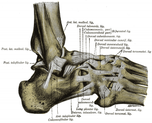
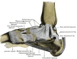
The Ankle Joint, also known as the Talocrural Articulation, is a synovial hinge joint connecting the distal ends of the tibia and fibula in the lower limb with the proximal end of the talus. The ankle joint is maintained by the shape of the talus and its tight fit between the tibia and fibula. In the neutral position, there are strong bony constraints. With increasing plantar flexion, the bony constraints are decreased and the ligaments are more susceptible to strain and injury. The articulation between the tibia and the talus bears more weight than that between the smaller fibula and the talus. [1]
| [2] | [3] |
Anatomy[edit | edit source]
Articulating Surfaces[edit | edit source]
- Trochlea of Talus
- Malleolar Mortis formed by Tibia & Fibula
- Lateral & Medial Malleolus
Joint Capsule[edit | edit source]
The articular capsule surrounds the joints, and is attached, above, to the borders of the articular surfaces of the tibia and malleoli; and below, to the talus around its upper articular surface. The joint capsule anteriorly is a broad, thin, fibrous layer, posteriorly the fibres are thin and run mainly transversely blending with the transverse ligament and laterally the capsule is thickened, and attaches to the hollow on the medial surface of the lateral malleolus. The synovial membrane extends superiorly between Tibia & Fibula as far as the Interosseous Tibiofibular Ligament.[4]
Ligaments[edit | edit source]
Lateral Ligaments of Ankle[edit | edit source]
Reinforce Joint Laterally through three ligaments. These ligaments stabilize the ankle, and serve as a guide to direct ankle motion by attaching the lateral malleolus to the bones below the ankle joint. They are responsible for resistance against inversion and internal rotation stress. [4]
|
LIGAMENT |
DESCRIPTION | PROXIMAL ATTACHMENT | DISTAL ATTACHMENT | ROLE |
|---|---|---|---|---|
|
Anterior Talofibular Ligament (ATFL) |
Flat Weak Band that extends Anteriomedially. Most commonly damaged ligament of the ankle. |
Lateral Malleolus | Neck of Talus |
Restrain anterior displacement of the talus in respect to the fibula and tibia. Resists Inversion in planterflexion. |
|
Posterior Talofibular Ligament (PTFL) |
Thick, fairly strong band that runs horizontally medially. This ligament is under greater strain in full dorsiflexion of ankle. Rarely injured because bony stability protects ligaments when ankle in dorsiflexion. |
Malleolar Fossa of Fibula | Lateral Tubercle of Talus |
Forms the back wall of the recipient socket for the talus' trochlea. Resists posterior displacement of the talus. |
|
Calcaneofibular Ligament (CFL) |
Round cord that passes posterioinferiorly | Tip of Lateral Malleolus | Lateral Surface of Calcaneus |
Aids Talofibular stability during Dorsiflexion. Restrain inversion of the calcaneus with respect to the fibula. Prevent Talar tilt into Inversion. |
Medial Ligaments of Ankle[edit | edit source]
Known collectively as the Deltoid Ligament the medial ligaments of the ankle attaches proximally to the Medial Malleolus and fan out to attach distally to the Talus, Calcaneus and Navicular via four adjacent and continuous parts. The deltoid ligament is triangular in shape and consists of a superficial and deep layer which connect the talus to the medial malleolus. It reinforces the joint capsule medially. Stabilise’s the ankle joint during eversion of the foot and prevents subluxation of the ankle joint. [4]
|
LIGAMENTS |
DESCRIPTION | PROXIMAL ATTACHMENT | DISTAL ATTACHMENT | ROLE |
|---|---|---|---|---|
|
Anterior Tibiotalar Ligament |
Medial Malleolus |
Head of Talus |
Reinforces Ankle Joint. Control Plantarflexion & Eversion | |
|
Posterior Tibiotalar Ligament |
Talus Posteriorly | Control Dorsiflexion | ||
|
Tibionavicular Ligament |
Forms most anterior part of the Deltoid Ligament |
Dorsomedial Aspect of Navicular | Reinforces Ankle Joint | |
|
Tibiocalcaneal Ligament |
Very thin ligament | Sustentaculum Tali | Reinforces Ankle Joint |
Muscles[edit | edit source]
Plantarflexion[edit | edit source]
Muscles which contribute to Plantarflexion
|
MUSCLE |
ACTION | PROXIMAL ATTACHMENT | DISTAL ATTACHMENT | INNERVATION |
|---|---|---|---|---|
|
POSTERIOR COMPARTMENT | ||||
|
SUPERFICIAL | ||||
| Gastrocnemius |
Plantarflexion when Knee Extended Flexion Knee Raises Heel during Walking |
Lateral Head: Lateral Aspect of Lateral Femoral Condyle Medial Head: Popliteal Surface of Femur Superior to Medial Femoral Condyle |
Posterior Surface Calcaneus via Calcaneal Tendon (Achilles Tendon) |
Tibial Nerve S1-S2 |
| Soleus |
Plantarflexion Steadies Leg on Foot |
Posterior Aspect of Head Fibula Superior ¼ Posterior Surface Tibia Soleal Line & Medial Border Tibia | ||
| Plantaris |
Weakly Assists Gastrocnemius in Plantarflexion |
Inferior end Lateral Supracondylar Line of Femur Oblique Popliteal Ligament | ||
|
DEEP | ||||
| Tibialis Posterior |
Plantarflexion Inversion Supports Medial Longitudinal Arch |
Interosseous Membrane Posterior Surface Tibia inferior to Soleal Line Posterior Surface Fibula |
Navicular Tuberosity Cuneiform Cuboid Bases of Metatarsals 2-4 |
Tibial Nerve L4-L5 |
| Flexor Digitorum Longus |
Plantarflexion Flexion Lateral Four Digits Supports Longitudinal Arch |
Medial Part Posterior Surface Tibia inferior to Soleal Line Broad Tendon to Fibula |
Base Distal Phalanges Digits 2-4 |
Tibial Nerve S2-S3 |
| Flexor Hallucis Longus |
Weak Plantarflexion Flexion Big Toe at all Joints Supports Medial Longitudinal Arch |
Inferior 2/3 Posterior Surface Fibula Inferior Part Interosseous Membrane |
Base Distal Phalanx of Big Toe | |
|
LATERAL COMPARTMENT | ||||
|
Peroneus Brevis |
Weak Plantarflexion Eversion |
Inferior 2/3 of Lateral Surface Tibia |
Dorsal Surface Tuberosity of Base 5th Metatarsal |
Superficial Peroneal Nerve (Superficial Fibular Nerve) L5 - S2 |
|
Peroneus Longus |
Weak Plantarflexion Eversion Supports Transverse Arch |
Head & Superior 2/3 of Lateral Surface Tibia |
Base 1st Metatarsal Medial Cuniform | |
Dorsiflexion[edit | edit source]
Muscles which contribute to Dorsiflexion
|
MUSCLE |
ACTION | PROXIMAL ATTACHMENT | DISTAL ATTACHMENT | INNERVATION |
|
ANTERIOR COMPARTMENT | ||||
|
Tibialis Anterior |
Dorsiflexion Inversion Supports Medial Longitudinal Arch |
Lateral Condyle Tibia Superior ½ Lateral Surface Tibia Interosseous Membrane |
Medial & Inferior Surfaces Medial Cuniform Base of 1st Metatarsal |
Deep Peroneal Nerve (Deep Fibular Nerve) L4-L5 |
|
Extensor Digitorum Longus |
Dorsiflexion Extends Lateral Four Digits |
Lateral Condyle Tibia Superior ¾ Anterior Surface Interosseous Membrane |
Middle & Distal Phalanges of Lateral Four Digits |
Deep Peroneal Nerve (Deep Fibular Nerve) L5-S1 |
|
Extensor Hallucis Longus |
Dorsiflexion Extends Big Toe |
Middle Part Anterior Surface Fibula Interosseous Membrane |
Dorsal Aspect of Base Distal Phalanx of Big Toe | |
|
Peroneus Tertius |
Dorsiflexion Aids Eversion |
Inferior 1/3 Anterior Surface Fibula Interosseous Membrane |
Dorsum Base 5th Metatarsal | |
Blood Supply[edit | edit source]
Derived from Malleolar Branches of:
- Peroneal Artery
- Tibial Artery
Nerve Supply[edit | edit source]
- Common Peroneal Nerve
- Tibial Nerve
Function[edit | edit source]
Motions Available[edit | edit source]
- Talocrural Joint is a uniaxial hinge joint which has just 1° of Motion
- Dorsiflexion 0° - 20°
- Plantarflexion 0° - 50°
Closed Packed Position[edit | edit source]
- Maximum Dorsiflexion
Open Packed Position[edit | edit source]
- 10° Plantarflexion
Structures Limiting Movement[edit | edit source]
| Movement | Limiting Structures |
|---|---|
| Plantarflexion Posterior & Lateral Compartment |
Anterior Talofibular Ligamanet Anterior Part of Medial Ligament Anterior Joint Capsule Tension Contact of Talus with Tibia Dorsiflexor Tension |
| Dorsiflexion Anterior Compartment |
Medial Ligament Calcaneofibular Ligament Posterior Talofibular Ligament Posterior Joint Capsule Tension Contact of Talus with Tibia Plantarflexors Tension |
Clinical Examination[edit | edit source]
Assessment[edit | edit source]
Special Tests[edit | edit source]
- Kaltenborn Ankle & Foot Examination
- Anterior Drawer of the Ankle
- Ligament Tests
- Squeeze Test
- Talar Tilt Test
- Kleiger Test
Clinical Predicition Rules[edit | edit source]
- Ottawa Ankle Rules to rule in/out radiography of the ankle after trauma
Outcome Measures[edit | edit source]
- Foot and Disability Index&is a 34-item self report questionnaire divided into two subscales: the Foot and Ankle Disability Index and the Foot and Ankle Disability Index Sport
Pathology/Injury[edit | edit source]
- Ankle Arthrodesis
- Ankle Impingement
- Ankle Osteoarthritis
- Ankle Osteochondral Lesions
- Ankle Sprain
- Ankle and Foot Fractures
- Ankle and Foot Arthropathies
- Chronic Ankle Instability
Physiotherapeutic Techniques[edit | edit source]
Manual Therapy[edit | edit source]
- Talocrural Joint Posterior Glide to Promote Dorsiflexion
- Talocrural Joint Anterior Glide to Promote Plantarflexion
- Talocrural Joint Distal Distraction [5][5]
Balance Retraining[edit | edit source]
Procedures[edit | edit source]
Resources[edit | edit source]
- Anatomy of the Ankle Ligaments: A Pictorial Essay - In this pictorial essay, the ligaments around the ankle are grouped, depending on their anatomic orientation, and each of the ankle ligaments is discussed in detail.
References[edit | edit source]
- ↑ Allen F. Anderson Sports Medicine. Anatomy Ankle Available from: http://www.drallenfanderson.com/ankle/anatomy [last accessed 20/03/2015]
- ↑ Anatomy Zone. Ankle Joint - 3D Anatomy Tutorial. Available from: https://www.youtube.com/watch?v=lPLdoFQlZXQ [last accessed 19/03/2015]
- ↑ AnimatedBiomedical. Ankle Joint, Bones of the Foot - 3D Medical Animation. Available from: https://www.youtube.com/watch?v=X-eAXKS4pJM [last accessed 19/03/2015]
- ↑ 4.0 4.1 4.2 Moore KL, Agur AMR, Dalley AF. Essential Clinial Anatomy. Baltimore: Lippincott Williams and Wilkins, 2011.
- ↑ PT Haven. Talocrural Joint Distal Distraction. Available from: http://www.pthaven.com/page/show/162347-talocrural-joint-distal-distraction [last accessed 19/03/2015]


