When refering to evidence in academic writing, you should always try to reference the primary (original) source. That is usually the journal article where the information was first stated. In most cases Physiopedia articles are a secondary source and so should not be used as references. Physiopedia articles are best used to find the original sources of information (see the references list at the bottom of the article).
If you believe that this Physiopedia article is the primary source for the information you are refering to, you can use the button below to access a related citation statement.
The integumentary system is the largest organ of the body that forms a physical barrier between the external environment and the internal environment that it serves to protect and maintain.
In addition to its barrier function, this system performs many intricate functions such as body temperature regulation, cell fluid maintenance, synthesis of Vitamin D, and detection of stimuli. The various components of this system work in conjunction to carry out these functions[1].
General Function
The integumentary system has several functions that provide several purposes[2]:
Physical protection: The integumentary is the covering of the human body and its' most apparent function is physical protection: skin - a tightly knit network of cells, with each layer contributing to its strength. The epidermis has an outermost layer created by layers of dead keratin that can withstand wear and tear of the outer environment, the dermis provides the epidermis with blood supply and has nerves that bring danger to attention amongst other functions; hypodermis provides physical cushioning to any mechanical trauma through adipose storage; glands secrete protective films throughout the body; nails protect the digits; hairs throughout the body filter harmful particles from entering the eyes, ears, nose, etc.
Immunity: The skin is the body’s first line of defense acting as a physical barrier preventing direct entry of pathogens. Antimicrobial peptides (AMPs) and lipids on the skin also act as a biomolecular barrier that disrupts bacterial membranes. Resident immune cells, both myeloid and lymphoid cells are present in the skin, and some, eg Langerhans cells or dermal dendritic cells, can travel to the periphery and activate the greater immune system[1]
Wound healing: When our body undergoes trauma with a resulting injury, the integumentary system orchestrates the wound healing process through hemostasis, inflammation, proliferation, and remodeling.[1][1]
Thermoregulation: The skin has a large surface area that is highly vascularized, which allows it to conserve and release heat through vasoconstriction and vasodilation, respectively[1].
Vitamin D synthesis: The primary sources of vitamin D are sun exposure and oral intake (crucial for bone health)[1].
Sensation- Skin innervation is by various types of sensory nerve endings that discriminate pain, temperature, touch, and vibration. Each type of receptor and nerve fiber varies in its adaptive and conductive speeds, leading to a wide range of signals that can be integrated to create an understanding of the external environment and help the body to react appropriately[1].
Organ Systems Involved
Skin
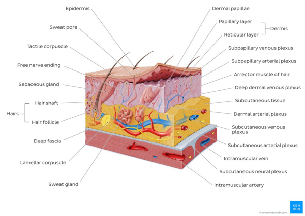
Overview of the integumentary systemAccounts for about 16% of your total body weight. Its surface area covers between 1.5-2m2 [3]
Made up of two layers—the superficial epidermis and the deeper dermis.
Epidermis:
Tough, outer layer that acts as the first line of defense against the external environment
Regenerates from stem cells located in the basal layer that grow up towards the corneum. The epidermis itself is devoid of blood supply and derives its nutrition from the underlying dermis
Composed of stratified squamous epithelial cells that further break down into four to five layers (see image R). From superficial to deep, the primary layers are the
Stratum corneum
Stratum granulosum
Stratum spinosum
Stratum basale
In the palms and soles where the skin is thicker, there is an additional layer of skin between the stratum corneum and stratum granulosum called the stratum lucidum.
Dermis
Underlying connective tissue framework that supports the epidermis
The dermis as a whole contains blood and lymph vessels, nerves, sweat glands, hair follicles, and various other structures embedded within the connective tissue.
Further subdivides into two layers
Superficial papillary dermis - forms finger-like projections into the epidermis, known as dermal papillae, and consists of highly vascularized, loose connective tissue.
Deep reticular layer - has dense connective tissue that forms a strong network[1].
The hypodermis lies between the dermis and underlying organs.
Commonly referred to as subcutaneous tissue
Composed of loose areolar tissue and adipose tissue.
Provides additional cushion and insulation through its function of fat storage and connects the skin to underlying structures such as muscle[1].
Hair
Hair is a component of the integumentary system and extends downward into the dermal layer where it sits in the hair follicle.
The presence of hair is a primary differentiator of mammals as a unique class of organisms.
In humans, it is a cherished and highly visible indicator of health, youth, and even class.
It has a sensory function, protects from cold and UV radiation.
Areas of clinical significance include diseases of hair loss, excess, alterations due to nutritional deficiencies, infectious causes, and effects of drug reactions[5]
Nail
Nails form as layers of keratin and appear at the dorsal tips of the fingers and toes.
Nails function to protect the fingers and toes while increasing the precision of movements and enhancing sensation.
Pathophysiology: Onychomycosis (fungal infection,common clinical presentation involves nail discoloration, subungual hyperkeratosis, onycholysis, and splitting or destruction of the nail plate), Pitting (presents in conditions such as psoriasis, eczema) Koilonychia (spoon nail, been associated with iron deficiency anemia but can be due to idiopathic changes) Clubbing (the most common manifestation of hypertrophic osteoarthropathy and correlates with many systemic conditions).[1]
Associated Glands
Four types of exocrine glands within human skin—Sweat, sebaceous, ceruminous, and mammary glands.
Sweat glands, are further divided into eccrine and apocrine glands.
Eccrine glands are distributed throughout the body and primarily produce serous fluid to regulate body temperature.
Apocrine glands are present in the axilla and pubic area and produce milky protein-rich sweat. These glands are responsible for odor as bacteria break down the secreted organic substances.
Sebaceous glands are part of the pilosebaceous unit, which includes the hair, hair follicle, and arrector pili muscle.
Secretes an oily substance called sebum, a mixture of lipids that forms a thin film on the skin.
This layer adds a protective layer, prevents fluid loss, and also plays an antimicrobial role[1].
The integumentary system provides numerous functions necessary for human life while also maintaining an optimal internal environment for other critical components to thrive.
When there is an imbalance in this system, many disorders can manifest.
The integumentary system also acts as a reflection of underlying pathologies eg showing jaundice with liver disfunction, displaying petechiae with thrombocytopenia; decreased skin turgor with dehydration.
It is a system that can provide many external clues regarding an individual’s physiological state and is a vital component of a complete clinical picture[1].