Subtalar Joint Arthritis: Difference between revisions
No edit summary |
No edit summary |
||
| (37 intermediate revisions by 5 users not shown) | |||
| Line 1: | Line 1: | ||
<div class=" | <div class="editorbox"> '''Original Editor '''- [[User:Nehal Khater|Nehal Khater]] '''Top Contributors''' - {{Special:Contributors/{{FULLPAGENAME}}}}</div> | ||
== Anatomy == | |||
[[File:SubtalarJoint.PNG|thumb|250x250px|Subtalar Joint|alt=]] | |||
The [[Subtalar Joint|subtalar joint]] also named the talocalcaneal joint, is articulating between two tarsal bones the [[talus]] and [[calcaneus]] bones. There are three facets on each of the talus and calcaneus. The posterior talocalcaneal articulation represents the largest component of the subtalar joint.<ref>Ficke J, Byerly DW. [https://www.ncbi.nlm.nih.gov/books/NBK546698/#_article-21883_s2_ Anatomy, Bony Pelvis and Lower Limb, Foot.] InStatPearls [Internet] 2021 Aug 11. StatPearls Publishing. Accessed 7 June 2022.</ref>The malalignment of the subtalar joint can lead to primary osteoarthritis of the ankle joint in the long run. See more here [[Subtalar Joint|Subtalar joint]] | |||
== Function and Biomechanics of Subtalar joint == | |||
The Subtalar joint is necessary for: | |||
== | |||
* The function of the [[Ankle and Foot|foot and the ankle]] and [[gait]].<ref>Jastifer JR, Gustafson PA. [https://www.sciencedirect.com/science/article/abs/pii/S0958259214000613 The subtalar joint: biomechanics and functional representations in the literature.] The foot. 2014 Dec 1;24(4):203-9. Accessed 15 June 2022.</ref> | |||
* Transmission of rotations from the leg and the ankle to the foot.<ref name=":0">Stagni R, Leardini A, O'Connor JJ, Giannini S. [https://journals.sagepub.com/doi/abs/10.1177/107110070302400505 Role of passive structures in the mobility and stability of the human subtalar joint: a literature review.] Foot & ankle international. 2003 May;24(5):402-9. Accessed 15 June 2022.</ref> | |||
= | * Shock absorption during the early [[Gait Definitions|stance phase]].<ref name=":0" /> | ||
The Subtalar joint movements are inversion-eversion<ref>Krähenbühl N, Horn-Lang T, Hintermann B, Knupp M. [https://eor.bioscientifica.com/view/journals/eor/2/7/2058-5241.2.160050.xml The subtalar joint: a complex mechanism.] EFORT open reviews. 2017 Jul 6;2(7):309-16. Accessed 15 June 2022.</ref> and supination-pronation (three-dimensional motions)<ref>Brockett CL, Chapman GJ. [https://www.sciencedirect.com/science/article/pii/S1877132716300483 Biomechanics of the ankle.] Orthopaedics and trauma. 2016 Jun 1;30(3):232-8. Accessed 15 June 2022.</ref>. Pronation combines dorsiflexion, abduction, and eversion, and supination combines plantarflexion, adduction, and inversion<ref>Peña Fernández M, Hoxha D, Chan O, Mordecai S, Blunn GW, Tozzi G, Goldberg A. [https://www.nature.com/articles/s41598-020-57912-z Centre of rotation of the human subtalar joint using weight-bearing clinical computed tomography.] Scientific reports. 2020 Jan 23;10(1):1-4. Accessed 15 June 2022.</ref>. | |||
== | === Main ligaments of Subtalar Joint === | ||
* Anterior talocalcaneal ligament | |||
* Posterior talocalcaneal ligament | |||
* Lateral talocalcaneal ligament | |||
* Medial talocalcaneal ligament | |||
== | === Nonweight bearing Subtalar Joint Motion === | ||
In non-weight bearing positions, the movement is described as the motion of the distal segment which is the calcaneus. The calcaneus is moving on the relatively stationary talus and the lower leg. '''Supination''' is a combination of '''adduction''', '''inversion''' and '''plantar flexion'''. '''Pronation''' is a combination of '''abduction''', '''eversion,''' and '''dorsiflexion'''. | |||
The most easily observed coupled motions of the calcaneus are inversion and eversion. During inversion, there is varus movement of the calcaneus and during eversion, there is the valgus movement of the calcaneus. | |||
== | === Weight-bearing Subtalar Motion === | ||
The movement is the same as in the non-weight bearing position but the description of which segment is moving is different. The motion of the proximal segment to the distal segment is described. | |||
== Etiology == | |||
* [[File:Subtalar dislocation.jpg|thumb|203x203px|Subtalar dislocation]][[Subtalar Dislocation|Subtalar Joint Dislocation]]<ref>Flippin M, Fallat LM. [https://www.sciencedirect.com/science/article/abs/pii/S1067251618303909 Open talar neck fracture with medial subtalar joint dislocation: a case report.] The Journal of Foot and Ankle Surgery. 2019 Mar 1;58(2):392-7. Accessed 28, June 2022.</ref>. | |||
* Subtalar joint instability. | |||
* Malalignment of the joint. | |||
* [[Rheumatoid Arthritis|Rheumatoid arthritis]]. | |||
* [[Fracture]] of calcaneus/talus. | |||
* Supramalleolar deformities.<ref>Kanemitsu M, Nakasa T, Ikuta Y, Ota Y, Sumii J, Nekomoto A, Sakurai S, Adachi N. [https://pubmed.ncbi.nlm.nih.gov/34823970/ Characteristic Bone Morphology Change of the Subtalar Joint in Severe Varus Ankle Osteoarthritis.] The Journal of Foot and Ankle Surgery. 2022 May 1;61(3):627-32. Accessed 31, August 2022.</ref> | |||
* Primary [[osteoarthritis]] | |||
* Infection: [[Septic (Infectious) Arthritis|septic arthritis]] | |||
* Deposition disease: [[gout]], pseudogout | |||
* [[Tarsal Coalition|Tarsal coalition]]<ref>Radiopedia Subtalar arthritis Available: https://radiopaedia.org/articles/subtalar-arthritis<nowiki/>(accessed 9.9.2022)</ref> | |||
== Clinical Signs Of Osteoarthritis == | |||
* Coarse Crepitus. | |||
* Bony Enlargement. | |||
* Joint line tenderness. | |||
* Reduced joint [[Range of Motion|range of motion]].<ref>Abhishek A, Doherty M. Diagnosis and clinical presentation of osteoarthritis. Rheumatic Disease Clinics. 2013 Feb 1;39(1):45-66.</ref> | |||
== Treatment == | |||
Diseases such as arthritis can influence the Ankle range of motion<ref>Brockett CL, Chapman GJ. [https://pubmed.ncbi.nlm.nih.gov/27594929/ Biomechanics of the ankle]. Orthopaedics and trauma. 2016 Jun 1;30(3):232-8. Accessed 31 August 2022.</ref>, so it is important to do Subtalar ROM exercises (inversion-eversion and supination-pronation). | |||
== | === Physiotherapy Management === | ||
===== Maitland Mobilisation of Subtalar Joint ===== | |||
Subtalar joint distraction | |||
Subtalar joint medial glide & lateral glide | |||
===== Passive and Active ROM exercises ===== | |||
Active and Passive Supination and Pronation exercises along with inversion and eversion.{{#ev:youtube|GJCWxx_3oiE|300}}<ref>Subtalar Joint | Passive Range of Motion. Available from: https://www.youtube.com/watch?v=GJCWxx_3oiE [last accessed 31/8/2022]</ref> | |||
== | {{#ev:youtube|v=JzOZl2YbzeA}} | ||
== | |||
< | |||
== References == | == References == | ||
<references /> | |||
[[Category:Osteoarthritis]] | |||
[[Category:Rheumatology]] | |||
[[Category:Foot - Conditions]] | |||
[[Category: | |||
Latest revision as of 03:34, 3 September 2023
Anatomy[edit | edit source]
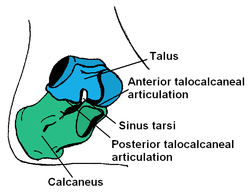
The subtalar joint also named the talocalcaneal joint, is articulating between two tarsal bones the talus and calcaneus bones. There are three facets on each of the talus and calcaneus. The posterior talocalcaneal articulation represents the largest component of the subtalar joint.[1]The malalignment of the subtalar joint can lead to primary osteoarthritis of the ankle joint in the long run. See more here Subtalar joint
Function and Biomechanics of Subtalar joint[edit | edit source]
The Subtalar joint is necessary for:
- The function of the foot and the ankle and gait.[2]
- Transmission of rotations from the leg and the ankle to the foot.[3]
- Shock absorption during the early stance phase.[3]
The Subtalar joint movements are inversion-eversion[4] and supination-pronation (three-dimensional motions)[5]. Pronation combines dorsiflexion, abduction, and eversion, and supination combines plantarflexion, adduction, and inversion[6].
Main ligaments of Subtalar Joint[edit | edit source]
- Anterior talocalcaneal ligament
- Posterior talocalcaneal ligament
- Lateral talocalcaneal ligament
- Medial talocalcaneal ligament
Nonweight bearing Subtalar Joint Motion[edit | edit source]
In non-weight bearing positions, the movement is described as the motion of the distal segment which is the calcaneus. The calcaneus is moving on the relatively stationary talus and the lower leg. Supination is a combination of adduction, inversion and plantar flexion. Pronation is a combination of abduction, eversion, and dorsiflexion.
The most easily observed coupled motions of the calcaneus are inversion and eversion. During inversion, there is varus movement of the calcaneus and during eversion, there is the valgus movement of the calcaneus.
Weight-bearing Subtalar Motion[edit | edit source]
The movement is the same as in the non-weight bearing position but the description of which segment is moving is different. The motion of the proximal segment to the distal segment is described.
Etiology[edit | edit source]
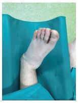
- Subtalar joint instability.
- Malalignment of the joint.
- Fracture of calcaneus/talus.
- Supramalleolar deformities.[8]
- Primary osteoarthritis
- Infection: septic arthritis
- Deposition disease: gout, pseudogout
- Tarsal coalition[9]
Clinical Signs Of Osteoarthritis[edit | edit source]
- Coarse Crepitus.
- Bony Enlargement.
- Joint line tenderness.
- Reduced joint range of motion.[10]
Treatment[edit | edit source]
Diseases such as arthritis can influence the Ankle range of motion[11], so it is important to do Subtalar ROM exercises (inversion-eversion and supination-pronation).
Physiotherapy Management[edit | edit source]
Maitland Mobilisation of Subtalar Joint[edit | edit source]
Subtalar joint distraction
Subtalar joint medial glide & lateral glide
Passive and Active ROM exercises[edit | edit source]
Active and Passive Supination and Pronation exercises along with inversion and eversion.
References[edit | edit source]
- ↑ Ficke J, Byerly DW. Anatomy, Bony Pelvis and Lower Limb, Foot. InStatPearls [Internet] 2021 Aug 11. StatPearls Publishing. Accessed 7 June 2022.
- ↑ Jastifer JR, Gustafson PA. The subtalar joint: biomechanics and functional representations in the literature. The foot. 2014 Dec 1;24(4):203-9. Accessed 15 June 2022.
- ↑ 3.0 3.1 Stagni R, Leardini A, O'Connor JJ, Giannini S. Role of passive structures in the mobility and stability of the human subtalar joint: a literature review. Foot & ankle international. 2003 May;24(5):402-9. Accessed 15 June 2022.
- ↑ Krähenbühl N, Horn-Lang T, Hintermann B, Knupp M. The subtalar joint: a complex mechanism. EFORT open reviews. 2017 Jul 6;2(7):309-16. Accessed 15 June 2022.
- ↑ Brockett CL, Chapman GJ. Biomechanics of the ankle. Orthopaedics and trauma. 2016 Jun 1;30(3):232-8. Accessed 15 June 2022.
- ↑ Peña Fernández M, Hoxha D, Chan O, Mordecai S, Blunn GW, Tozzi G, Goldberg A. Centre of rotation of the human subtalar joint using weight-bearing clinical computed tomography. Scientific reports. 2020 Jan 23;10(1):1-4. Accessed 15 June 2022.
- ↑ Flippin M, Fallat LM. Open talar neck fracture with medial subtalar joint dislocation: a case report. The Journal of Foot and Ankle Surgery. 2019 Mar 1;58(2):392-7. Accessed 28, June 2022.
- ↑ Kanemitsu M, Nakasa T, Ikuta Y, Ota Y, Sumii J, Nekomoto A, Sakurai S, Adachi N. Characteristic Bone Morphology Change of the Subtalar Joint in Severe Varus Ankle Osteoarthritis. The Journal of Foot and Ankle Surgery. 2022 May 1;61(3):627-32. Accessed 31, August 2022.
- ↑ Radiopedia Subtalar arthritis Available: https://radiopaedia.org/articles/subtalar-arthritis(accessed 9.9.2022)
- ↑ Abhishek A, Doherty M. Diagnosis and clinical presentation of osteoarthritis. Rheumatic Disease Clinics. 2013 Feb 1;39(1):45-66.
- ↑ Brockett CL, Chapman GJ. Biomechanics of the ankle. Orthopaedics and trauma. 2016 Jun 1;30(3):232-8. Accessed 31 August 2022.
- ↑ Subtalar Joint | Passive Range of Motion. Available from: https://www.youtube.com/watch?v=GJCWxx_3oiE [last accessed 31/8/2022]


