Spondylolisthesis
Original Editors - Margo De Mesmaeker
Top Contributors - Maëlle Cormond, Simisola Ajeyalemi, Evi Peeters, Margo De Mesmaeker, Kim Jackson, Admin, Chrysolite Jyothi Kommu, Kenneth de Becker, Shaimaa Eldib, Lucinda hampton, Mike Myracle, Rachael Lowe, Evan Thomas, Mariam Hashem, Carlos De Coster, Johnathan Fahrner, Aminat Abolade, Camille Linussio, Shreya Pavaskar, Claire Knott, Rucha Gadgil, Laura Ritchie, Jess Bell, Kirenga Bamurange Liliane and Scott Buxton
Search Strategy[edit | edit source]
• Databases: PubMed, Web of Knowledge, Google Scholar, Cochraine Library, PEDro, Medscape
• Tools used in PubMed: advanced search, MeSH terms.
• Keywords: Spondylolisthesis (adding terms to narrow the search like 'diagnostics' or 'etiology' etc. )
• MeSH Terms: Spondylolistheses, Spondylisthesis, Spondylistheses
Definition/Description[edit | edit source]
The term spondylolisthesis is derived from the Greek words spondylo = vertebra, and olisthesis = translation.[52](LE: 5) Spondylolisthesis is defined as a translation of one vertebra over the adjacent caudal vertebra. This can be a translation in the anterior (anterolisthesis) or posterior direction (retrolysthesis) or, in more serious cases, anterior-caudal direction.[1](LE: 2A) [2](LE: 1A) It is classified on the basis of etiology into the following six types by Wiltse: Dysplastic (congenital), isthmic, degenerative, traumatic, pathologic and iatrogenic spondylolisthesis.[1](LE: 2A) [3]
Video illustration: https://www.youtube.com/watch?v=DlJM2kLGwUI
Clinically Relevant Anatomy[edit | edit source]
http://www.spine-health.com/video/degenerative-spondylolisthesis-video
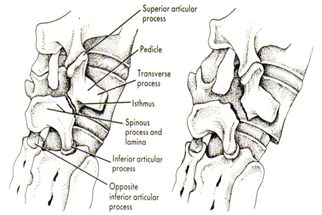
Spondylolysthesis, regardless of the type, is mostly common preceded by spondylolysis (other causes : see 4. Epidemiology / Etiology). This pathology involves a fractured pars interarticularis of the lumbar vertebrae, also called the isthmus [1] (LOE: 2A). This affects the supporting structural integrity of the vertebrae, wich could lead to slippage of the corpus of the vertebrae, called spondylolysthesis. This in turn leads to one of the most obvious manifestations of lumbar instability. This slippage can occure in 2 directions : most commonly in anterior translation, called anterolisthesis, or a backward transletion, called retrolisthesis. [3] (LOE: 1B)
In addition, ligamentous structures, including the iliolumbar ligament, between L5 and the sacrum, are stronger than those between L4 and L5, by which the vertebral slip commonly develops in L4 [2] (LOE: 1A).
A study of Dai L.Y. analysided the correlation between disc degeneration and the age (1) duration and severety of clinical symptomps (2) and grade of vertebral slip (3). The disc degeneration on subsegmental level was highly significantly related to age and duration of clinical symptoms. Although it was not related to the severity of clinical symptoms or the grade of vertebral slip. [21] (LOE: 2B)
Figure 1: Scottie dog [1] (LE :1B).
Epidemiology /Etiology[edit | edit source]
In literature there are different classificationsystems, developed to help in the decision making process for the diagnostic workup and treatment plan, regarding the etiology, terminology, subtypes of spondylolysis and spondylolisthesis, and treatment.
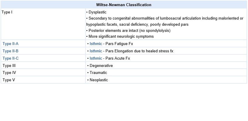
A. Wiltse Classification:
One of the most commonly used classification systems to convey the etiology of a spondylolisthesis is the Wiltse Classification:
• Type 1 : Congenital spondylolisthesis
An elongation of the pars interarticularis can be seen in congenital spondylolisthesis, in which the pars lesion is due to a congenital anomaly of the L5-S1 facet articulation. As the slip progresses, the pars elongates in response to the deformity. Therefore, with an elongated pars, it is important to evaluate the lumbosacral facets to properly classify the lesion.[57](LE: 2A)
The symptoms usually develop during the adolescent growth period.[10](LE: 5)
The symptoms usually develop during the adolescent growth period.[28] (LOE: 5)
• Type 2: Isthmic spondylolisthesis
Isthmic spondylolisthesis, or spondylolisthesis due to a lesion of the pars interarticularis, is a common source of pain and disability in both pediatric and adult population.
The basic lesion in isthmic spondylolisthesis is in the pars interarticularis and mainly appears at the lumbosacral level (L5-S1).[6](LE: 2B) It is characterized by high lordosis angles and lordotic wedging of the affected vertebra (L5) and very high L4-5 intervertebral disc wedging.[7](LE: 3B) [8](LE: 2B)
Isthmic spondylosithesis is typically considered as a pediatric condition.[20] (LOE: 1A)
Spondylolisthesis is mostly often caused by spondyloslysis. Spondylolysis is considerd a stressfracture caused by an eccesive amount of mechanical stress that affects the isthmus. This part of the vertebrae forms the connection between the corpus and the facet joints, at the back of the vertebrae. Therefore, a load for the facet joints result in a stressor for the isthmus. The stress on the pars interarticularis is the highest with extension and rotation. Anterior pelvic tilt, abdominal muscle weakness and hamstring thightness magnify these biomechanical forces. [40] (LOE: 2A)
http://www.physio-pedia.com/Spondylolysis_in_Young_Athletes
<span />
Wiltse et al. divided this category into three subtypes: lThe lytic lesion of the pars (Type II-A) is the most common cause of spondylolisthesis and is termed spondylolysis. This defect is present in 6% of the population by young adulthood. The elongation of the pars interarticularis (Type II-B) is thought to be due to repetitive microfractures with subsequent healing in an elongated position. Elongation of the pars can also be seen in congenital spondylolisthesis.
An acute fracture of the pars (Type II-C), the third subtype, resulting from a single traumatic episode. Wiltse et al. suggested that this type of isthmic spondylolisthesis could also be classified as traumatic spondyloslisthesis.[57](LE: 2A)
Isthmic spondylosithesis is typically considered as a pediatric condition.[20](LE: 1A) But Saraste (1987) demonstrated that the onset of symptoms tends to occur after childhood, with a mean age at presentation of 20 years.[21](LE: 2B)
• Type 3 : Degenerative spondylolisthesis
Degenerative spondylolisthesis is most common in adults.[54](LE: 2A)
In this type the L4–L5 vertebral space is affected 6 to 9 times more commonly than other spinal levels.[22](LE: 2B) [10](LE: 5) It is characterized by a significant constriction of the cauda equina, combined with a diminished cross-sectional area of the vertebral canal, thickening and buckling of the ligamentum flavum and hypertrophy of adjacent facet joints.[6](LE: 2B) It is also a common condition in the elderly (>50 years).
The main causes are: [23](LE: 3A) [24](LE: 5) [25](LE: 1A)
• Disc degeneration;
• Facet joint arthrosis;
• Malfunction of the ligamentous stabilizing component;
• Ineffectual muscular stabilization.
Degenerative spondylolisthesis is believed to result from chronic intersegmental instability. Degenerative changes to both the facet joints and the intervertebral disk cause the slip. Sagittal orientation of the facet joints and facet tropism also have been related to the development of degenerative spondylolisthesis. [57](LE: 2A)
• Type 4: Traumatic spondylolisthesis
Traumatic spondylolisthesis is caused by a fracture in a region other than the pars. This fracture leads to slippage of the vertebrae.[57](LE: 2A)
• Type 5: pathological spondylolisthesis
Pathological spondylolisthesis is due to generalized or localized musculoskeletal processes affecting the posterior elements and causing instability.[57](LE: 2A)
Diffuse or local disease compromises the usual structure integrity that prevents slippage.[58](LE: 3B)
• Type 6: iatrogenic spondylolisthesis
Iatrogenic spondylolisthesis results from excessive removal of the posterior elements after laminectomy.[57](LE: 2A)
The incidence of spondylolisthesis varies considerably depending on ethnicity, sex, family history, relevant disease and sports activity.[10](LE: 5) Several epidemiological studies have revealed that the incidence of symptomatic listhesis in Caucasian populations varies from 4 to 6% [11](LE: 2A) [12](LE: 2A), but rises as high as 26% in secluded Eskimo populations [13](LE: 2A) and varies from 19 to 69% among first-degree relatives of the affected patients.[14](LE: 2A)
Depending on the studies conducted, this percentage can vary. [15](LE:2B) [16](LE: 2B) [17](LE: 2B) [18](LE: 2B) [19](LE: 2B)
Potential risk factors are: [26](LE: 3A)
• Increasing age;
• Female sex;
• Pregnancies;
• African-American ethnicity;
• Generalized joint laxity
• anatomical predisposition (sagitally oriented facet joints, hyperlordosis, high pelvic incidence)
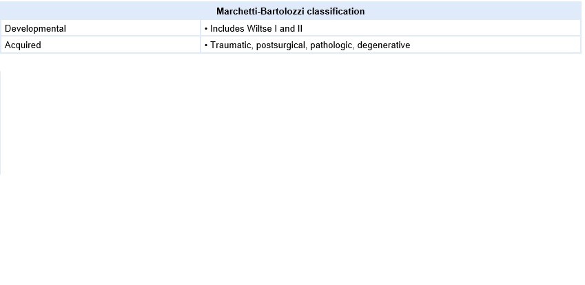
B. Marchetti-Bartolozzi Classification [58] (LOE: 2C)
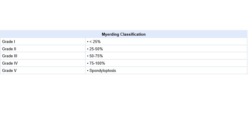
C. Myerding Classification [82] (LOE:2C)
Characteristics/Clinical Presentation[edit | edit source]
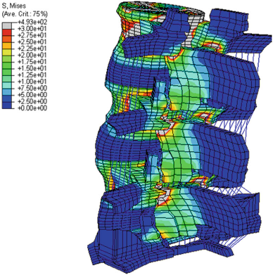
Figure 2: Highest stress during various lumbar motions is found at the pars interarticularis, as shown in a threedimensional finite element moedel [2] (LE: 2B)
Symptoms and findings in spondylolisthesis are: [68](LE: 2A)
- Low-back Pain and leg pain
- Pain
- Trophic changes
- Atrophy of the muscles, muscle weakness
- Tense hamstrings, hamstrings spasms
- Disturbance in patterns
- Diminished ROM (spine)
- Disturbances in coördination and balance
- Neurological symptoms (possible evolution towards cauda equine syndrome)
- Dull pain, typically situated in the lumbosacral region after exercise, especially with an extension of the lumbar spine
Patients usually report that their symptoms vary as a function of mechanical loads (such as in going from supine to erect position) and pain frequently worsens over the course of the day (figure 3). Radiation into the posterolateral thighs is also common and is independent of neurologic signs and symptoms. The pain could be diffuse in the lower extremities, involving the L5 and/or L4 roots unilaterally or bilaterally, but generally bilaterally [8] [3] (LE: 5).
Symptoms decrease with sitting or standing with lumbar flexion and with lying. As symptoms worsen patients are more and more limited in their activities and walking distance. This relationship is known as neurogenic intermittend claudication [39] (LOE: 1B)
In addition to these findings, each type of spondylolythesis has its own characteristics
Spondylolisthesis can occur with other disorders and seems to have a link with some of them:
Several studies support a positive association between spina bifida occulta and spondylolysthesis [6] (LE: 2A). This high association may not be due to mechanical factors but to genetic factors [4] (LE: 2C).
http://www.physio-pedia.com/Spina_Bifida_Occulta
- Cerebral palsy [7] (LE: 2B);
A number of studies proved the association between cerebral palsy and spondylolysthesis, certainly in athetoid cerebral palsy (60%) [7] (LE:2B).
https://www.physio-pedia.com/Cerebral_Palsy_Introduction
Ogilvie and Sherman reported a 50% incidence of spondylolysthesis in 18 patients with Scheuermann’s disease [8] (LE: 2B). Greene et al. found spondylolysthesis (grade I or II) at L5-S1 in 32% of patients with Scheuermann’s disease [9](LE: 2B).
http://www.physio-pedia.com/Scheuermanns_Disease
- Scoliosis [6] (LE: 2A).
Fisk et al. reported that the incidence in 539 patients with ideopathic scoliosis was 6.2%, which corresponded to that found in the general population [10] (LE: 2B). But the relation between scoliosis and spondylolysthesis has not been clarified [6] (LE:2A).
http://www.physio-pedia.com/Scoliosis
- Spinal stenosis [53](LE: 2A);
http://www.physio-pedia.com/Spinal_Stenosis
As mentioned in etiology and epidemiology, spondylolisthesis (isthmic type) remains asymptomatic in the pediatric population due to skeletal immaturity. [20] (LOE: 1A)
Differential Diagnosis[edit | edit source]
- Spondylolysis [11] (LE: 1A) [12] (LE: 4)
- Metastatic disease [13] (LE: 2B)
- Low back pain [14] (LE: 1C)
- Osteoarthritis [14] (LE: 1C) http://www.physio-pedia.com/Osteoarthritis
- Neuroforaminal stenosis [14] (LE: 1C)
- Spinal Stenosis [14] (LE: 1C)
Diagnostic Procedures[edit | edit source]
According to Jerrad MD. et al. an early diagnosis, i.e. in the first month after the first symptomps, enlarges the likelihood of the formation of a bony callus. Panteliadis et al. concluded that the formation of a bony unit is not inevitable for a good clinical outcome of therapy. As it happens a fibrocartilaginous callus can also be sufficient for normal functioning and pain reduction, and can meet the requirements of an atlete. [49] (LOE: 5)
Radiographic examination provides the best diagnostic information when spondylolisthesis (or spondylosis) is suspected. Standard lumbar anteroposterior and lateral views are needed, but for a better look at the problem oblique views are essential to visualize the pars interarticularis directly. These views may demonstrate a pars interarticularis abnormality, which is depicted as a defect in the collar of the ‘‘Scotty dog.’’ Radiographic evaluation should not be an isolated clinical examination. It should be correlated with further examination such as history, physical examination found further below.[37] (LOE : 4)
In order to determine spinal balance (especially in the sagittal plane) and the associated deformity, full-length radiographs of the spine are essential [43] (LOE : 3A)
Radiological assessments are required in order to make the diagnosis clear and to determine the grade and prognosis of spondylolisthesis. [70] ( LOE:2B) Most commonly used clinical imaging is X-ray, CT and MRI. [36] (LOE: 1A)
Although guidelines for spondylolisthesis concerning X-ray, MRI and CT remain elusive [47] (LOE : 1B), the following recommendations were drafted by the The North American Spine Society (NASS) concerning the diagnosis of adult isthmic spondylolisthesis.[11] (LOE: 2A)
The most appropriate physical examination findings consistent with the diagnosis of isthmic spondylolisthesis in adult patients:
1. There is insufficient evidence to make a recommendation for or against the use of palpation in the physical exam diagnosis of adult patients with isthmic spondylolisthesis.
Grade of Recommendation: I (Insufficient Evidence)
2. Approximately half of adult patients with symptomatic isthmic spondylolisthesis will have a positive straight leg test on examination.
Grade of Recommendation: B (Suggested)
The most appropriate diagnostic tests for adult isthmic spondylolisthesis:
There is a relative paucity of high-quality studies on imaging in adult patients with isthmic spondylolisthesis.
• It is the opinion of the work group that in adult patients with history and physical examination findings consistent with isthmic spondylolisthesis, standing plain radiographs, with or without oblique views or dynamic radiographs, be considered as the most appropriate, non-invasive test to confirm the presence of isthmic spondylolisthesis.
• In the absence of a reliable diagnosis on plain radiographs, computed tomography scan is considered the most reliable diagnostic test to diagnose a defect of the pars interarticularis.
• In adult patients with radiculopathy, magnetic resonance imaging should be considered.
Work Group Consensus Statement
1. MRI is suggested to identify neuroforaminal stenosis in adult patients with isthmic spondylolisthesis [13–15] (LOE: 2A). Grade of Recommendation: B (Suggested)
2. There is insufficient evidence to make a recommendation for or against the use of magnetic resonance imaging to differentiate isthmic versus degenerative spondylolisthesis in adult patients [15] (LOE: 2B). Grade of Recommendation: I (Insufficient Evidence)
3. There is insufficient evidence to make a recommendation for or against the use of discography to evaluate adult patients with isthmic spondylolisthesis [16] (LOE: 2B). Grade of Recommendation: I (Insufficient Evidence)
4. Computed tomography may be considered as an option to diagnose isthmic spondylolisthesis in adult patients [18] (LOE 2B). Grade of Recommendation: C (May Be Considered; Option)
5. There is insufficient evidence to make a recommendation for or against the use of single-photon emission computed tomography (SPECT) in evaluating isthmic spondylolisthesis in adult patients [19] (LOE: 2B) . Grade of Recommendation: I (Insufficient Evidence)
• X-ray
Overall X-ray of the spine and lumbosacral X-ray are seen as the golden standard for diagnosis. [38] (LOE: 1C) There are multiple views used with the most common one being the anteroposterior, lateral and oblique views. [36] (LOE: 1A) Multiple characteristics can be seen, such as the degree of the slip or the slip angle. The most prominent sign remains the defect of the pars interarticularis, or more commonly named the broken collar or neck of the “Scottie Dog”. [9] (LOE: 1B)
• CT and MRI
Advanced imaging techniques like MRI and CT have to be used when neurological symptoms are present, and when surgical intervention is indicated.[70] (LOE:2B) CT and MRI, which give an accurate localization and a better illustration of the lesion [35] (LOE: 1A), are taken when one of the following signs are present: [6] (LOE: 2B)
• Significant and progressing neurologic claudication [6] (LOE: 2B)
• Radiculopathies and the clinical suspicion that another condition may be causative [6] (LOE: 2B)
• Bladder or bowel complaints [6] (LOE: 2B)
• Metastatic disease [38] (LOE: 1C)
Something one should bare in mind is that defects in the pars interarticularis may not appear on a CT. That’s the case for 85% of patients according to Congerni et al. [37] (LOE: 4)
And are used for one of the following reasons:
• Evaluating an atypical presentation including pre-lysis [9] (LOE: 1B)
• Determining the condition of the intervertebral disc [36] (LOE: 1A)
• Evaluating vertebral slipping and possible neural element compression [38] (LOE: 1C)
CT and MRI give the best visualization of bone morphology and are therefore, most often used to check the alignment of the facet joints and their degenerative changes. [36] (LOE: 1A) Images resulting from CT and MRI are the most sensitive and specific when a pars fracture is present. [9] (LOE: 1B) Myelography can be used together with CT, but nowadays MRI is used instead. [6] (LOE: 2B)
[56] (LOE : 1B)
Outcome Measures[edit | edit source]
• Disability: Oswestry Disability Index, the SF-36 Physical Functioning scale, the Quebec Back Pain Disability Scale [50] (LOE: 2B)
www.physio-pedia.com/Oswestry_Disability_Index
• Dysfunctional thoughts: Short Form of the Medical Outcomes Study (SF-36) [76] (LOE: 1A)
• Pain: pain Numerical Rating Scale, VAS [76] (LOE: 1A)
www.physio-pedia.com/Visual_Analogue_Scale
• Quality of life: Short-Form Health Survey [76] (LOE: 1A)
• Kinesiophobia and Catastrophising: Tampa Scale for Kinesiophobia, Pain Catastrophising Scale [76] (LOE: 1B)
• Radiculopathy [76] (LOE: 1B)
www.physio-pedia.com/Radiculopathy
www.physio-pedia.com/Lumbar_Radiculopathy
[1]www.physio-pedia.com/Cervical_Radiculopathy
www.physio-pedia.com/Thoracic_radiculopathy
• Grade of slippage: Classifying an isthmic spondylolisthesis into low- versus high grade has important implications regarding treatment decisions. High-grade spondylolisthesis (HGS) is defined as greater than 50% slippage of a spinal vertebral body relative to an adjacent vertebral body as per Meyerding classification, and most often affects the alignment of the L5 and S1 vertebral bodies. [77] (LOE: 2B)
• The grade of disc degeneration below the spondylolytic defect (MRI): study’s suggest that the grade of disc degeneration below the spondylolytic defect highly significantly correlates with the duration of clinical symptoms (p < 0.01), although it was not associated with the severity of clinical symptoms or the grade of vertebral slip (p > 0.05). The results suggest that the degree of disc degeneration at the spondylolytic level is a more important determinant for choice of the fusion procedure than the age of the patients or the grade of vertebral slip. [78] (LOE: 1B)
Examination[edit | edit source]
History
Specific questions should be asked to the patient referring to:
• Pain [38](LOE: 1C)
• Location
• Severity
• Duration
• Quality (for example: tingling, burning,.. sensations)
• Exacerbating factors
• Alleviating factors
• Leisure activities [38](LOE: 1C)
• Occupational risks [38](LOE: 1C)
• Pain changes throughout day: difference morning compared to evening/night?
General [3] (LOE: 1B)
• Initially resting and avoiding movements like lifting, bending and sports.
• Analgesics and NSAIDs reduce musculoskeletal pain and have an anti-inflammatory effect on nerve root and joint irritation.
• Epidural steroid injections can be used to relieve low back pain, lower extremity pain related to radiculopathy and neurogenic claudication.
• A brace may be useful to decrease segmental spinal instability and pain. [2] (LOE: 1A)
• Pain, usually in the lower-limb, is invariably the chief symptom. It is important to correlate this with the findings coming from the diagnostic procedures. [43] (LOE: 3A).
Medical Management[edit | edit source]
General [13] (LE: 2B), [15] (LE: 2B)
- Initially resting and avoiding movements like lifting, bending and sports.
- Analgesics and NSAIDs reduce musculoskeletal pain and have an anti-inflammatory effect on nerve root and joint irritation.
- Epidural steroid injections can be used to relieve low back pain, lower extremity pain related to radiculopathy and neurogenic claudication.
- A brace may be useful to decrease segmental spinal instability and pain. [16] (LE: 3B)
Surgery
Patients with chronic and disabling symptoms, who fail to respond to conservative management may be referred for surgery.[63](LE: 1B)
When the condition of spinal instability is very severe, a surgical intervention may be necessary to attach the vertebras together.[63](LE: 1B) It can help the patient to reduce pain, improve spinal function and increase the quality of life. The goal of surgery is to stabilize the segment with listhesis, decompress the neural elements, reconstruction of the disc space height and restoration of normal sagittal alignment.[1](LE: 2A) [41](LE: 2C) The grade of listhesis can be reduced to some extent, but complete reduction is rarely achieved.[41](LE:2C) There is a wide variety of complications, such as neurological complications, vascular injury, instrument failure, and infections.[1](LE: 2A) [6](LE: 3B) [39](LE: 2B)
Only perform a surgical intervention after a careful analysis of indications and outcomes.[63](LE: 1B)
When evaluating a patient, many factors, such as age, degree of slip and risk of slip progression, must be considered. Also a thorough evaluation of social and physiological factors should be undertaken. Therefore each patient's treatment program should be individualized to achieve optimal outcome.[1](LE: 2A) [41](LE: 2C)
Indications [6](LE: 2B) [1](LE: 2A) [42](LE: 1A)
- Neurologic signs / neurogenic claudication / radiculopathy (unresponsive to conservative measures)
- Myelopathy
- High-grade slip (>50%)
- Type 1 (congenital) and 2 (isthmic) slips with evidence of instability, progression of listhesis, or lack of response to conservative measures
- Type 3 (degenerative) listhesis with gross instability and incapacitating pain
- Bladder or bowel symptoms (especially in type 3)
- Traumatic spondylolisthesis
- Iatrogenic spondylolisthesis
- Postural deformity and gait abnormality
Contra-indications [1](LE: 2A)
- Poor medical health
- High operative risk (higher risk than potential benefits)
- High risk of hemorrhage: Anticoagulation with warfarin, or antiplatelet therapy
- Smoking
There are several different options for surgical treatment; one of them is fusion (e.g. posterolateral fusion). The aim of fusion is to reduce pain by reducing the motion of the segment. Other treatment options include decompression (Gill laminectomy), supplemental instrumentation and supplemental anterior column support. Controversies exist about the effectiveness of these treatment options that can be used separately or in any combination.[6](LE: 3B) [39](LE: 1A)
The same principles are maintained with children: conservative treatment is recommended and proven to be effective. Instrumented posterolateral fusion is indicated in patients with persistent symptoms and for iatrogenic cases. [58] (LE: 3A)
Physical Therapy Management[edit | edit source]
A systematic review [47] (LOE: 1A) including 10 articles from which 5 were RCT, concluded that no consensus could be reached on the role of nonoperative care vs. surgery due to the heterogeneity of the different studies reported.
Spondylolisthesis should be treated first with conservative therapy, which includes physical therapy, rest, medication and braces.[43](LE: 3A) [70](LE 2B) Non-operative treatment should be the initial course of action in most cases of degenerative spondylolisthesis and symptomatic isthmic spondylolisthesis, with or without neurologic symptoms. [6](LE: 3A) [70](LE 2B) Children or young adults with a high-grade dysplastic or isthmic spondylolisthesis or adults with any type of spondylolisthesis, who do not respond to non-operative care, should consider surgery.
Traumatic spondylolisthesis can be treated successfully using conservative methods, but most authors suggested it would result in posttraumatic translational instability or chronic low back pain.[44](LE: 3A) Exercises should be done on a daily basis.[6](LE: 2B)
- Exercise therapy
There is strong evidence for exercise therapy, which consists of strengthening exercises of the deep abdominal musculature.[46](LE: 1B) [45](LE: 1A). In addition, isometric and isotonic exercises may be beneficial for strengthening of the main muscle groups of the trunk, which stabilize the spine. These techniques may also play a role in pain reduction.[46](LE: 1B) [45](LE: 1A) [47](LE: 1B)
In order to improve the patient’s mobility, physical therapy includes stretching exercises of the hamstrings, hip flexors and lumbar paraspinal muscles.[46](LE: 1B) [45](LE: 1A)
Furthermore endurance training is effective for chronic low back pain.[45](LE: 1A) [72](LE: 2A) The objective of stretching and strengthening is to decrease the extension forces on the lumbar spine, due to agonist muscle tightness, antagonist weakness, or both, which may result in decreased lumbar lordosis.[46](LE: 1B) [45](LE: 1A) Rehabilitation programs should be designed to improve muscle balance rather than muscle strength alone.[69](LE: 2B)
There is evidence that suggests that specific stabilization exercises and core stability exercises can be useful in reducing pain and disability in chronic low back pain in patient with spondylolisthesis.[59](LE: 1A) [66](LE: 2B) [68](LE: 2A)
When using core stability exercises a therapist can use different modalities: [68](LE: 2B)
- Movements in closed-chain-kinetics
- Renewing of the motion-pattern
- Antilordotic movement patterns of the spine
- Elastic band exercises in the lying position
- Gait training
- Brace-gymnastics
- Stretching exercises
- Sensomotoric training on unstable devices
- Functional electric stimulation
- Walking in all variations
- Underwater therapy
- Balance training
- Coordinative skills
Exercises should be done on a daily basis [13] (LE: 2B).
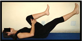
Figure 3: Strengthening of the deep abdominal muscles.
Alternating legs, with leg extension while exhaling, maintaining contraction of transverse abdominis, paravertebral and pelvic floor muscles [17] (LE: 1B).
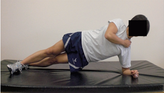
Figure 4: Horizontal side support exercise for core stability [18] (LE: 1B).
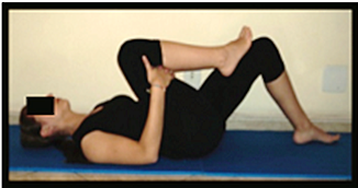
Figure 5: Stretching of the erector spine muscles.
Flexing of the hip, toward the chest [17] (LE: 1B).
- Lumbosacral braces or corset
In healthy subjects it has been found that the lumbosacral brace can improve the sitting position of the patient. The fact that there was wear of the brace, indicates that the brace has an important function in the sitting position.[64](LE: 2B) Lumbar supports also reduce the numbers of days when low back pain occurs.[65](LE: 1B) According to Prateepavanich et al., a lumbosacral corset can be used to improve walking distance and to reduce pain in daily activities [51](LE: 2B), but it does not reduce the shift of the vertebra.[6](LE:2B) It is a good aid during the painful periods but should be discontinued when the patients' complaints are reduced.
- Posture and lifting techniques
Special attention has to be given to posture and proper lifting techniques [48](LE: 3A), wherein the physiotherapist has an important educational role.[46](LE: 1B) [45](LE: 1A) Lifting techniques is effective for chronic low back pain.[45](LE: 1A) [72](LE: 2A)
- Management of catastrophising and kinesiophobia
Physical therapy treatment in combination with management of catastrophising and kinesiophobia gave good results. The disability, pain, dysfunctional thoughts were significant reduced.[63](LE: 1B)
- Alternative cardiovascular exercise
Athletes with spondylolysis and first-degree spondylolisthesis can take part in all sports activities. However, attention should be given to those kinds of sport where recurring trauma resulting from repeated flexion, hyperextension and twisting is usually undertaken (e.g. gymnastics, aerobics, swimming in the dolphin technique). Athletes with a grade 2, 3 or 4 can also participate in all the sport activities but have to do this with a special and individually adapted directives.[68](LE:2A)
Low aerobic impact sports are highly recommended. [20] (LOE: 1A) Sports that certainly can be practiced are walking, swimming and cross-training. Although these activities will not improve the shift, these sports are a good alternative for cardiovascular exercises.[6] (LOE:2B) Impact sports like running should not be done in order to avoid wear. The adolescent athlete or manual laborer should avoid hyperextension and/or contact sports. [48] (LOE: 3A) Bicycling is also a good cardiovascular exercise, because it promotes spine flexion.
• Massage therapy
A case report showed that the the onset of low back pain was delayed during walking/standing over the course of treatment, hyperlordosis decreased, and hypertonicity of iliopsoas and quadrates lumborum muscles decreased. Bilateral net reduction of illial rotation was achieved, but with irregular changes. These results were inconclusive but brings into question the role of hip flexor and spinal extensor muscles in normalizing postural misalignment associated with spondylolisthesis. [73] (LOE: 4)
• Pulsed radiofrequency (PRF)
Results of a cohort study demonstrated that the application of PRF might be more effective than steroid and bupivacaine injection in decreasing back pain due to degenerative facet pain and improvement in function of patients with degenerative spondylolisthesis. [74] (LOE: 1B)
Key Research[edit | edit source]
• Steven, S.A. et al, (20mc10). Contemporary management of isthmic spondylolisthesis: pediatric and adult. The Spine Journal, 2010, 530-543. (Level of evidence: 1A)
• Van Tulder M.W. et al, Conservative treatment of acute and chronic nonspecific low back pain. A systematic review of randomized controlled trials of the most common interventions, Spine, 1997; 22(18): 2128-2156 f(Level of evidence 1A)
• Paulo H Ferreira et al.; Specific stabilisation exercise for spinal and pelvic pain: A systematic review; Australian Journal of Physiotherapy 2006 Vol. 52 ( level of evidence 1A)
Recources[edit | edit source]
Clinical bottom line[edit | edit source]
Spondylolisthesis is defined as a translation of one vertebra over the adjacent caudal vertebra. This can be a translation in the anterior (anterolisthesis) or posterior direction (retrolysthesis) or, in more serious cases, anterior-caudal direction. It is classified on the basis of etiology into the following six types by Wiltse: Dysplastic (congenital), isthmic, degenerative, traumatic, pathologic and iatrogenic spondylolisthesis.
Radiographic examination provides the best diagnostic information when spondylolisthesis (or spondylosis) is suspected. Spondylolisthesis should be treated first with conservative therapy. When this fails, and the patients deals with chronic and disabling symptoms, surgery is referred. When the condition of spinal instability is very severe, a surgical intervention may be necessary to attach the vertebras together. This can be a fusion or Gill laminectomy.
[edit | edit source]
• Bilateral tubular minimally invasive approach for decompression, reduction and fixation in lumbosacral lythic spondylolisthesis.
• In situ posterolateral and fibular interbody fusion in high grade spondylolysthesis.
• A case of nail-patella syndrome associated with thyrotoxicosis.
• Computed tomorgaphy diagnostic of lumbar spondylolysis.
• A case of lumbar sciatica in a patient with spondylolysis and spondylolysthesis and underlying misdiagnosed brucellar discitis.
• [Role of surgery in spinal degenerative disease. Analysis of systematic reviews on surgical and conservative treatments from an evidence-based approach].
• [Lumbar spine injuries in pediatric and adolescent athletes].
• [Lumbar interbody fusion with threaded titanium cages. Results on 222 cases].
• [Measurement of the lumbosacral angle and its clinical significance].
• [Morbid anatomy and pathogenesis of spondylolysis and spondylolysthesis in childhood (author's transl)].
References[edit | edit source]
1. Amir Vokshoor et al., Spondylolisthesis, Spondylolysis, and Spondylosis. Medscape, updated Sep 10, 2014, Consulted on Oct 20, 2014 (Level of evidence 2A)
2. Tebet, M.A., Currents concepts on the sagittal balance and classification of spondylolysis and spondylolisthesis. Revista Brasileira de Ortopedia, 2014, 49 (1), 3-12. (Level of evidence 1A)
3. O’sullivan RCT + Iguchi T. et al., Lumbar multilevel degenerative spondylolisthesis: radiological evaluation and factors related to anterolisthesis and retrolisthesis, J Spinal Disord Tech. 2002. Apr;15(2):93-9 (Level of evidence 1B)
4. Garet M. et al., Nonoperative treatment in lumbar spondylolysis and spondylolisthesis: a systematic review, Sports Health, 2013 May; 5(3):225-32 (Level of evidence 3A)
5. Funao H. et al., Comparative study of spinopelvic sagittal alignment between patients with and without degenerative spondylolisthesis, Eur Spine J, 2012; 21:2181-2187 (Level of evidence 3B)
6. Kalichman L. et al, Diagnosis and conservative management of degenerative lumbar spondylolisthesis, Eur Spine J, 2008; 17:327-335 (Level of evidence 2B)
7. Been E. et al, Geometry of the vertebral bodies and the intervertebral discs in lumbar segments adjacent to spondylolysis and spondylolisthesis: pilot study, Eur Spine J, 2011; 20:1159-1165 (Level of evidence 3B)
8. Labelle H. et al., Spino-pelvic alignment after surgical correction for developmental spondylolisthesis, Eur. Spine J, 2008; 17:1170-1176 (Level of evidence 2B)
9. Foreman P. et al, L5 spondylolysis/spondylolisthesis: a comprehensive review with an anatomic focus, Childs Nerv Syst. 2013;29(2):209-16 (Level of evidence 1B)
10. Sakai T. et al., Incidence and etiology of lumbar spondylolysis: review of the literature. Journal of orthopedic sience,2010 May;15(3):281-8. (Level of evidence: 1A)
11. McTimoney, C.A. et al, Current evaluation and management of spondylolysis and spondylolisthesis. Current Sports Medicine Reports, 2003, 2, (1), 41–46. (Level of evdidence: 2A)
12. Taillard, W.F., Etiology of spondylolisthesis. Clinical Orthopaedics and Related Research, 1976, 117, 30–39. (Level of evidence: 2A)
13. Stewart, T., The age incidence of neural arch defects in Alaskan natives, considered from the standpoint of etiology. The American Journal of Bone and Joint Surgery, 1953, 35, 937–950. (Level of evidence: 2A)
14. Lonstein, J.E., Spondylolisthesis in children Cause, natural history, and management. Spine, 1999, 24, (24), 2640–2648. (Level of evidence: 2A)
15. Sonne-Holm, S. et al, Lumbar spondylolysis: a life long dynamic condition? A cross sectional survey of 4,151 adults. European Spine Journal, 2007, 16, 821-828. (Level of evidence: 2B)
16. Amato, M. et al, Spondylolysis of the lumbar spine: demonstration of defects and laminal fragmentation. Radiology, 1984, 153, 627-629. (Level of evidence: 2B)
17. Weil, Y. et al, Correlation between pre-employment screening X-ray finding of spondylolysis and sickness absenteeism due to low back pain among policemen of the Israeli police force. Spine (Phila Pa 1976), 2004, 29, 2168-72. (Level of evidence: 2B)
18. Kalichman, L. et al, Spondylolysis and spondylolisthesis: prevalence and association with low back pain in the adult community-based population. Spine (Phila Pa 1976), 2009, 34, 199-205. (Level of evidence: 2B)
19. Belfi, L.M. et al, Computed tomography evaluation of spondylolysis and spondylolisthesis in asymptomatic patients. Spine (Phila Pa 1976), 2006, 31, E907-10. (Level of evidence: 2B)
20. Steven, S.A. et al, (20mc10). Contemporary management of isthmic spondylolisthesis: pediatric and adult. The Spine Journal, 2010, 530-543. (Level of evidence: 1A)
21. Iguchi T. et al., Lumbar multilevel degenerative spondylolisthesis: radiological evaluation and factors related to anterolisthesis and retrolisthesis, J Spinal Disord Tech. 2002. Apr;15(2):93-9 (Level of evidence 2B)
22. Dai LY et al., Disc degeneration in patients with lumbar spondylolysis. J Spinal Disord. 2000 Dec;13(6):478-86 [2B]
23. Sheng-Dan, J. et al, Degenerative cervical spondylolisthesis: a systematic review. International Orthopaedics (SICOT), 2011, 35, 869-875. (Level of evidence: 3A)
24. Vibert, B.T. et al, Treatment of instability and spondylolisthesis: surgical versus nonsurgical treatment. Clinical Orthopaedics and Related Research, 2006, 443, 222–227. (Level of evidence: 5)
25. Sengupta, D.K. et al, Degenerative spondylolisthesis: review of current trends and controversies. Spine, 2005, 30(Suppl), S71–S81. (Level of evidence: 1A)
26. N.J. Rosenberg. Degenerative spondylolisthesis. Predisposing factors. The journal of Bone and Joint Surgery (1975) 57:467-474. (level of evidence 1C)
27. Mays, S., Spondylolysis, spondylolisthesis, and lumbo-sacral morphology in a medieval English skeletal population. American Journal of Physical Anthropolgy, 2006, 131, 352–62. (Level of evidence: 2B)
28. Frymoyer, J.W.,Degenerative spondylolisthesis. In: Andersson GBJ, McNeill TW (eds) Lumbar spinal stenosis. Mosby Year Book, St Louis, 1992 (Level of Evidence: 5)
29. Sairyo, K. et al, Biomechanical comparison of lumbar spine with or without spina bifida occulta: a finite element analysis. Spinal Cord,2006, 44, 440–4. (Level of evidence: 2C)
30. Burkus, J.K., Unilateral spondylolysis associated with spina bifida occulta and nerve root compression. Spine (Phila Pa 1976), 1990, 15, 555–9. (Level of evidence: 3B)
31. Toshinori, S. et al, Incidence and etiology of lumbar spondylolysis: review of the literature. Journal of Orthopaedic Science, 2010, 15, 281-288. (Level of evidence: 2A)
32. Sakai, T. et al, Lumbar spinal disorders in patients with athetoid cerebral palsy: a clinical and biomechanical study. Spine (Phila Pa 1976), 2006, 31, E66–70. (Level of evidence: 2B)
33. Ogilvie, J.W. et al, Spondylolysis in Scheuermann’s disease. Spine (Phila Pa 1976), 1987, 12, 251–3. (Level of evidence: 2B)
34. Greene, T.L. et al, Back pain and vertebral changes simulating Scheuermann’s disease. Journal of Pediatrics Orthopedics, 1985, 5, 1–7. (Level of evidence: 2B)
35. Fisk JR et al, Scoliosis, spondylolysis, and spondylolisthesis: their relationship as reviewed in 539 patients. Spine (Phila Pa 1976) 1978;3:234–45. (Level of evidence 2B)
36. Tsirikos AI et al, Spondylolysis and spondylolisthesis in children and adolescents. J Bone Joint Surg Br. 2010 Jun;92(6):751-9. doi: 10.1302/0301-620X.92B6.23014 (Level of evidence 1A)
37. Nissenbaum J. et al, Differential diagnosis of spondylolysis in a patient with chronic low back pain. J Orthop Sports Phys Ther. 2005 May;35(5):319-26. (Level of evidence 4)
38. Metzger R. et al, Spondylolysis and spondylolisthesis: what the primary care provider should know. J Am Assoc Nurse Pract. 2014 Jan;26(1):5-12. doi: 10.1002/2327-6924.12083. (Level of evidence 1C)
39. Phalen GS. et al, Spondylolisthesis and tight hamstrings. J Bone Joint Surg, 1961, 43:505–512 (Level of evidence 1B)
40. Standaert C.J., Herring S.A., Cole A.J., and Stratton S.A.The lumbar spine and sports. The low back pain handbook, 2003, 385-404.) (Level of evidence 2A)
41. Chang Hyun Oh et al., Slip Reduction Rate between Minimal Invasive and Conventional Unilateral Transforaminal Interbody Fusion in Patients with Low-Grade Isthmic Spondylolisthesis, Korean J Spine, 2013 (Level of evidence 2C)
42. Sairyo K., Decompression Surgery For Lumbar Spondylolysis Without Fusion: A Review Article, The Internet Journal of Spine Surgery, 2005. (Level of evidence 1A)
43. Serena S. Hu et al., Spondylolisthesis and Spondylolysis, J Bone Joint Surg Am, 2008 Mar 01;90(3):656-671 (Level of evidence 3A)
44. Tang S., Treating Traumatic Lumbosacral Spondylolisthesis Using Posterior Lumbar Interbody Fusion with three years follow up. Pakistan Journal of Medical Sciences 2014;30(5):1137-1140. (Level of evidence 4)
45. Van Tulder M.W. et al, Conservative treatment of acute and chronic nonspecific low back pain. A systematic review of randomized controlled trials of the most common interventions, Spine, 1997; 22(18): 2128-2156 f(Level of evidence 1A)
46. Jerrad MD. Et al., Bony Healing in a Patient with Bilateral L5 Spondylolysis. Current Sports Medicine Reports, (2005) 35-37. (Level of evidence 5)
47. Sinaki M. et al. Lumbar spondylolisthesis: retrospective comparison and three year follow-up of two conservative treatment programs. Arch Phys Med Rehabil 1989 70:594-598. (level of evidence 1B)
48. Agabegi S.S. et al, Contemporary management of isthmic spondylolisthesis: pediatric and adult, The Spine Journal, 2010; 10:530-543 (Level of evidence 3A)
49. Panteliadis et al., Athletic Population with Spondylolysis: Review of Outcomes following Surgical Repair or Conservative Management. Global spine J., (2016) 615-625. (Level of evidence 5)
50. Davidson, M. & Keating, J.L., A comparison of five low back disability questionnaires: reliability and responsiveness. Phys Ther. 2002 Jan;82(1):8-24 (Level of evidence 2B)
51. Prateepavanich P. et al., The effectiveness of lumbosacral corset in symptomatic degenerative lumbar spinal stenosis, J Med Assoc Thai., 2001; 84(4):572-6. (Level of evidence 2B)
52. D. Winkel, orthopedische geneeskunde en manuele therapie: de wervelkolom deel 2. (Level of evidence 5)
53. Anderson T. et al, Degenerative spondylolisthesis is associated with low spinal bone density: a comparative study between spinal stenosis and degenerative spondylolisthesis, Biomed Res Int. 2013; 123847 (Level of evidence: 2A)
54. Gille O. et al, degenerative lumbar spondylolisthesis: cohort of 670 patients, and proposal of a new classification, 2014 (Level of evidence: 2A)
55. Ferrari S. et al., Clinical presentation and physiotherapy treatment of 4 patients with low back pain and isthmic spondylolisthesis, 2012 (Level of evidence: 2B)
56. Biely et al., clinical instabitlity of the lumbar spine: diagnosis and intervention., orthopedic practice vol. 18;3;06 (Level of evidence: 1B)
57. Wiltse LL. Classification, Terminology and Measurements in Spondylolisthesis. Iowa Orthop J. 1981; 1: 52–57. (Level of evidence: 2A)
58. Leonidou, Andreas, et al. "Treatment for spondylolysis and spondylolisthesis in children." Journal of Orthopaedic Surgery 23.3 (2015): 379. (LE:3A)
59. Paulo H Ferreira et al.; Specific stabilisation exercise for spinal and pelvic pain: A systematic review; Australian Journal of Physiotherapy 2006 Vol. 52 ( level of evidence 1A)
60. Matthew Garet et al.; Nonoperative Treatment in Lumbar Spondylolysis and Spondylolisthesis: A Systematic Review; SPORTS HEALTH; 2013; vol. 5 no. 3 (Level of evidence 2A)
61. Farzad Omidi-Kashani et al.; Radiologic and Clinical Outcomes of Surgery in High Grade Spondylolisthesis Treated with Temporary Distraction Rod; Clin Orthop Surg. 2015 Mar; 7(1): 85–90 (level of evidence 2B)
62. Masci L. et al, Use of the one‐legged hyperextension test and magnetic resonance imaging in the diagnosis of active spondylolysis, Br J Sports Med. 2006 Nov (level of evidence 2B)
63. Monticone M. et al, Management of catastrophising and kinesiophobia improves rehabilitation after fusion for lumbar spondylolisthesis and stenosis. A randomised controlled trial , Eur Spine J. 2014 Jan (level of evidence 1B)
64. Mathias M. et al, In healthy subjects, the sitting position can be used to validate the postural effects induced by wearing a lumbar lordosis brace. , Ann Phys Rehabil Med. 2010 Oct (level of evidence 2B)
65. Roelofs PD. et al, Lumbar supports to prevent recurrent low back pain among home care workers: a randomized trial., 2007 Nov 20 (level of evidence 1B)
66. Nava-Bringas, Effects of a stabilization exercise program in functionality and pain in patients with degenerative spondylolisthesis; Journal of Back and Musculoskeletal Rehabilitation, vol. 27, no. 1, pp. 41-46, 2014 (level of evidence 2B)
67. Stephen Maya et al; Is surgery more effective than non-surgical treatment for spinal stenosis, and which non-surgical treatment is more effective? A systematic review; Physiotherapy 99 (2013) 12–20 (level of evidence 2B)
68. Antony Wicker et al; Spondylolysis and spondylolisthesis in sports; International SportMed Journal, Vol. 9 No.2, 2008 pp.74-7 (level of evidence 2B)
69. Nava-Bringasa T.I. et al; Association of strength, muscle balance, and atrophy with pain and function in patients with degenerative spondylolisthesis; Journal of Back and Musculoskeletal Rehabilitation 27 (2014) 371–376 (level of evidence 2B)
70. Kalpakcioglua B. et al; Determination of spondylolisthesis in low back pain by clinical evaluation; Journal of Back and Musculoskeletal Rehabilitation 22 (2009) 27–32 ( level of evidence 2B)
71. Kristen E. Radcliff et al.; Surgical Management of Spondylolysis and Spondylolisthesis in Athletes: Indications and Return to Play.; Curr. Sports Med. Rep., Vol. 8, No. 1, pp. 350, 2009 ( level of evidence 2B)
72. Margaret L. McNeely et al.; A systematic review of physiotherapy for spondylolysis and spondylolisthesis; Manual Therapy (2003) 8(2), 80–91. ( level of evidence 1B)
73. Halpin, Shannon. "Case report: The effects of massage therapy on lumbar spondylolisthesis." Journal of bodywork and movement therapies 16.1 (2012): 115-123. (level of evidence: 4)
74. Hashemi, Masoud, et al. "Effect of pulsed radiofrequency in treatment of facet-joint origin back pain in patients with degenerative spondylolisthesis." European Spine Journal 23.9 (2014): 1927-1932. (level of evidence 2B)
75. Burton, D. et al., Diagnosis and treatment of adult isthmic spondylolysthesis. North American Spine Society (2014) Evidence based clinical guidelines for multidisciplinary spine care (LE 1A)
76. Monticone M. et al., Management of catastrophising and kinesiophobia improves rehabilitation after fusion for lumbar spondylolisthesis and stenosis. A randomised controlled trial. Eur Spine J. (2014) Jan 23(1):87-95. (LE 1B)
77. Saraste, H., Long-term clinical, radiological follow-up of spondylolysis and spondylolisthesis. The Journal of Pediatric Orthophedy, 1987, 7, 631–8. (Level of evidence: 2B)
78. Dai L.Y., Disc degeneration in patients with lumbar spondylolysis. J Spinal Disord (2000), Dec; 13(6):478-86. (LE: 1B)
79. Lenke L.G. & Bridwell K.H., Evaluation and surgical treatment of high-grade isthmic dysplastic spondylolisthesis. Instr Course Lect (2003) 52:525-32. (LE 2C)
80. Saraste, H., Long-term clinical, radiological follow-up of spondylolysis and spondylolisthesis. The Journal of Pediatric Orthophedy, 1987, 7, 631–8. (LE: 2B)
81. Marc-Thiong et al. reliability and development of a new classification of lumbosacral spondylolisthesis. BioMed central, 10/12/2008, 3:19 (LOE: 2C)
82. Niggemann et al. Spondylolysis and isthmic spondylolisthesis: impact of vertebral hypoplasia on the use of the Meyerding Classification, The British Journal of Radiology, 85 (2012), 368-362 (LOE: 2C)
- ↑ Foreman P. et al, L5 spondylolysis/spondylolisthesis: a comprehensive review with an anatomic focus, Childs Nerv Syst. 2013;29(2):209-16 (Level of evidence 1B)
- ↑ 2.0 2.1 Mays, S. (2006). Spondylolysis, spondylolisthesis, and lumbo-sacral morphology in a medieval English skeletal population. American Journal of Physical Anthropolgy, 131, 352–62. (Level of evidence: 2B)
- ↑ Frymoyer, J.W. (1992). Degenerative spondylolisthesis. In: Andersson GBJ, McNeill TW (eds) Lumbar spinal stenosis. Mosby Year Book, St Louis. (Level of Evidence: 5)
- ↑ 4.0 4.1 Sairyo, K., Goel, V.K., Vadapalli, S., Vishnubhotla, S.L., Biyani, A., Ebraheim, N. et al. (2006). Biomechanical comparison of lumbar spine with or without spina bifida occulta: a finite element analysis. Spinal Cord, 44, 440–4. (Level of evidence: 2C)
- ↑ Burkus, J.K. (1990). Unilateral spondylolysis associated with spina bifida occulta and nerve root compression. Spine (Phila Pa 1976), 15, 555–9. (Level of evidence: 3B)
- ↑ 6.0 6.1 6.2 Toshinori, S, Koichi, S., Naoto, S., Hirofumi, K., Natsuo, Y. (2010). Incidence and etiology of lumbar spondylolysis: review of the literature. Journal of Orthopaedic Science, 15, 281-288. (Level of evidence: 2A)
- ↑ 7.0 7.1 Sakai, T., Yamada, H., Nakamura, T., Nanamori, K., Kawasaki, Y., Hanaoka, N. et al. (2006). Lumbar spinal disorders in patients with athetoid cerebral palsy: a clinical and biomechanical study. Spine (Phila Pa 1976), 31, E66–70. (Level of evidence: 2B)
- ↑ 8.0 8.1 Ogilvie, J.W., Sherman, J. (1987). Spondylolysis in Scheuermann’s disease. Spine (Phila Pa 1976), 12, 251–3. (Level of evidence: 2B)
- ↑ 9.0 9.1 Greene, T.L., Hensinger, R.N., Hunter, L.Y. (1985). Back pain and vertebral changes simulating Scheuermann’s disease. Journal of Pediatrics Orthopedics, 5, 1–7. (Level of evidence: 2B)
- ↑ Fisk JR, Moe JH, Winter RB. Scoliosis, spondylolysis, and spondylolisthesis: their relationship as reviewed in 539 patients. Spine (Phila Pa 1976) 1978;3:234–45. (Level of evidence 2B)
- ↑ Tsirikos AI, Garrido EG. Spondylolysis and spondylolisthesis in children and adolescents. J Bone Joint Surg Br. 2010 Jun;92(6):751-9. doi: 10.1302/0301-620X.92B6.23014 (Level of evidence 1A)
- ↑ Thein-Nissenbaum J, Boissonnault WG. Differential diagnosis of spondylolysis in a patient with chronic low back pain. J Orthop Sports Phys Ther. 2005 May;35(5):319-26. (Level of evidence 4)
- ↑ 13.0 13.1 13.2 Cite error: Invalid
<ref>tag; no text was provided for refs namedKalichman - ↑ 14.0 14.1 14.2 14.3 Metzger R, Chaney S. Spondylolysis and spondylolisthesis: what the primary care provider should know. J Am Assoc Nurse Pract. 2014 Jan;26(1):5-12. doi: 10.1002/2327-6924.12083. (Level of evidence 1C)
- ↑ James N. Weinstein, Surgical versus Nonsurgical Treatment for Lumbar Degenerative Spondylolisthesis,New England Journal of Medicine. May 2007; 356:2257-2270. (Level of evidence 2B)
- ↑ Cite error: Invalid
<ref>tag; no text was provided for refs namedFunao - ↑ 17.0 17.1 Garcia A.N. et al., Effectiveness of back school versus McKenzie exercises in patients with chronic nonspecific low back pain : a randomized controlled trial, Phys Ther, 2013; 93(6):729-47. (Level of evidence 1B)
- ↑ Childs J.D. et al, Effects of traditional sit-up training versus core stabilization exercises on short-term musculoskeletal injuries in US Army soldiers: a cluster randomized trial, Phys Ther, 2010; 90 (10): 1404-12. (Level of evidence 1B)
