Pectineus Muscle: Difference between revisions
Kim Jackson (talk | contribs) mNo edit summary |
Kim Jackson (talk | contribs) No edit summary |
||
| (17 intermediate revisions by 4 users not shown) | |||
| Line 3: | Line 3: | ||
'''Top Contributors''' - {{Special:Contributors/{{FULLPAGENAME}}}} </div> | '''Top Contributors''' - {{Special:Contributors/{{FULLPAGENAME}}}} </div> | ||
== | == Introduction == | ||
[[File: | [[File:Pectineus 3D.gif|right|frameless]] | ||
The Pectineus muscle assists in hip adduction and flexion, and is one of the muscles located on the medial thigh, alongside a group of four primary large muscles. These primary muscles include the [[Adductor Longus]], [[Adductor Brevis]], [[Adductor Magnus]], and [[Gracilis]] muscles, which primarily function in hip adduction.. | |||
Activities that use this muscle include: running, skating, kicking a soccer ball, playing basketball.<ref>very well health Pectineus muscle Available: https://www.verywellhealth.com/pectineus-muscle-anatomy-5084562<nowiki/>(accessed 15.1.2022)</ref> | |||
An adductor strain can occur in this muscle during sporting activities or in a fatigues muscle. Treatment often includes physiotherapy. | |||
== | Image 1: Pectineus, red outline | ||
== Anatomy == | |||
[[File:Pectineus attachments.png|right|frameless|346x346px]] | |||
Pectineus can be classified in the medial compartment of thigh(when the function is emphasized) or the anterior compartment of thigh (when the nerve is emphasized)<ref name=":3">Mosby's [https://en.wikipedia.org/wiki/Mosby%27s_Medical,_Nursing_%26_Allied_Health_Dictionary Medical, Nursing & Allied Health Dictionary, Fourth Edition,] ., 1994</ref>. | |||
'''Origin''': pectineal line (pecten pubis) and adjacent bone of pelvis | |||
'''Insertion''': oblique line extending from base of lesser trochanter to linea aspera on posterior surface of femur | |||
Image 2: Pectineus of right side - outline and attachment-areas. | |||
'''Action''': adducts and flexes thigh at hip joint | |||
'''Arterial supply''': superficial part by medial circumflex femoral artery and deep part by the anterior branch of obturator artery<ref>Radiopedia [https://radiopaedia.org/articles/pectineus-muscle Pectineus] Available:https://radiopaedia.org/articles/pectineus-muscle (accessed 15.1.2022)</ref> | |||
'''Nerve supply''' | |||
The pectineus is considered a transitional muscle between the anterior thigh and medial thigh; this is due to innervation mainly from the [[Femoral Nerve|femoral nerve]] and also sometimes from the [[Obturator Nerve|obturator nerve]]. <ref>Glenister R, Sharma S. [https://www.statpearls.com/articlelibrary/viewarticle/32250/ Anatomy, bony pelvis and lower limb, hip.] StatPearls [Internet]. 2021 Jul 26. Available: https://www.statpearls.com/articlelibrary/viewarticle/32250/<nowiki/>(accessed 15.1.2022)</ref> | |||
== Relation == | == Relation == | ||
The muscle lies in the frontal plane | [[File:Pectineus m.png|right|frameless|500x500px]] | ||
The muscle lies in the frontal plane | |||
The anterior surface of pectineus forms the medial part of the floor | * Medial to adductor longus. | ||
* Laterally, it is related to the [[Psoas Major|psoas major]] muscle and the medial circumflex femoral artery and vein. | |||
* The anterior surface of pectineus forms the medial part of the floor of femoral traingle together with adductor longus. | |||
* This surface of pectineus is covered with the deep layer of fascia lata that separates it from the femoral artery, femoral vein and great saphenous vein that course through the femoral triangle. | |||
* Posterior to pectineus are the [[Adductor Magnus|adductor magnus]], [[Adductor Brevis|adductor brevis]] and [[Obturator Externus|obturator externus]] muscles, and the anterior branch of [[Obturator Nerve|obturator nerve]].<ref name=":0">Moore,. [https://www.amazon.com/Clinically-Oriented-Anatomy-Keith-Moore/dp/1451119453 .Clinically Oriented Anatomy (7th ed).] (2014) </ref><ref name=":1">Standring,. [https://archive.org/details/graysanatomyanat0000unse/page/518 Gray's Antomy (41tst ed.)]. (2016)</ref> | |||
Image 3: Pectineus muscle. human anatomy | |||
== Action == | == Action == | ||
Due to the course of its fibers, pectineus both flexes and adducts the thigh at the [[Hip Anatomy|hip joint]] when it contracts. When the lower limb is in the anatomical position, contraction of the muscle first causes flexion to occur at the hip joint. This flexion can go as far as the thigh being at a 45 degree angle to the hip joint<ref name=":2">Palastanga, | Due to the course of its fibers, pectineus both flexes and adducts the thigh at the [[Hip Anatomy|hip joint]] when it contracts. When the lower limb is in the anatomical position, contraction of the muscle first causes flexion to occur at the hip joint. This flexion can go as far as the thigh being at a 45 degree angle to the hip joint<ref name=":2">Palastanga,. . [https://www.google.com/books?hl=ar&lr=&id=OAU6AwAAQBAJ&oi=fnd&pg=PP1&ots=PAIz85VqHG&sig=Qs6yoirp87GPNMobqyI-_qGz2ZM Anatomy and human movement: structure and function (6th ed.).](2012)</ref>. | ||
At that point, the angulation of the fibers is such that the contracted muscle fibers now pull the thigh towards the midline, producing thigh adduction<ref name=":2" />. | At that point, the angulation of the fibers is such that the contracted muscle fibers now pull the thigh towards the midline, producing thigh adduction<ref name=":2" />. | ||
== Injury == | == Injury == | ||
The pectineus muscle can become injured by overstretching; specifically, by stretching a leg or legs too far out to the side or front of the body. Pectineus injuries can also be caused by rapid movements like kicking or sprinting, changing directions too quickly while running, or even by sitting with a leg crossed for too long. | [[File:Soccer injury.jpeg|right|frameless]] | ||
The pectineus muscle can become injured by overstretching; specifically, by stretching a leg or legs too far out to the side or front of the body. Pectineus injuries can also be caused by rapid movements like kicking or sprinting, changing directions too quickly while running, or even by sitting with a leg crossed for too long.<ref name=":1" /> | |||
See [[Groin Strain]] and [[Assessment of Athletes with Groin Pain]] | |||
== Symptoms of the injury == | == Symptoms of the injury == | ||
| Line 53: | Line 63: | ||
<references /> | <references /> | ||
[[Category:Anatomy]] | [[Category:Anatomy]] | ||
[[Category:Musculoskeletal/Orthopaedics]] | [[Category:Musculoskeletal/Orthopaedics]] | ||
[[Category:Hip]] | [[Category:Hip]] | ||
[[Category:Muscles]] | |||
[[Category:Hip - Anatomy]] | |||
[[Category:Hip - Muscles]] | |||
Latest revision as of 13:19, 3 October 2023
Introduction[edit | edit source]
The Pectineus muscle assists in hip adduction and flexion, and is one of the muscles located on the medial thigh, alongside a group of four primary large muscles. These primary muscles include the Adductor Longus, Adductor Brevis, Adductor Magnus, and Gracilis muscles, which primarily function in hip adduction..
Activities that use this muscle include: running, skating, kicking a soccer ball, playing basketball.[1]
An adductor strain can occur in this muscle during sporting activities or in a fatigues muscle. Treatment often includes physiotherapy.
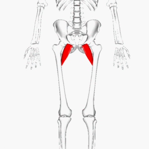
Image 1: Pectineus, red outline
Anatomy[edit | edit source]
Pectineus can be classified in the medial compartment of thigh(when the function is emphasized) or the anterior compartment of thigh (when the nerve is emphasized)[2].
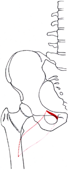
Origin: pectineal line (pecten pubis) and adjacent bone of pelvis
Insertion: oblique line extending from base of lesser trochanter to linea aspera on posterior surface of femur
Image 2: Pectineus of right side - outline and attachment-areas.
Action: adducts and flexes thigh at hip joint
Arterial supply: superficial part by medial circumflex femoral artery and deep part by the anterior branch of obturator artery[3]
Nerve supply
The pectineus is considered a transitional muscle between the anterior thigh and medial thigh; this is due to innervation mainly from the femoral nerve and also sometimes from the obturator nerve. [4]
Relation[edit | edit source]
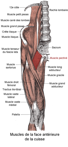
The muscle lies in the frontal plane
- Medial to adductor longus.
- Laterally, it is related to the psoas major muscle and the medial circumflex femoral artery and vein.
- The anterior surface of pectineus forms the medial part of the floor of femoral traingle together with adductor longus.
- This surface of pectineus is covered with the deep layer of fascia lata that separates it from the femoral artery, femoral vein and great saphenous vein that course through the femoral triangle.
- Posterior to pectineus are the adductor magnus, adductor brevis and obturator externus muscles, and the anterior branch of obturator nerve.[5][6]
Image 3: Pectineus muscle. human anatomy
Action[edit | edit source]
Due to the course of its fibers, pectineus both flexes and adducts the thigh at the hip joint when it contracts. When the lower limb is in the anatomical position, contraction of the muscle first causes flexion to occur at the hip joint. This flexion can go as far as the thigh being at a 45 degree angle to the hip joint[7].
At that point, the angulation of the fibers is such that the contracted muscle fibers now pull the thigh towards the midline, producing thigh adduction[7].
Injury[edit | edit source]
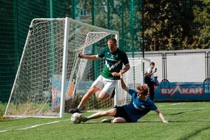
The pectineus muscle can become injured by overstretching; specifically, by stretching a leg or legs too far out to the side or front of the body. Pectineus injuries can also be caused by rapid movements like kicking or sprinting, changing directions too quickly while running, or even by sitting with a leg crossed for too long.[6]
See Groin Strain and Assessment of Athletes with Groin Pain
Symptoms of the injury[edit | edit source]
The most common symptom of an injured pectineus muscle is pain. Other may include bruising,swelling,tenderness and stiffness[5].
Treatment[edit | edit source]
Treatment of pectinus muscle injury is ask patient to protect the injured from movements and objects that might cause further injury; minimize activities that use the pectinus muscle, like walking and running, in order to allow the muscle time to heal; and ice the injury to decrease and prevent swelling, as well as help decrease any pain.icing shouhd be performed 15:20 minutes every few hours[5].
References[edit | edit source]
- ↑ very well health Pectineus muscle Available: https://www.verywellhealth.com/pectineus-muscle-anatomy-5084562(accessed 15.1.2022)
- ↑ Mosby's Medical, Nursing & Allied Health Dictionary, Fourth Edition, ., 1994
- ↑ Radiopedia Pectineus Available:https://radiopaedia.org/articles/pectineus-muscle (accessed 15.1.2022)
- ↑ Glenister R, Sharma S. Anatomy, bony pelvis and lower limb, hip. StatPearls [Internet]. 2021 Jul 26. Available: https://www.statpearls.com/articlelibrary/viewarticle/32250/(accessed 15.1.2022)
- ↑ 5.0 5.1 5.2 Moore,. .Clinically Oriented Anatomy (7th ed). (2014)
- ↑ 6.0 6.1 Standring,. Gray's Antomy (41tst ed.). (2016)
- ↑ 7.0 7.1 Palastanga,. . Anatomy and human movement: structure and function (6th ed.).(2012)


