Iliopsoas: Difference between revisions
No edit summary |
(reference and citations added. Info updated) |
||
| Line 6: | Line 6: | ||
== Introduction == | == Introduction == | ||
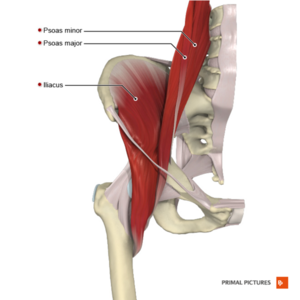
[[File:Iliopsoas.png|thumb|A compound muscle composed of the iliac and psoas muscles]] | [[File:Iliopsoas.png|thumb|A compound muscle composed of the iliac and psoas muscles]] | ||
The iliopsoas muscle complex is made up of three muscles that | The iliopsoas muscle complex is made up of three muscles that includes the [[iliacus]], [[Psoas Major|psoas major]] and [[Psoas Minor|psoas minor]]. This complex muscle system can function as a unit or as separate muscles.<ref name=":1">Bordoni B, Varacallo M. [https://www.ncbi.nlm.nih.gov/books/NBK531508/ Anatomy, bony pelvis and lower limb, Iliopsoas Muscle]. StatPearls [Internet]. 2021 Jul 21. Available:https://www.ncbi.nlm.nih.gov/books/NBK531508/ (accessed 15.2.2022)</ref>. Psoas minor is only present in 60% to 65% of individuals<ref name=":1" /><ref name=":2">Anderson CN. [https://toa.com/storage/wysiwyg/Anderson%20Iliopsoas%20Pathology%20Diagnosis%20and%20Treatment.pdf Iliopsoas: pathology, diagnosis, and treatment.] Clinics in sports medicine. 2016 Jul 1;35(3):419-33.</ref>. | ||
The iliopsoas muscle is the | The iliopsoas muscle is the primary [[Hip Flexors|hip flexor]] and assists in external rotation of the [[Hip|hip joint]], playing an important role in maintaining the strength and integrity of the [[Hip Anatomy|hip joint]]. It is essential for correct standing or sitting lumbar posture and plays a critical role during walking and running<ref name=":1" />. | ||
The [[fascia]] covering the iliopsoas muscle creates multiple fascial connections, relating the muscle with different viscera and muscle areas<ref name=":1" /> | The [[fascia]] covering the iliopsoas muscle creates multiple fascial connections, relating the muscle with different viscera and muscle areas<ref name=":1" /><ref name=":0">Physiopedia [[Iliopsoas Tendinopathy]] Available:https://www.physio-pedia.com/Iliopsoas_Tendinopathy?utm_source=physiopedia&utm_medium=related_articles&utm_campaign=ongoing_internal (accessed 15.2.2022)</ref>. | ||
== Anatomy == | == Anatomy == | ||
The iliopsoas musculotendinous unit is part of the inner muscles of the hip and forms part of the posterior [[Abdominal Muscles|abdominal wall]], | The iliopsoas musculotendinous unit is part of the inner muscles of the hip. It lies posteriorly at the retroperitoneum level and forms part of the posterior [[Abdominal Muscles|abdominal wall]] <ref name=":1" />. There are many anatomical variations of the iliopsoas muscle, with the most common origin and insertion listed below<ref name=":1" /><ref name=":2" />. | ||
'''Origin''': | '''Origin''': | ||
Psoas major: The transverse processes and lateral surfaces of the vertebral bodies of L1 - L4<ref name=":1" /> or T12 - L5<ref name=":2" /> and the path involves the intervertebral discs<ref name=":1" /><ref name=":2" />. | |||
Psaos minor: It originates from T12 and L1 and lies anteriorly to the psoas major<ref name=":1" /><ref name=":2" />. | |||
Iliacus: It originates on the upper two-thirds of the iliac fossa and the lateral parts of the wing of the sacrum. | |||
''' | '''Insertion''': | ||
''' | Psoas major and iliacus: The psoas major and iliacus join together, pass under the inguinal ligament and insert onto the femoral lesser trochanter<ref name=":1" /><ref name=":2" />. | ||
Psaos minor: It inserts onto the iliopectineal eminence after converging with the iliac fascia and the psoas major tendon<ref name=":1" /><ref name=":2" />. | |||
'''Bursa''': The iliopsoas or iliopectineal bursa lies between the bony surfaces of the pelvis and proximal femur and the musculotendinous unit. It is the largest bursa in humans. It has an average length of 5 to 6 cm and width of 3 cm and extends from the iliopectineal eminence to the lower portion of the femoral head<ref name=":2" />. | |||
'''Innervation''': | |||
Psaos major and minor: short collateral branches of L1 to L3<ref name=":1" /> | |||
Iliacus: femoral nerve or terminal nerve of L1 to L4<ref name=":1" /> | |||
'''Vascular supply''': Common iliac artery and external iliac artery<ref name=":1" /> | |||
== Clinical Relevance == | == Clinical Relevance == | ||
Injury to the iliopsoas may cause hip pain and limited mobility. | Injury to the iliopsoas may cause hip pain and limited mobility. | ||
[[Snapping Hip Syndrome|Snapping hip syndrome]] | [[Snapping Hip Syndrome|<u>Snapping hip syndrome</u>]] | ||
[[Iliopsoas Tendinopathy|<u>Iliopsoas tendinopathy</u>]] refers to a condition that affects the insertion of the muscle on the femur, and can occur with repetitive hip flexion and other deficits of the [[Biomechanics|biomechanical]] system resulting in chronic degenerative changes of the tendon. | |||
Impingement of the Iliopsoas Tendon: Following an operation to replace the femoral head, movement of the artificial head may during a hip extension press against the surrounding soft tissues, including the tendon of the iliopsoas complex. The surgeon decides the course of action. | Impingement of the Iliopsoas Tendon: Following an operation to replace the femoral head, movement of the artificial head may during a hip extension press against the surrounding soft tissues, including the tendon of the iliopsoas complex. The surgeon decides the course of action. | ||
[[Iliopsoas Bursitis|Iliopsoas Bursitis:]] Bursitis that involves the tendon of the iliopsoas complex is an inflammation that enlarges the volume of the bursa and produces pain on movement. | [[Iliopsoas Bursitis|<u>Iliopsoas Bursitis:</u>]] Bursitis that involves the tendon of the iliopsoas complex is an inflammation that enlarges the volume of the bursa and produces pain on movement. | ||
In pediatrics, in the presence of spasticity eg cerebral palsy and the presence of important contractures, surgery is performed with distal tenotomy. This reduces the difficulty of walking and enables a posture that makes the child independent when possible. | In pediatrics, in the presence of spasticity eg cerebral palsy and the presence of important contractures, surgery is performed with distal tenotomy. This reduces the difficulty of walking and enables a posture that makes the child independent when possible. | ||
Revision as of 13:18, 14 August 2023
Original Editor - Lucinda hampton
Top Contributors - Lucinda hampton, Wendy Snyders, Ewa Jaraczewska, Ahmed M Diab and Vidya Acharya
Introduction[edit | edit source]
The iliopsoas muscle complex is made up of three muscles that includes the iliacus, psoas major and psoas minor. This complex muscle system can function as a unit or as separate muscles.[1]. Psoas minor is only present in 60% to 65% of individuals[1][2].
The iliopsoas muscle is the primary hip flexor and assists in external rotation of the hip joint, playing an important role in maintaining the strength and integrity of the hip joint. It is essential for correct standing or sitting lumbar posture and plays a critical role during walking and running[1].
The fascia covering the iliopsoas muscle creates multiple fascial connections, relating the muscle with different viscera and muscle areas[1][3].
Anatomy[edit | edit source]
The iliopsoas musculotendinous unit is part of the inner muscles of the hip. It lies posteriorly at the retroperitoneum level and forms part of the posterior abdominal wall [1]. There are many anatomical variations of the iliopsoas muscle, with the most common origin and insertion listed below[1][2].
Origin:
Psoas major: The transverse processes and lateral surfaces of the vertebral bodies of L1 - L4[1] or T12 - L5[2] and the path involves the intervertebral discs[1][2].
Psaos minor: It originates from T12 and L1 and lies anteriorly to the psoas major[1][2].
Iliacus: It originates on the upper two-thirds of the iliac fossa and the lateral parts of the wing of the sacrum.
Insertion:
Psoas major and iliacus: The psoas major and iliacus join together, pass under the inguinal ligament and insert onto the femoral lesser trochanter[1][2].
Psaos minor: It inserts onto the iliopectineal eminence after converging with the iliac fascia and the psoas major tendon[1][2].
Bursa: The iliopsoas or iliopectineal bursa lies between the bony surfaces of the pelvis and proximal femur and the musculotendinous unit. It is the largest bursa in humans. It has an average length of 5 to 6 cm and width of 3 cm and extends from the iliopectineal eminence to the lower portion of the femoral head[2].
Innervation:
Psaos major and minor: short collateral branches of L1 to L3[1]
Iliacus: femoral nerve or terminal nerve of L1 to L4[1]
Vascular supply: Common iliac artery and external iliac artery[1]
Clinical Relevance[edit | edit source]
Injury to the iliopsoas may cause hip pain and limited mobility.
Iliopsoas tendinopathy refers to a condition that affects the insertion of the muscle on the femur, and can occur with repetitive hip flexion and other deficits of the biomechanical system resulting in chronic degenerative changes of the tendon.
Impingement of the Iliopsoas Tendon: Following an operation to replace the femoral head, movement of the artificial head may during a hip extension press against the surrounding soft tissues, including the tendon of the iliopsoas complex. The surgeon decides the course of action.
Iliopsoas Bursitis: Bursitis that involves the tendon of the iliopsoas complex is an inflammation that enlarges the volume of the bursa and produces pain on movement.
In pediatrics, in the presence of spasticity eg cerebral palsy and the presence of important contractures, surgery is performed with distal tenotomy. This reduces the difficulty of walking and enables a posture that makes the child independent when possible.
The iliopsoas muscle can cause compression of the femoral nerve and cause knee pain. Before deciding on surgical treatment and releasing of the femoral nerve, the patient should learn stretching exercises to reduce the tension generated by the muscle. Generally, if the patient can follow the physiotherapy indications, the use of surgery can be avoided.
Hypertrophy of the muscle may be responsible for the compression of the femoral nerve and cause knee pain. Before deciding on surgical treatment to release the femoral nerve, the patient should learn stretching exercises to reduce the tension generated by the muscle. Generally, if the patient can follow the physiotherapy indications, the use of surgery can be avoided. [1]
References[edit | edit source]
- ↑ 1.00 1.01 1.02 1.03 1.04 1.05 1.06 1.07 1.08 1.09 1.10 1.11 1.12 1.13 1.14 Bordoni B, Varacallo M. Anatomy, bony pelvis and lower limb, Iliopsoas Muscle. StatPearls [Internet]. 2021 Jul 21. Available:https://www.ncbi.nlm.nih.gov/books/NBK531508/ (accessed 15.2.2022)
- ↑ 2.0 2.1 2.2 2.3 2.4 2.5 2.6 2.7 Anderson CN. Iliopsoas: pathology, diagnosis, and treatment. Clinics in sports medicine. 2016 Jul 1;35(3):419-33.
- ↑ Physiopedia Iliopsoas Tendinopathy Available:https://www.physio-pedia.com/Iliopsoas_Tendinopathy?utm_source=physiopedia&utm_medium=related_articles&utm_campaign=ongoing_internal (accessed 15.2.2022)


