Hip Dislocation
Original Editor - Annelies Noppe
Top Contributors - Annelies Noppe, Leana Louw, Lucinda hampton, Kim Jackson, Lokiru Paul, WikiSysop, Vidya Acharya, Anas Mohamed and Kirenga Bamurange Liliane
Introduction[edit | edit source]
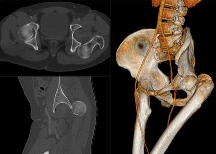
Hip dislocations are relatively rare, are be congenital or acquired and account for ~5% of all joint dislocations The native hip joint (as opposed to prosthetic hip) is inherently stable and needs a huge amount of force to cause dislocation, such as in a motor vehicle accidents.[1]
Hip dislocation can be classified as:
- Posterior dislocation (most common ~85%). Caused by combined forces of: hip flexion, adduction, and internal rotation.
- Anterior dislocation (~10%). Caused by combined forces of : hyper-abduction with the extension.[1]
- Central dislocation (always occurring with Acetabulum Fracture)[2]
Below is a good 5 minute video on hip dislocations.
Etiology[edit | edit source]
Acquired Dislocation: Motor vehicle collisions accounting for >50% of dislocations.[2] Another common mechanism is falling from a height. Hip dislocations are thus rarely isolated, and often goes together with other injuries or fractures. With hip dislocations, the soft tissue around the hip, such as the muscles, ligaments and labrum are also damaged. Neural injuries may also be present. Fractures to the acetabulum and femur head is most commonly associated with traumatic hip dislocations.
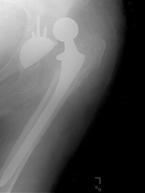
Total hip replacement (THR) dislocation is a complication of THR usually occurring due to patient noncomplicance with post-operative precautions, implant malposition, or soft-tissue deficiency. This type of dislocation normally caused by less trauma, usually falls or turning, moving into the contra-indicated positions, and putting stress on the capsule that was cut to do the replacement surgery..[3][2] For more on THR dislocation see link.
Congenital Hip Dislocation have been appraised and are now viewed as part of the spectrum of developmental dysplasia of the hip. See link for information on this.
Clinical Presentation[edit | edit source]
The patient's history commonly involve a description of a significant "clunk" or "popping" followed instantly by severe pain. A physical deformity with ipsilateral shortening/hip flexion, adduction and internal rotation will be visible. Inability to walk results from of pain and swelling With the separation of the femur head from the acetabulum, surrounding muscles and tendons can be damaged as well. Subsequent knee injuries might also be present.
- Other common features include: leg length discrepancy: hip immobility with [4] reduced hip range of motion.[1][4]
- Signs of possible vascular or sciatic nerve injury: Local hematoma. Painful buttock, posterior thigh, and leg. Altered sensation in posterior leg and foot. Weakness or total loss of dorsiflexion (peroneal branch) or plantar flexion (tibial branch). Diminished or absent deep tendon reflexes at the ankle.[1]
Differential Diagnosis[edit | edit source]
- Hip dysplasia
- Hip sublaxation
Diagnostic Procedures[edit | edit source]
- X-rays: AP pelvis and lateral
- To confirm dislocation and successful relocation
- Assess for associated fractures
- Progression of hip dysplasia
- CT:
- To rule out concomitant injuries in traumatic dislocations (e.g. acetabulum or femur head fractures)
- Clearance of lumbar spine[5]
Complications[edit | edit source]
Immediate:[6]
- Associated soft tissue injuries
- Neural injuries, especially to the sciatic nerve in posterior dislocations (present in about 10% of traumatic dislocations)
- Fractures, mostly to the femur head or acetabulum (mostly posterior wall)
Long term:[7]
- Avascular necrosis:
- Incidence of 1.7-40% is reducable to 0-10% if relocation is done within 6 hours post traumatic dislocation[8]
- Post-traumatic osteoarthritis
- Chronic dislocations
- Leg length discrepancy
Examination[edit | edit source]
- Observation:
- Hip dislocations can often be diagnosed by just looking at the hip. The hip will be shortend, in external rotation, slight flexion and adduction in the more common posterior dislocations.
- Imaging (as explained above)
- Neurological assessment (to determine any associated neural injuries)
Medical Management[edit | edit source]
The management of hip dislocations may be operative and non-operative. Many studies note that time to reduction is crucial as the longer the hip is dislocated, the greater the risk of avascular necrosis in the native hip.
Non-surgical[edit | edit source]
Closed relocation of the hip is done by a traction force performed in the opposite direction of the dislocation, with the hip in 90° flexion. This should preferably be done under general or regional anesthesia and muscle relaxation to prevent greater damage to cartilage and soft tissue.[9] It may also be done in under anaesthetics in theater.[6] After the relocation, the stability of the hip should be tested very carefully. A period of bed rest might be recommended depending on the stability of the hip and the extent of the soft tissue injuries.
Surgical[edit | edit source]
Indications:
- Failed conservative relocation
- Instability following conservative relocation
- Associated fractures of the femur head or acetabulum
- Loose bone fragments in joint space after relocation
Hip arthroscopy can be used to evaluate intra-articular fractures and chondral injuries and to remove intra-articular fragments, Hip replacement surgery can also be considered if optimal stability is not achieved with relocation and fixation of the associated injuries.[7] Dislocation following hip replacement surgery might indicate revision surgery to ensure the stability of the hip in the long run.
Open reduction indications:[7]
- Used with challenging relocations or if any obstructions (e.g. loose fragments/soft tissue) is limiting closed reduction
- Deteriorating neurological signs following closed reduction (especially sciatic nerve function following posterior dislocation)
- Cases with proximal femur fractures, where manipulation of the leg is contra-indicated
Physiotherapy Management[edit | edit source]
It is important to take the time frames for soft tissue healing (and bone healing in cases with associated fractures) into consideration with rehabilitation following a hip dislocation. The orthopaedic surgeon will give guidance on weight bearing restrictions that might be present following the medical management of the hip. Full rehabilitation following hip dislocation can take 2-3 months.[6]
- Gait re-education: Initially with mobility assistive devices (walking frame/crutches) to limit weight bearing, and progression thereof
- Improve hip range of motion: Especially extension in children after the use of a brace/splint/harness that kept the hip in flexion
- Strengthening of muscles around the hip, with special focus on hip stabilizers
- Stretching
- Joint mobilization
- Graded return to activity/sport
See rehabilitation resources below.
Resources[edit | edit source]
- Rehabilitation Guidelines for Surgical Hip Dislocation
- Surgical Hip Dislocation Rehabilitation Protocol
- Hip Surgical Dislocation Guidelines
Clinical Bottom Line[edit | edit source]
Hip dislocations are classified into congenial and acquired. Congenital hip dislocations, or developmental hip dysplasia can be successfully managed in children, but might cause problems later in life, when total hip replacement surgery might be indicated to improve function, leg length discrepancies and pain. Acquired, or traumatic hip dislocations are medical emergencies, and treatment should be sought as soon as possible. Relocation should ideally occur within 6 hours from the dislocation, in order to reduce complications. Traumatic dislocations are reduced either open or closed, and open or arthroscopy surgery might be indicated in cases with associated fractures. Physiotherapy plays an important role in the rehabilitation following a hip dislocation, in order to get the patients back to their previous level of function, and to prevent further dislocations.
References[edit | edit source]
- ↑ 1.0 1.1 1.2 1.3 Masiewicz S, Mabrouk A, Johnson DE. Posterior hip dislocation.Available:https://www.ncbi.nlm.nih.gov/books/NBK459319/ (accessed 7.1.2023)
- ↑ 2.0 2.1 2.2 Radiopedia Hip dislocation Available:https://radiopaedia.org/articles/hip-dislocation (accessed 7.1.2023)
- ↑ Orthobullets THA Dislocation Available:https://www.orthobullets.com/recon/5012/tha-dislocation (accessed 7.1.2022)
- ↑ 4.0 4.1 Hung NN. Traumatic hip dislocation in children. Journal of Pediatric Orthopaedics B 2012;21(6):542-51.
- ↑ Larson DE. Gezin en gezondheid. Cambium BV:Zeewolde, 1995.
- ↑ 6.0 6.1 6.2 Ortho Info. Developmental Dislocation (Dysplasia) of the Hip (DDH). Available from: https://orthoinfo.aaos.org/en/diseases--conditions/developmental-dislocation-dysplasia-of-the-hip-ddh (accessed 08/08/2020).
- ↑ 7.0 7.1 7.2 Lima LC, Nascimento RA, Almeida VM, Façanha Filho FA. Epidemiology of traumatic hip dislocation in patients treated in Ceará, Brazil. Acta ortopedica brasileira 2014;22(3):151-4.
- ↑ Bucholz R, Heckman JD. Rockwood e Green fraturas em adultos. In: Rockwood e Green fraturas em adultos, 2006: pp. 2263-2263.
- ↑ Medscape. Hip dislocation. Available from: https://emedicine.medscape.com/article/86930-overview (accessed 09/08/2020).


