Coronary Ligaments of the Knee: Difference between revisions
Chloe Waller (talk | contribs) (Updated description) |
Chloe Waller (talk | contribs) (Added picture) |
||
| (7 intermediate revisions by the same user not shown) | |||
| Line 1: | Line 1: | ||
'''Original Editor '''-[[User:Simisola Ajeyalemi |Simisola Ajeyalemi]] and[[User:Rewan Elsayed Elkanafany |Rewan Elsayed Elkanafany]] | '''Original Editor '''-[[User:Simisola Ajeyalemi |Simisola Ajeyalemi]] and[[User:Rewan Elsayed Elkanafany |Rewan Elsayed Elkanafany]] | ||
'''Top Contributors''' - {{Special:Contributors/{{FULLPAGENAME}}}} | '''Top Contributors''' - {{Special:Contributors/{{FULLPAGENAME}}}} | ||
== Description == | == Description == | ||
[[File:Meniscotibial ligament.jpg|alt=| | [[File:Meniscotibial ligament.jpg|alt=|thumb]] | ||
The coronary ligaments, also known as the '''meniscotibial ligaments,''' are part of the fibrous capsule of the [[Knee|knee joint]]. They are made up of the medial coronary ligament and the lateral coronary ligament. They connect the inferior edges of the [[Meniscal Lesions|menisci]] to the of the [[Tibia|tibial plateau]]<ref>Guy S, Ferreira A, Carrozzo A, Delaloye JR, Cavaignac E, Vieira TD, Sonnery-Cottet B. [https://pubmed.ncbi.nlm.nih.gov/35155104/ Isolated Meniscotibial Ligament Rupture: The Medial Meniscus "Belt Lesion".] Arthrosc Tech. 2022 Jan 13;11(2):e133-e138. </ref>. | The coronary [[Ligament|ligaments]], also known as the '''meniscotibial ligaments,''' are part of the fibrous capsule of the [[Knee|knee joint]]. They are made up of the medial coronary ligament and the lateral coronary ligament. They connect the inferior edges of the [[Meniscal Lesions|menisci]] to the of the [[Tibia|tibial plateau]]<ref>Guy S, Ferreira A, Carrozzo A, Delaloye JR, Cavaignac E, Vieira TD, Sonnery-Cottet B. [https://pubmed.ncbi.nlm.nih.gov/35155104/ Isolated Meniscotibial Ligament Rupture: The Medial Meniscus "Belt Lesion".] Arthrosc Tech. 2022 Jan 13;11(2):e133-e138. </ref>. | ||
== Function == | == Function == | ||
The coronary ligaments support rotational stability of the knee and prevent anterior tibial translation<ref>Feger J, Knipe H, Knipe H, et al. Meniscotibial ligaments. Available from: https://radiopaedia.org/articles/meniscotibial-ligaments<nowiki/>(Accessed on 21 Nov 2022)</ref>. | The coronary ligaments support rotational stability of the knee and prevent anterior [[Tibia|tibial]] translation<ref>Feger J, Knipe H, Knipe H, et al. Meniscotibial ligaments. Available from: https://radiopaedia.org/articles/meniscotibial-ligaments<nowiki/>(Accessed on 21 Nov 2022)</ref>. | ||
== Clinical relevance == | == Clinical relevance == | ||
Coronary ligament injuries can lead to other knee joint pathology and negatively alter the [[biomechanics]] of the knee<ref name=":1">Smith PA, Humpherys JL, Stannard JP, Cook JL. [https://pubmed.ncbi.nlm.nih.gov/31914474/ Impact of Medial Meniscotibial Ligament Disruption Compared to Peripheral Medial Meniscal Tear on Knee Biomechanics]. J Knee Surg. 2021 Jun;34(7):784-792</ref>. Lesions to the coronary ligaments can increase rotational instability<ref name=":0">A. Peltier, T. Lording, L. Maubisson, R. Ballis, P. Neyret, S. Lustig. [https://pubmed.ncbi.nlm.nih.gov/26264383/ The role of the meniscotibial ligament in posteromedial rotational knee stability] Knee Surg Sports Traumatol Arthrosc 2015 23:2967–2973</ref>. They can also cause increased strain on the [[Anterior Cruciate Ligament|anterior cruciate ligament]] or menisci<ref>Krych AJ, Bernard CD, Leland DP, Camp CL, Johnson AC, Finnoff JT, Stuart MJ. [https://pubmed.ncbi.nlm.nih.gov/31332493/ Isolated meniscus extrusion associated with meniscotibial ligament abnormality.] Knee Surg Sports Traumatol Arthrosc. 2020 Nov;28(11):3599-3605.</ref><ref>Smith PA, Humpherys JL, Stannard JP, Cook JL. [https://pubmed.ncbi.nlm.nih.gov/31914474/ Impact of Medial Meniscotibial Ligament Disruption Compared to Peripheral Medial Meniscal Tear on Knee Biomechanics.] J Knee Surg. 2021 Jun;34(7):784-792.</ref> . This leads to risk of meniscal extrusion or other injury to these structures<ref>Mariani PP, Torre G, Battaglia MJ. [https://pubmed.ncbi.nlm.nih.gov/35123034/ The post-traumatic meniscal extrusion, sign of meniscotibial ligament injury. A case series.] Orthop Traumatol Surg Res. 2022 May;108(3):103226.</ref>. Research suggests coronary ligament injuries are under diagnosed and untreated, as they present similar to meniscus pathologies<ref name=":1" />. | |||
== Assessment == | |||
Common signs and symptoms of coronary ligament injury include<ref name=":2">Otten MHC. Pain in the Coronary Ligaments of the Knee: How Regenerative Therapies Can Help. Available from: https://cellaxys.com/pain-in-the-coronary-ligaments-of-the-knee-how-regenerative-therapies-can-help/ (Accessed 22/11/2022).</ref>: | |||
* Tenderness along the lateral and medial aspects of the knee joint line. | |||
* Feeling of instability. | |||
* Sharp pain during knee flexion or rotational movements. | |||
* Usually have full range of motion but feel discomfort at end of the range flexion and extension. | |||
Swelling is not common in these injuries. | |||
After getting a thorough [[Subjective Assessment|history]], the physical examination should include: | |||
* Palpation of the knee. | |||
* [[McMurrays Test|McMurray's Test]] | |||
If required, [[MRI Scans|MRI scans]] can be used to confirm the diagnosis. | |||
== Treatment == | == Treatment == | ||
[[File:Knee physio.jpg|thumb|300x300px]] | |||
Treatment will vary according to the injury, the severity and if other structures have been affected. | |||
First line treatment for coronary ligaments includes [[RICE]], [[NSAIDs|medicines]] such as painkillers or anti-inflammatories and physiotherapy<ref name=":2" />. Physiotherapy will be tailored to improving biomechanics and strengthen the surrounding muscles, especially the [[Quadriceps Muscle|quadriceps]]. | |||
Surgery may be required if the injury is severe, to promote stability of the knee, or in order to protect other structures from further strain<ref name=":1" />. One study found repair of medial coronary ligaments reduced meniscal extrusion, which may improve meniscal function and reduce the risk for [[osteoarthritis]]<ref>Paletta GA Jr, Crane DM, Konicek J, Piepenbrink M, Higgins LD, Milner JD, Wijdicks CA. [https://pubmed.ncbi.nlm.nih.gov/32775474/ Surgical Treatment of Meniscal Extrusion: A Biomechanical Study on the Role of the Medial Meniscotibial Ligaments With Early Clinical Validation]. Orthop J Sports Med. 2020 Jul 29;8(7):2325967120936672.</ref>. | |||
== References == | == References == | ||
Latest revision as of 12:43, 22 November 2022
Original Editor -Simisola Ajeyalemi andRewan Elsayed Elkanafany
Top Contributors - Chloe Waller, Simisola Ajeyalemi, Rewan Elsayed Elkanafany, Kim Jackson and Tien Yunn How
Description[edit | edit source]
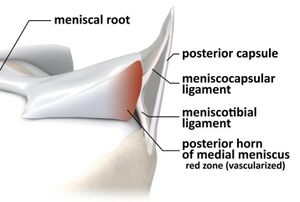
The coronary ligaments, also known as the meniscotibial ligaments, are part of the fibrous capsule of the knee joint. They are made up of the medial coronary ligament and the lateral coronary ligament. They connect the inferior edges of the menisci to the of the tibial plateau[1].
Function[edit | edit source]
The coronary ligaments support rotational stability of the knee and prevent anterior tibial translation[2].
Clinical relevance[edit | edit source]
Coronary ligament injuries can lead to other knee joint pathology and negatively alter the biomechanics of the knee[3]. Lesions to the coronary ligaments can increase rotational instability[4]. They can also cause increased strain on the anterior cruciate ligament or menisci[5][6] . This leads to risk of meniscal extrusion or other injury to these structures[7]. Research suggests coronary ligament injuries are under diagnosed and untreated, as they present similar to meniscus pathologies[3].
Assessment[edit | edit source]
Common signs and symptoms of coronary ligament injury include[8]:
- Tenderness along the lateral and medial aspects of the knee joint line.
- Feeling of instability.
- Sharp pain during knee flexion or rotational movements.
- Usually have full range of motion but feel discomfort at end of the range flexion and extension.
Swelling is not common in these injuries.
After getting a thorough history, the physical examination should include:
- Palpation of the knee.
- McMurray's Test
If required, MRI scans can be used to confirm the diagnosis.
Treatment[edit | edit source]
Treatment will vary according to the injury, the severity and if other structures have been affected.
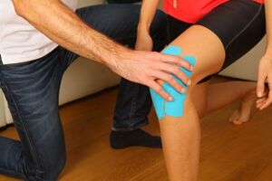
First line treatment for coronary ligaments includes RICE, medicines such as painkillers or anti-inflammatories and physiotherapy[8]. Physiotherapy will be tailored to improving biomechanics and strengthen the surrounding muscles, especially the quadriceps.
Surgery may be required if the injury is severe, to promote stability of the knee, or in order to protect other structures from further strain[3]. One study found repair of medial coronary ligaments reduced meniscal extrusion, which may improve meniscal function and reduce the risk for osteoarthritis[9].
References[edit | edit source]
- ↑ Guy S, Ferreira A, Carrozzo A, Delaloye JR, Cavaignac E, Vieira TD, Sonnery-Cottet B. Isolated Meniscotibial Ligament Rupture: The Medial Meniscus "Belt Lesion". Arthrosc Tech. 2022 Jan 13;11(2):e133-e138.
- ↑ Feger J, Knipe H, Knipe H, et al. Meniscotibial ligaments. Available from: https://radiopaedia.org/articles/meniscotibial-ligaments(Accessed on 21 Nov 2022)
- ↑ 3.0 3.1 3.2 Smith PA, Humpherys JL, Stannard JP, Cook JL. Impact of Medial Meniscotibial Ligament Disruption Compared to Peripheral Medial Meniscal Tear on Knee Biomechanics. J Knee Surg. 2021 Jun;34(7):784-792
- ↑ A. Peltier, T. Lording, L. Maubisson, R. Ballis, P. Neyret, S. Lustig. The role of the meniscotibial ligament in posteromedial rotational knee stability Knee Surg Sports Traumatol Arthrosc 2015 23:2967–2973
- ↑ Krych AJ, Bernard CD, Leland DP, Camp CL, Johnson AC, Finnoff JT, Stuart MJ. Isolated meniscus extrusion associated with meniscotibial ligament abnormality. Knee Surg Sports Traumatol Arthrosc. 2020 Nov;28(11):3599-3605.
- ↑ Smith PA, Humpherys JL, Stannard JP, Cook JL. Impact of Medial Meniscotibial Ligament Disruption Compared to Peripheral Medial Meniscal Tear on Knee Biomechanics. J Knee Surg. 2021 Jun;34(7):784-792.
- ↑ Mariani PP, Torre G, Battaglia MJ. The post-traumatic meniscal extrusion, sign of meniscotibial ligament injury. A case series. Orthop Traumatol Surg Res. 2022 May;108(3):103226.
- ↑ 8.0 8.1 Otten MHC. Pain in the Coronary Ligaments of the Knee: How Regenerative Therapies Can Help. Available from: https://cellaxys.com/pain-in-the-coronary-ligaments-of-the-knee-how-regenerative-therapies-can-help/ (Accessed 22/11/2022).
- ↑ Paletta GA Jr, Crane DM, Konicek J, Piepenbrink M, Higgins LD, Milner JD, Wijdicks CA. Surgical Treatment of Meniscal Extrusion: A Biomechanical Study on the Role of the Medial Meniscotibial Ligaments With Early Clinical Validation. Orthop J Sports Med. 2020 Jul 29;8(7):2325967120936672.


