Cervical Anatomy: Difference between revisions
No edit summary |
No edit summary |
||
| Line 62: | Line 62: | ||
== Blood Supply == | == Blood Supply == | ||
Distal to their bifurcation from the subclavian arteries, the vertebral arteries course cranially to the base of the skull. They progress past the C7 vertebrae anteriorly before entering the transverse foramen of C6. After exiting the C2 transverse foramen, the arteries bend laterally to enter the more widely placed transverse foramen of C1 and then revert medially over the superior aspect of the arch of the atlas before entering the base of the skull. Main tributaries of the vertebral arteries, the cervical radicular arteries, supply blood directly to the cervical vertebral bodies. | Distal to their bifurcation from the subclavian arteries, the vertebral arteries course cranially to the base of the skull. They progress past the C7 vertebrae anteriorly before entering the transverse foramen of C6. After exiting the C2 transverse foramen, the arteries bend laterally to enter the more widely placed transverse foramen of C1 and then revert medially over the superior aspect of the arch of the atlas before entering the base of the skull. Main tributaries of the vertebral arteries, the cervical radicular arteries, supply blood directly to the cervical vertebral bodies. | ||
Also located in the cervical spine region are the carotid arteries. The common carotids bifurcate into the internal and external carotids at the C3 vertebral level. Despite their association with the neck, they do not perfuse any structures in the cervical spine.<ref name=":0" /> | |||
== Nerves == | == Nerves == | ||
Revision as of 23:22, 28 January 2020
This article or area is currently under construction and may only be partially complete. Please come back soon to see the finished work! (28/01/2020)
Original Editor - Your name will be added here if you created the original content for this page.
Top Contributors - Lucinda hampton, Manisha Shrestha, Kim Jackson, Robin Tacchetti and Abbey Wright
Introduction[edit | edit source]
An expert understanding of cervical anatomy is critical to physiotherapists working in this region. An understanding of this anatomy is essential for assessment and treatment of cervical spine problems. This page is a review of cervical spine osteology, ligaments, muscles, and neurovascular structures.
The cervical spine, comprised of seven cervical vertebrae referred to as C1 to C7, is divided into two major segments: the craniocervical junction (CCJ) and the subaxial spine. The CCJ includes the occiput and the two most cranial cervical vertebrae known as the atlas (C1) and the axis (C2). The subaxial spine includes the five most caudal cervical vertebrae (C3-C5).[1] The cervical spine’s major functions include supporting and cushioning loads to the head/neck while allowing for rotation, and protecting the spinal cord extending from the brain.[2]
Vertebrae[edit | edit source]
Intervertebral Disc[edit | edit source]
Joints[edit | edit source]
Ligaments[edit | edit source]
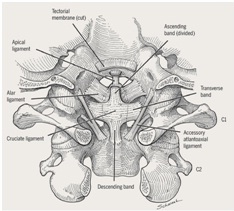
There are six major ligaments to consider in the cervical spine. The majority of these ligaments are present throughout the entire vertebral column.
Present throughout Vertebral Column
- Anterior and posterior longitudinal ligaments – long ligaments that run the length of the vertebral column, covering the vertebral bodies and intervertebral discs.
- Ligamentum flavum – connects the laminae of adjacent vertebrae.
- Interspinous ligament – connects the spinous processes of adjacent vertebrae.
Unique to Cervical Spine
- Nuchal ligament – a continuation of the supraspinous ligament. It attaches to the tips of the spinous processes from C1-C7, and provides the proximal attachment for the rhomboids and trapezius.
- Transverse ligament of the atlas – connects the lateral masses of the atlas, and in doing so anchors the dens in place.
(Note: Some texts consider the interspinous ligament to be part of the nuchal ligament).[3]
Muscles[edit | edit source]
The cervical vertebrae serve as the origination and insertion points for a host of muscles that support but also enable movement of the head and neck. The musculature of the neck is comprised of a number of different muscle groups. They can be divided into anterior, lateral and posterior groups based on their position in the neck. They are further divided into more specific groups based on a number of determinants; including depth, precise location, and function.
1.Anterior neck muscles
Superficial muscles - platysma, sternocleidomastoid, subclavius
Suprahyoids - digastric, mylohyoid, geniohyoid, stylohyoid
Infrahyoids - sternohyoid, sternothyroid, thyrohyoid, omohyoid
Scalenes - anterior, middle and posterior scalene muscles
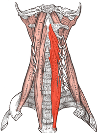
2.Lateral (prevertebral) neck muscles
Rectus capitis, Longus capitis, Longus colli
3.Posterior neck muscles
Superficial muscles - splenius capitis, splenius cervicis
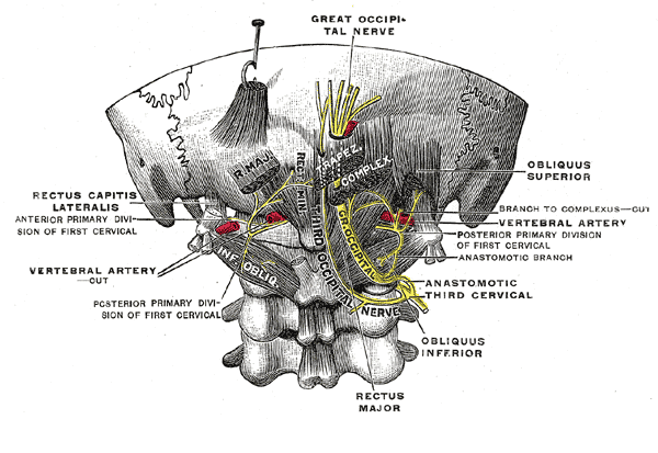
Suboccipital muscles - rectus capitis posterior major, rectus capitis posterior minor, obliquus capitis inferior, obliquus capitis superior
Transversospinalis muscles - semispinalis capitis, semispinalis cervicis, rotatores cervicis, interspinales, intertransversarii.[4]
Blood Supply[edit | edit source]
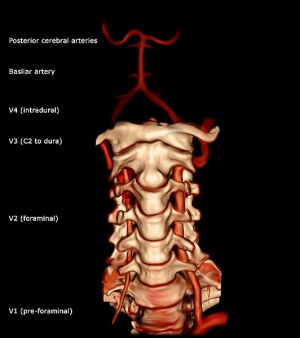
Distal to their bifurcation from the subclavian arteries, the vertebral arteries course cranially to the base of the skull. They progress past the C7 vertebrae anteriorly before entering the transverse foramen of C6. After exiting the C2 transverse foramen, the arteries bend laterally to enter the more widely placed transverse foramen of C1 and then revert medially over the superior aspect of the arch of the atlas before entering the base of the skull. Main tributaries of the vertebral arteries, the cervical radicular arteries, supply blood directly to the cervical vertebral bodies.
Also located in the cervical spine region are the carotid arteries. The common carotids bifurcate into the internal and external carotids at the C3 vertebral level. Despite their association with the neck, they do not perfuse any structures in the cervical spine.[1]
Nerves[edit | edit source]
The cervical spine functions as bony protection of the spinal cord as it exits the cranium.
Despite the presence of seven cervical vertebrae, there are eight pairs of cervical nerves, termed C1 to C8. C1 through C7 exit the spine cranially to its associated vertebrae, while C8 exits caudally to C7. [1]
The transverse foramina of the cervical vertebrae provide a passageway by which the vertebral artery, vein and sympathetic nerves can pass. The only exception to this is C7 – where the vertebral artery passes around the vertebra, instead of through the transverse foramen.[3]
References[edit | edit source]
- ↑ 1.0 1.1 1.2 Kaiser JT, Lugo-Pico JG. Anatomy, Head and Neck, Cervical Vertebrae. Available from: https://www.ncbi.nlm.nih.gov/books/NBK539734/ (last accessed 28.1.2020)
- ↑ Frost BA, Camarero-Espinosa S, Foster EJ. Materials for the spine: anatomy, problems, and solutions. Materials. 2019 Jan;12(2):253. Available from:https://www.ncbi.nlm.nih.gov/pmc/articles/PMC6356370/ (last accessed 28.1.2020)
- ↑ 3.0 3.1 Teach me anatomy The cervical Spine Available from:https://teachmeanatomy.info/neck/bones/cervical-spine/ (last accessed 28.1.2020)
- ↑ Ken Hub Muscles of the neck Available from: https://www.kenhub.com/en/library/anatomy/muscles-of-the-neck-an-overview (last accessed 28.1.2020)


