When refering to evidence in academic writing, you should always try to reference the primary (original) source. That is usually the journal article where the information was first stated. In most cases Physiopedia articles are a secondary source and so should not be used as references. Physiopedia articles are best used to find the original sources of information (see the references list at the bottom of the article).
If you believe that this Physiopedia article is the primary source for the information you are refering to, you can use the button below to access a related citation statement.
Cardiac muscle (or myocardium) makes up the thick middle layer of the heart. It is one of three types of muscle in the body, along with skeletal and smooth muscle.
The myocardium is surrounded by a thin outer layer called the epicardium (AKA visceral pericardium) and an inner endocardium.
Cardiomyocytes are the individual cells that make up the cardiac muscle[1].
Cardiac muscle tissue is only found in your heart, where it performs coordinated contractions that allow your heart to pump blood through your circulatory system.[2]
Cellular Level
At the cellular level, the cardiac muscle looks like a wall of a house. The wall is made up of bricks of cardiomyocytes, and the mortar surrounding the bricks is called the extracellular matrix. The fibroblasts are the supporting cells that produce the extracellular matrix.
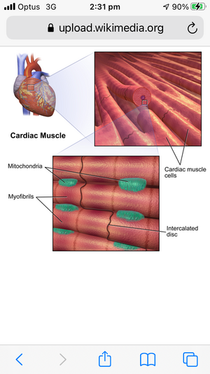
The cardiac muscle cells are interconnected through specialed intercalated discs.[1] Intercalated discs are small connections that join cardiac muscle cells (cardiomyocytes) to each other. Gap junctions are part of the intercalated discs. When one cardiac muscle cell is stimulated to contract, a gap junction transfers the stimulation to the next cardiac cell. This allows the myofibrils to contract together in a fluid synchronized fashion to enable the heart to work as a pump.[1]
Like gap junctions, desmosomes are also found within intercalated discs. The function of desmosomes is to adhere cells together. They are found in high numbers in tissues that are subject to a lot of mechanical forces. Eg, many are found in the epidermis, which is the outer layer of skin, and the myocardium, which is muscle tissue in the heart. They help hold the cardiac muscle fibers together during a contraction.[3]
Cardiomyocytes are rectangular, branching cells that typically contain only one centrally-located nucleus. The nucleus is the “control center” of a cell. It contains all of the cell’s genetic material. While skeletal muscle cells can have multiple nuclei, cardiac muscle cells typically only have one nucleus[2].
Cardiomyocytes contain many mitochondria to produce large amounts of adenosine triphosphate (ATP) and myoglobin to store oxygen to meet the demands of muscle contraction, and has an extensive cardiac capillary network.[4]
Pacemaker Cells
A distinct feature of cardiac muscle fibers is that they have their own auto rhythmicity.
Unlike smooth or skeletal muscle which require neural input for contraction, cardiac fibers have their own pacemaker cells like the sinoatrial (SA) node that spontaneously depolarizes. These depolarizations occur at a consistent pace, but the pacemaker cells can also receive input from the autonomic nervous system to decrease or increase the heart rate depending on the requirements of the body.[4]
The pacemaker cells are connected to other cardiac muscle cells, allowing them to pass along signals. This results in a wave of contractions of your cardiac muscle, which creates your heartbeat[2]
Nerves
The autonomic nervous system (ANS) is a significant regulator of contractility, heart rate, stroke volume, and cardiac output. Parasympathetic innervation is provided from the right and left vagus nerves (CN X). Sympathetic innervation comes from fibers of the sympathetic trunk arising from the upper segments of the thoracic spinal cord. Afferent nerves also provide the central nervous system with feedback on blood pressure, blood chemistry, and to relay pain sensation from the heart[1].
Blood Supply
The process of contraction and relaxation requires a constant supply of oxygen and nutrients to meet the energy demands of cardiac muscle. Blood supply is delivered to the myocardium by coronary arteries, which are the first branches of the aortic root. Blood is drained away by the cardiac veins through the coronary sinus into the right atrium. There are left and right coronary arteries.[1]See Coronary Artery
Pathophysiology
Diseases affecting cardiac muscle have a tremendous impact on health worldwide, with ischemic heart disease being the leading cause of morbidity and mortality in the world (as measured by disability-adjusted life-years, DALYs). Any insult or injury to cardiac muscle can have grave consequences as cardiac muscle cells have minimal regeneration capabilities. However, ongoing research into the use of stem cells to grow cardiac muscle is an area of ongoing research. Below is a few examples of cardiac muscle related health issues.
Ischemic syndromes include angina pectoris, acute myocardial infarction, and sudden cardiac death. Damage to the myocardium from ischemia leads to irreversible loss of cardiac function.
Damage to the heart muscle can also occur with a insufficient blood supply eg: as in myocarditis (inflammation of the heart muscle), which is most commonly due to viral infection.
Education Role: Exercise and Health Promotion. As physiotherapists we are in a good position to advise people on how to keep their heart and cardiovascular system as healthy as possible. Some of the tips for heart health include:
As with many other muscles in your body, exercise can strengthen your cardiac muscle. Exercise can also help reduce your risk of developing cardiomyopathy and make your heart work more efficiently.
The WHO recommends at least 150 minutes of moderate exercise per week. To reach this goal, try to get about 30 minutes of exercise five days a week.
As for the type of exercise, cardio workouts are named for their cardiac muscle benefits. Regular cardio exercise can help lower your blood pressure, reduce your heart rate, and make your heart pump more effectively. Common types of cardio exercises include walking, running, biking, and swimming[2]
A heart healthy diet is a pattern of food you eat over days, weeks and months.
Quitting smoking decreases your risk of heart attack and stroke almost straight away.