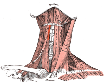
Infrahyoid Muscles
Original Editor - User Name
Top Contributors - Areeba Raja, Manisha Shrestha and Nupur Smit Shah
Introduction[edit | edit source]
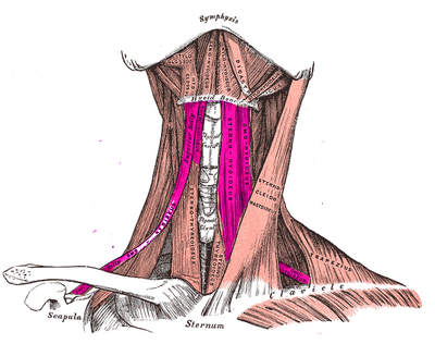
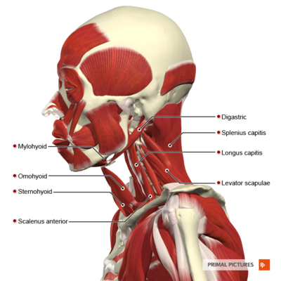
Infrahyoid muscles are also known as “strap muscles” which connect hyoid, sternum, clavicle and scapula. They are located below the hyoid bone on the anterolateral surface of the thyroid gland and are involved in movements of the hyoid bone and thyroid cartilage during vocalization, swallowing and mastication. They are composed of four paired muscles, organized into two layers as following :[1][2][3][4][5][6]
- Superficial layer
- Deep layer
Superficial layer[edit | edit source]
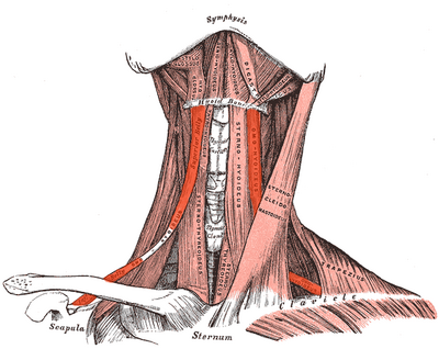
The superficial layer consists of the following muscles:[2]
- Sternohyoid
- Omohyoid
Sternohyoid[edit | edit source]
It is a thin, narrow strap and lies anterior to the remaining strap muscles and the thyroid gland.
Origin[edit | edit source]
It originates from the posterior surface of the medial end of the clavicle, the posterior sternoclavicular ligament and the upper posterior aspect of the manubrium sterni.
Insertion[edit | edit source]
It inserts at the inferior border of the body of the hyoid bone.
Nerve Supply[edit | edit source]
It is innervated by branches of ansa cervicalis (C1, C2, and C3).
Arterial Supply[edit | edit source]
It arterial supply comes from the branches of superior thyroid artery.
Action[edit | edit source]
It depresses the hyoid bone after it has been elevated.
Variation[edit | edit source]
It may be absent or double, augmented by clavicle slip (cleidohyoid), or interrupted by a tendinous intersection.[1][2]
Omohyoid[edit | edit source]
The omohyoid muscle is found deep in the sternocleidomastoid and consist of two bellies (superior and inferior) united at an angle by an intermediate tendon.
Superior Belly[edit | edit source]
Origin:[edit | edit source]
It begins at the intermediate tendon.
Insertion:[edit | edit source]
It inserts to the lower border of the hyoid bone.
Nerve Supply:[edit | edit source]
It is innervated by the superior ramus of the ansa cervicalis and from hypoglossal nerve.
variation[edit | edit source]
It can be absent or double; It is sometimes fused with sternohyoid.
Inferior Belly[edit | edit source]
Origin:[edit | edit source]
It arises from the upper border of the scapula near the scapular notch and occasionally from superior transverse scapular ligament.
Insertion:[edit | edit source]
It inserts into the intermediate tendon.
Nerve Supply:[edit | edit source]
It is supplied by ansa cervicalis (C1, C2 and C3).
Variation:[edit | edit source]
It can be absent or double; it may be attached directly to the clavicle.
Arterial Supply:[edit | edit source]
It is supplied by superior thyroid and lingual arteries.
Action:[edit | edit source]
- It depresses the hyoid bone after it has been elevated.
- It tenses the lower part of the deep cervical fascia in prolonged inspiratory effort, reducing the tendency for soft parts to be sucked inward.[1][2]
Deep layer[edit | edit source]
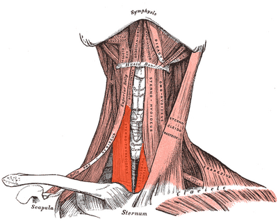
The deep layer consists of the following muscles:[2]
- Sternothyroid
- Thyrohyoid
Sternothyroid[edit | edit source]
It is shorter and wider than sternohyoid and contacts the anterior surface of the thyroid gland.
Origin:[edit | edit source]
It arises from the posterior surface of the manubrium sterni and from the posterior edge of the first rib.
Insertion:[edit | edit source]
It insert into the oblique line of the thyroid cartilage.
Nerve Supply:[edit | edit source]
It is innervated by branches of ansa cervicalis.
Arterial Supply:[edit | edit source]
It is supplied by superior thyroid and lingual arteries.
Action:[edit | edit source]
- It draws the larynx downwards after it has been elevated by swallowing or vocal movements.
- In low notes singing, this downward traction would be exerted with hyoid bone relatively fixed.
Variation:[edit | edit source]
An accessory belly can be present.[1][2][7]
Thyrohyoid[edit | edit source]
It is a small quadrilateral muscle that may be regarded as an upward continuation of the sternothyroid muscle.
Origin:[edit | edit source]
The oblique line on the lamina of the thyroid cartilage.
Insertion:[edit | edit source]
It attaches to the lower border of the greater cornu and adjacent part of the body of the hyoid bone.
Innervation:[edit | edit source]
It is innervated by fibers of the first cervical spinal nerve which branch of the hypoglossal nerve beyond the descendens hypoglossi.
Arterial Supply:[edit | edit source]
It is supplied by superior thyroid and lingual arteries.
Action[edit | edit source]
- It depresses the hyoid bone.
- When the high notes are sung it the thyrohyoid muscle pulls the larynx upwards with the hyoid bone stabilized.[1][2]
Clinical Relevance[edit | edit source]
Infrahyoid muscle paralysis : Trauma to the cervical spine can cause damage to the ansa cervicalis which in turn can result in paresis or paralysis of the infrahyoid muscles. Difficulty in swallowing, hoarseness in the voice or throat tightness can be present as a manifestation of the injury.[5]
Omohyoid Syndrome: It is a rare condition in which X- shaped neck mass protrudes laterally on swallowing. It is caused when the omohyoid muscle displaces the sternocleidomastoid muscle that lies on the top of it.[8][9][10]
Sternohyoid Syndrome: Laterally protruded mass is present similar to the omohyoid syndrome except the problematic muscle is sternohyoid and the mass has a different aspect when swallowing and at rest.[11]
Resources[edit | edit source]
References[edit | edit source]
- ↑ 1.0 1.1 1.2 1.3 1.4 Allen E, Fingeret A. Anatomy, head and neck, thyroid. StatPearls [Internet]. 2021 Jul 26.
- ↑ 2.0 2.1 2.2 2.3 2.4 2.5 2.6 Gray's anatomy : the anatomical basis of clinical practice. 2005. Neck. Muscles.
- ↑ Quadros LS, Bhat N, Babu A, D’souza AS. Anatomical variations in the ansa cervicalis and innervation of infrahyoid muscles. Int J Anat Res. 2013 Sep;1(2):69-74.
- ↑ Suzuki M, Sasaki M, Kamata K, Nakayama A, Shibamoto I, Tamada Y. Swallowing pattern classification method using multichannel surface EMG signals of suprahyoid and infrahyoid muscles. Advanced Biomedical Engineering. 2020;9:10-20.
- ↑ 5.0 5.1 Mnatsakanian A, Al Khalili Y. Anatomy, Head and Neck, Thyroid Muscles. StatPearls [Internet]. 2021 Jul 29.
- ↑ Fitzpatrick TH, Siccardi MA. Anatomy, Head and Neck, Adam's Apple. StatPearls [Internet]. 2021 Apr 14.
- ↑ Kang, Dong Wan, et al. “An Accessory Belly of the Sternothyroid Muscle on the Anterior Neck.” Surgical and Radiologic Anatomy, vol. 37, no. 2, 17 Apr. 2014, pp. 215–217, Accessed 2 Feb. 2022.
- ↑ Lee AD, Yu A, Young SB, Battaglia PJ, Ho CJ. Omohyoid muscle syndrome in a mixed martial arts athlete: a case report. Sports Health. 2015 Sep;7(5):458-62.
- ↑ Ye B. Omohyoid muscle syndrome: report of a case. Chinese Medical Journal. 1980;93(01):65-8.
- ↑ Wong DS, Li JH. The omohyoid sling syndrome. American journal of otolaryngology. 2000 Sep 1;21(5):318-22.
- ↑ Kim JS, Hong KH, Hong YT, Han BH. Sternohyoid muscle syndrome. American journal of otolaryngology. 2015 Mar 1;36(2):190-4.
