When refering to evidence in academic writing, you should always try to reference the primary (original) source. That is usually the journal article where the information was first stated. In most cases Physiopedia articles are a secondary source and so should not be used as references. Physiopedia articles are best used to find the original sources of information (see the references list at the bottom of the article).
If you believe that this Physiopedia article is the primary source for the information you are refering to, you can use the button below to access a related citation statement.
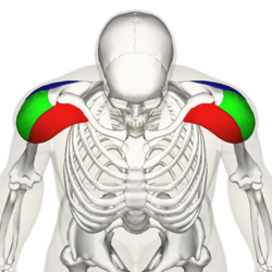
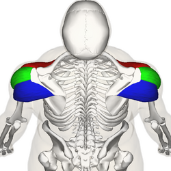
The Deltoid muscle is a large triangular-shaped muscle that lies over the glenohumeral joint and which gives the shoulder its rounded contour.
It is comprised of three distinct portions (anterior or clavicular, middle or acromial, and posterior or spinal)
Acts mainly as an abductor of the shoulder and stabiliser of the humeral head, as well as assists in forward elevation[1].
The deltoid is a very powerful muscle and is used in many ADLs (e.g. putting on clothes, carrying shopping bags, washing hair) and many athletic activities (e.g. netball, swimming, water polo).[2]
Deltoid receives its blood supply from the posterior circumflex humeral artery.
Function
An important function of the deltoid is the stabilisation of the shoulder joint preventing subluxation or even dislocation of the head of the humerus, particularly when carrying a load. Deltoid is the prime mover of shoulder abduction.
All heads of the deltoid work together to produce abduction of the Shoulder Joint. It helps lift the arm front, side, and backward. Deltoid pain can affect anyone who does repetitive overhead activities e.g. house painters, swimmers, waterpolo players, and pitchers. It also is active with carrying objects eg suitcases or reaching up for objects, to prevent subluxation/dislocation.
The deltoid compensates for the loss of strength in the rotator cuff.
Images 4 and 5: Deltoids in action in water polo; in carrying deltoid prevent subluxation of the shoulder joint.
Clinical Relevance
Deltoid pathologies can lead to functional disfunction of the shoulder complex
Tears are not infrequently associated with large or massive rotator cuff tears and may further jeopardise shoulder function.
Rotator cuff tears place more strain on the deltoid to prevent abduction motion loss. Fatigue or injury to the deltoid may result in a precipitous decline in abduction.[4]
A variety of other pathologies may affect the deltoid muscle including enthesitis, calcific tendinitis, myositis, infection, tumours, and chronic avulsion injury.
The deltoid muscle and its innervating axillary nerve may be injured during shoulder surgery, which may have disastrous functional consequences.
Axillary neuropathies lead to deltoid muscle dysfunction and cause denervation of the deltoid muscle.
Abnormalities of the deltoid may originate from nearby pathologies of the subdeltoid bursa, acromion, and distal clavicle.[1]
During dislocation of the shoulder or fracture of surgical neck of the humerus axillary nerve may be injured. The damage to axillary nerve leads to paralysis of the deltoid muscle.[5]
Palpation
Flex elbow to 90 degrees and have the patient abduct the shoulder against resistance.
Anterior Fibers: Deltoid palpated with the elbow extended, shoulder 90 degrees abduction, and then resist horizontal adduction.
Posterior Fibers: Position same as above and then resist horizontal abduction.
Length Tension Testing
Length Tension Testing
Anterior Deltoid- The position of the patient will be high sitting
The therapist will be standing behind the patient holding the testing shoulder
The primary function of the anterior deltoid is flexion, internal rotation, and horizontal adduction, to stretch this muscle, reverse the action by extension, external rotation, and horizontal abduction shoulder, without letting the torso rotate
While performing extension and external rotation, place your one hand on the patient forearm and with the other hand push the shoulder anteriorly from posterior, thereby lengthening the muscle
Posterior Deltoid- The position of the patient will be high sitting
The therapist will be standing in front of the patient initially and moving back to the patient later for a better length assessment
The primary function of the posterior deltoid is extension, external rotation, and horizontal abduction, to stretch this muscle, reverse the action by flexion, internal rotation, and horizontal adduction shoulder joint.
While performing flexion, internal rotation, and horizontal adduction, place one hand on the patient's shoulder and the other hand on the patient's forearm
Alternatively, perform flexion, internal rotation, and horizontal adduction shoulder by bringing the patient's shoulder against the chest and the therapist moving to the back of the patient by holding the patient's shoulder and forearm, lengthening the muscle.
Exercises
The following deltoid strengthening exercises are designed to improve the strength of the deltoid muscles
Isometric Deltoid Contraction
Standing next to a wall with back and elbow straight. Gently push the arm to the side against the wall as hard as possible and comfortably without pain. Hold for 5 seconds and repeat 10 times on each side provided it is pain-free.[6]
Progress
Suitable dumbbell weight, arm raises anterior, lateral, and posterior. Repeat 10 times on each side provided it is pain-free
Resistance Band Forward Raises, lateral raises, and pullbacks. Repeat 10 times on each side provided it is pain free[7]
Further progress
Lots of variations. eg Shoulder Diagonal PNF with a band; compound exercises eg forward arm raises with squats; high side plank walking.
Image: Shoulder Diagonal PNF with a band.
Resources
This video covers the anatomy of the deltoid in great detail [8]
↑Rispoli, Damian M.; Athwal, George S.; Sperling, John W.; Cofield, Robert H. (2009). "The anatomy of the deltoid insertion". J Shoulder Elbow Surg 18: 386–390