When refering to evidence in academic writing, you should always try to reference the primary (original) source. That is usually the journal article where the information was first stated. In most cases Physiopedia articles are a secondary source and so should not be used as references. Physiopedia articles are best used to find the original sources of information (see the references list at the bottom of the article).
If you believe that this Physiopedia article is the primary source for the information you are refering to, you can use the button below to access a related citation statement.
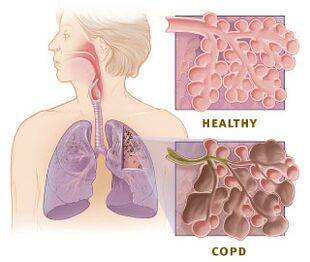
Chronic obstructive pulmonary disease (COPD) is a heterogeneous lung condition causing chronic respiratory symptoms and persistent, often progressive, airflow obstruction from airway and/or alveolar abnormalities.
Spirometry confirms COPD when the post-bronchodilator forced expiratory volume in one second to forced vital capacity (FEV₁/FVC) ratio falls below 0.70.
Risk factors for COPD, traditionally seen as a smoker’s disease, also include air pollution, occupational exposures, respiratory infections, impaired lung development and alpha-1 antitrypsin deficiency.
Physiotherapy for a COPD exacerbation includes dyspnoea management, airway clearance when sputum is retained, and early mobilisation through frequent, short functional sessions.
The Global Initiative for Chronic Obstructive Lung Disease (GOLD) defines COPD as “a heterogeneous lung condition characterized by chronic respiratory symptoms (dyspnea, cough, expectoration and/or exacerbations) due to abnormalities of the airways (bronchitis, bronchiolitis) and/or alveoli (emphysema) that cause persistent, often progressive, airflow obstruction.”[1]
It is a common condition and a leading cause of morbidity and mortality, but it is also preventable and treatable.[2][1] Although it cannot be cured, symptoms can improve with lifestyle changes (e.g. stopping smoking, avoiding air pollution), vaccination, medication, oxygen therapy and pulmonary rehabilitation.[3]
COPD is the third leading cause of death worldwide, causing an estimated 3.4 million deaths in 2023.[3] It has traditionally been seen as a smoker’s disease, but it results from interactions between a person’s genetics and various environmental exposures over their lifetime, which can damage the lungs or alter normal lung development and ageing. GOLD describes this as GETomics (gene–environment interactions over time).[1]
Smoking remains a major risk factor. It accounts for more than 70% of cases in high-income countries and around 30–40% in low- and middle-income countries.[1] Other environmental risk factors include household and outdoor air pollution, occupational exposures and respiratory infections, including childhood infections and tuberculosis. Premature birth, low birthweight and other factors that impair lung development can mean a person never reaches normal peak lung function, which increases their risk of COPD later in life. The best-documented genetic risk factor is alpha-1 antitrypsin deficiency. Asthma and ageing are also associated with COPD.[1]
Classifying COPD by Cause
Classifying COPD by cause (etiotype) has been proposed to raise awareness of non-smoking risk factors, improve detection of early and non-smoking-related COPD, and support research into treatments that target specific disease mechanisms.[5][6] GOLD combines the taxonomies proposed by the Lancet Commission on COPD and Celli et al. into the following classification:[1][5][6]
Genetically determined COPD (COPD-G): alpha-1 antitrypsin deficiency and other genetic variants
COPD due to abnormal lung development (COPD-D): for example, premature birth and low birthweight
Environmental COPD:
Cigarette smoking COPD (COPD-C): exposure to tobacco smoke, including second-hand smoke, vaping, e-cigarette use, or cannabis
Biomass and pollution exposure COPD (COPD-P): exposure to household pollution and occupational hazards
COPD due to infections (COPD-I): childhood infections, tuberculosis, HIV
COPD and asthma (COPD-A)
COPD of unknown cause (COPD-U)
COPD Pathological Process
Persistent airflow obstruction in COPD results from a combination of:[1]
small airways disease, which increases airway resistance
parenchymal destruction (emphysema), which reduces lung elastic recoil
Chronic inflammation is a key driver of these changes. Inhaled noxious particles and gases, such as cigarette smoke and air pollution, are thought to trigger an abnormal inflammatory response in the airways and lung parenchyma, which over time leads to small airway narrowing, mucus hypersecretion and alveolar wall destruction.[7][1]
These changes cause expiratory flow limitation and premature small airway closure, so air is trapped, and end-expiratory lung volume rises (hyperinflation), reducing inspiratory capacity.[1] Hyperinflation flattens the diaphragm, placing it at a mechanical disadvantage and increasing the work of breathing.[8] During exertion, faster breathing shortens expiratory time, so hyperinflation worsens (dynamic hyperinflation).
Structural changes in the airways, alveoli and pulmonary circulation also impair gas exchange. Ventilation–perfusion mismatch is the main cause of hypoxaemia, and hypercapnia becomes more likely as the disease progresses.[1][9]
COPD Clinical Presentation
The most characteristic symptom of COPD is persistent, progressive dyspnoea that is worse on exertion.[10] Chronic cough is often the first symptom. Up to 30% of people with COPD have cough with sputum production, but airflow obstruction can also be present without chronic dyspnoea or a productive cough.[1]
Other symptoms include recurrent wheeze, chest tightness and fatigue. People with COPD may also have recurrent lower respiratory tract infections, weight loss and loss of muscle mass.[1]
Physical signs are often absent in mild COPD and become more apparent as the disease progresses and during exacerbations. They include a barrel chest, pursed-lip breathing, tripod positioning (leaning forward with hands or elbows braced on the knees, a table, or the back of a chair), accessory muscle use and prolonged expiration. Hoover's sign (inward movement of the lower lateral rib margins during inspiration) is associated with air trapping and diaphragm dysfunction in severe COPD.[11]
COPD Diagnostic Tests
COPD should be considered in anyone with dyspnoea, chronic cough or sputum production, recurrent lower respiratory tract infections and/or exposure to risk factors. A diagnosis of COPD is confirmed by spirometry showing a post-bronchodilator FEV₁/FVC ratio below 0.70.[1][12]
Spirometry
Spirometry measures airflow during a forced expiration following a maximal inspiration. FEV₁ is the forced expiratory volume exhaled in the first second, and FVC (forced vital capacity) is the total volume exhaled. A reduced FEV₁/FVC ratio indicates airflow obstruction.[13]
Key Points
FEV₁ correlates only loosely with symptoms and function. A patient with a mildly reduced FEV₁ can still have significant breathlessness and reduced exercise tolerance, so spirometry should be interpreted alongside symptoms and validated questionnaires, such as the Modified Medical Research Council (mMRC) Dyspnea Scale and Chronic Airways Assessment Test (CAAT™, formerly the COPD Assessment Test, CAT™).[1][14]
The fixed 0.70 threshold is debated. Because FEV₁/FVC falls with age, a fixed ratio can overdiagnose COPD in older adults and underdiagnose it in younger adults, so some guidelines use the lower limit of normal instead.[14]
Specific lung volume measurements include total lung capacity (TLC), residual volume (RV) and functional residual capacity (FRC). Lung volumes can be measured using body plethysmography, helium dilution or nitrogen washout.[17]
Body plethysmography is the reference method in COPD because gas dilution methods (helium dilution and nitrogen washout) can underestimate lung volumes in COPD. Raised TLC, FRC or RV can indicate physiological changes associated with COPD, such as gas trapping or hyperinflation.[1][18]
Diffusing Capacity of the Lung for Carbon Monoxide (DLCO)
DLCO (also called the transfer factor of the lung for carbon monoxide or TLCO) measures how effectively carbon monoxide moves from the alveoli into the pulmonary capillary blood. Because oxygen follows a similar diffusion pathway, DLCO indicates how efficiently oxygen is transferred.[14][19]
COPD Severity Assessment
The initial assessment should consider the severity of airflow obstruction, symptom burden, exacerbation history, blood eosinophil count and multimorbidity.[1]
Spirometry Staging
GOLD grades the severity of airflow obstruction by post-bronchodilator FEV₁ % predicted, in people with FEV₁/FVC below 0.70.[1]
Table 1. Spirometry staging
GOLD grade
Severity
Post-bronchodilator FEV₁ (% predicted)
1
Mild
≥80%
2
Moderate
50–79%
3
Severe
30–49%
4
Very severe
<30%
Symptoms and Exacerbation History (ABE Groups)
Initial pharmacological treatment in patients with COPD is guided by the GOLD ABE classification and blood eosinophil count (BEC). Note that the ABE groups have replaced the older ABCD framework.[2][20]
In this framework, symptom burden and exacerbation history are assessed separately because, although related, they follow different courses and each has its own implications for prognosis and treatment. GOLD 2026 also lowered the exacerbation threshold, so a single moderate or severe exacerbation now places a patient in Group E. This reflects the evidence that even one exacerbation affects prognosis.[1][2]
Group A: no moderate or severe exacerbations in the previous year and mMRC of 0–1 and CAAT <10
Group B: no moderate or severe exacerbations in the previous year and mMRC ≥2 and/or CAAT ≥10
Group E: one or more moderate or severe exacerbations in the previous year
COPD Exacerbation
“An exacerbation of COPD is an acute event with symptoms worsening over a few days (up to 14 days) and characterized by increased dyspnea and/or cough and sputum that may be accompanied by tachypnea and/or tachycardia. Exacerbations are often associated with increased local and systemic inflammation caused by airway infection, pollution, or other insults to the lungs.”[1]
GOLD 2026 classifies exacerbations as mild, moderate or severe based on clinical and physiological findings at presentation, rather than on the treatment or care setting required (Table 2).[1]
Dyspnoea VAS < 5, respiratory rate < 24 breaths/min, heart rate < 95 bpm, resting SaO₂ ≥ 92% on room air (or person's usual oxygen) with a change ≤ 3% (when known), CRP < 10 mg/L (if available).
Moderate
At least three of the following: dyspnoea VAS ≥5, respiratory rate ≥24/min, heart rate ≥95/min, resting SaO₂ <92% on room air or usual oxygen and/or a change of >3% (when known), CRP ≥10 mg/L (if available). If taken, arterial blood gases may show hypoxaemia (PaO₂ 70-80 mmHg).
Severe
As for moderate, but if obtained, arterial blood gases showing hypoxaemia (PaO₂ ≤ 60 mmHg) and/or hypercapnia and acidosis (PaCO₂ >45 mmHg and pH <7.35).
The medical team must rule out conditions that mimic or worsen an exacerbation, such as pneumonia, pulmonary embolism, heart failure, arrhythmia or pneumothorax. Physiotherapists should be alert to signs that may indicate one of these conditions, such as new chest pain, unilateral leg swelling or signs of fluid overload, and escalate these to the medical team.[21]
Common COPD Medications
Table 3 lists the main classes of COPD medications.
Symptom relief; first-line treatment of exacerbations
Long-acting bronchodilators (LABA, LAMA)
Maintenance treatment
Inhaled corticosteroids (ICS), combined with long-acting bronchodilators
Maintenance treatment for selected patients with exacerbations and raised blood eosinophils
Systemic corticosteroids
Short course during exacerbations
Antibiotics
Exacerbations with signs of bacterial infection, such as increased sputum purulence
Add-on therapies
Selected patients who continue to have exacerbations despite maintenance treatment
Oxygen Therapy and COPD Exacerbations
During hospital admissions, the target SpO₂ for patients with COPD or others at risk of hypercapnia is typically 88–92%. Over-oxygenation can worsen CO₂ retention, so hypoxaemia should be treated without pushing oxygen saturation above the target range. SpO₂ often decreases on exertion, so the oxygen flow rate may need to be increased during activity to keep saturations within the target range, then reduced to the resting setting once the patient has recovered, to avoid over-oxygenation.[21][23][24]
Standard nasal cannulae are the preferred delivery method for most patients, with the flow rate adjusted to achieve the target saturation. Approximate FiO₂ values for each device are:[23]
Nasal cannulae: FiO₂ 24–35% at 1–4 L/min
Venturi mask: FiO₂ 24–60%
Simple face mask: FiO₂ 35–60% at 5–10 L/min - flows below 5 L/min should be avoided because of the risk of CO₂ rebreathing
High-flow nasal cannula (HFNC): FiO₂ 21–100% at flows of up to 60-70 L/min, depending on the device - the oxygen is heated and humidified, which may improve comfort and tolerance[25]
For acute respiratory failure, non-invasive ventilation (NIV) is the preferred initial form of ventilatory support in people hospitalised with a COPD exacerbation, and invasive ventilation may be needed if NIV fails.[1]
Physiotherapists must follow their local policy, national regulation and professional guidance on oxygen, and in many settings, may administer and adjust supplemental oxygen within institutional protocols and the oxygen prescription. Before mobilising, they should check the oxygen prescription and target saturation range, and compare current oxygen needs with the patient's baseline. A patient who normally uses no oxygen at rest but now needs it has had a significant change, and is likely to need more during activity.[21] Where local policy allows, a prescription based on a target saturation range rather than a fixed flow rate lets oxygen be titrated to the patient’s response during mobility.[21]
Physiotherapy for Patients Admitted with a COPD Exacerbation
Before seeing the patient, physiotherapists should review the chart for arterial blood gases (ABGs), SpO₂ and oxygen device, vital signs, medications and chest imaging, and establish the patient’s baseline oxygen use and function, so that current findings can be compared with what is normal for them.[21] At the bedside, assess respiratory rate, work of breathing, speech, sputum, mental status, strength and mobility. A rising respiratory rate, increasing accessory muscle use, inability to speak in full sentences, SpO₂ below target despite supplemental oxygen, or new drowsiness or confusion need urgent medical review. Mobility is also part of the assessment, essentially acting as a controlled cardiopulmonary stress test.[21]
Symptom Management
Commonly used techniques for managing dyspnoea include positioning, pursed-lip breathing, breathing control, pacing, energy conservation and planned recovery periods.[21] A handheld fan directed at the face may also be useful for some patients.[26]
It is important to start mobility as soon as the patient is clinically stable to limit deconditioning. The FITT-VP principles can guide exercise prescription during an acute COPD exacerbation:[21]
Frequency: consider several short bouts of mobility rather than one long session
Intensity: can be guided by RPE or dyspnoea scales (rather than heart rate)
Time: will likely be short sessions
Type: tends to be functional in an acute setting (walking, transfers, stairs)
Volume: accumulated through frequent, short sessions
Progression: distance, duration, number of sessions and task complexity should be progressed gradually, based on the patient's clinical response
Stopping or Modifying Exercise
During every session, the physiotherapist must consider whether to progress, modify or stop the activity, based on the patient’s response.
Modify the activity if symptoms or physiological responses suggest the dose is too high, for example by reducing speed or distance, increasing rest periods or adjusting oxygen within the prescribed target. Stop the activity if the patient develops new chest pain, severe respiratory distress, syncope, arrhythmia, significant haemodynamic instability, neurological or cognitive changes (including new confusion or drowsiness), or persistent physiological deterioration, or if SpO₂ falls below target and does not recover with oxygen adjustment.[21]
Discharge Planning
Discharge planning starts early in the admission. Physiotherapists assess whether the patient’s function, oxygen needs and home environment support a safe discharge. Referral to pulmonary rehabilitation should be considered after an exacerbation. Education before discharge should cover how to recognise early signs of a further exacerbation, inhaler technique, smoking cessation advice where relevant, breathing and pacing strategies, and returning gradually to activity.[1][21]
Outcome Measures
The choice of outcome measure depends on the clinical question, the patient’s stability and the time available. Outcome measures help quantify change, guide progression and inform discharge decisions.[21]
Breathlessness during activity can be rated with the modified Borg scale (CR10). The mMRC Dyspnoea Scale and CAAT describe breathlessness in daily life and the overall impact of COPD.[1]
↑National Institute for Health and Care Excellence (NICE), Chronic obstructive pulmonary disease in over 16s: diagnosis and management: NICE Guideline [NG115]. 2018. Available from: https://www.nice.org.uk/guidance/ng115 [Accessed 22 October 2026].
↑ 23.023.1Beasley R, Chien J, Douglas J, Eastlake L, Farah C, King G, et al. Thoracic Society of Australia and New Zealand oxygen guidelines for acute oxygen use in adults: ‘Swimming between the flags’. Respirology. 2015;20:1182–91.