When refering to evidence in academic writing, you should always try to reference the primary (original) source. That is usually the journal article where the information was first stated. In most cases Physiopedia articles are a secondary source and so should not be used as references. Physiopedia articles are best used to find the original sources of information (see the references list at the bottom of the article).
If you believe that this Physiopedia article is the primary source for the information you are refering to, you can use the button below to access a related citation statement.
Burn wound injuries place critical economic burden on healthcare infrastructures worldwide. They are also associated with high mortality rates due to severe complications. Infection is the most common complication in burn wound injuries. Thus, prompt and precise diagnosis is of critical importance for this patient population to prevent detrimental consequences and optimise healing outcomes.[1]
Burn wound injuries are at an increased infection risk for multiple reasons: (1) the body's physiological response to a burn injury presents with many of the same signs and symptoms of a developing infection, (2) the burn injury can impair many body systems which limits a patient's innate ability to fight off infection.[2]
Physiological Response to Burn Injuries
Burn wound injuries have both local and system consequences.[2] This requires the wound care professional to work closely with the interdisciplinary team to appropriately monitor and manage the patient in a holistic manner. Understanding the pathophysiological changes which occur after a burn wound injury will greatly improve the wound care professional's ability to prevent and/or manage infection.
Local response to burn wounds
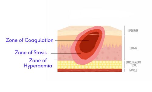
The local response involves the area of the burn injury and the tissue directly surrounding it. This is best described using Jackson's Burn Model which divides the wound into three zones.[3]
Jackson's Burn ModelZone of coagulation (also known as the zone of necrosis). This area sustains the most damage and suffers irreversible tissue loss.
Zone of stasis. This area is found around the zone of coagulation. It demonstrates decreased tissue perfusion and is, therefore, potentially salvageable with proper care. This zone can also suffer complete tissue loss with prolonged hypotension, infection, or oedema.
Zone of hyperaemia. This is the outermost zone of the injury. In this area, tissue perfusion is increased. This area will likely recover unless there is severe sepsis or prolonged hypoperfusion.
Capillary permeability is increased, leading to the loss of intravascular proteins and fluids into the interstitial compartment, which leads to oedema[2][4]
Myocardial contractility is decreased,[4] there is a decrease in cardiac output immediately post-burn
These changes, when combined with fluid loss from the burn wound, result in systemic hypotension and end organ hypoperfusion[3]
Local vascular compromise occurs in deep, partial, and full-thickness injuries where the arterial supply is either damaged or destroyed as a result of the burn wound[2]
Respiratory changes
Inflammatory mediators cause bronchoconstriction[4]
Basal metabolic rate can increase to three times its original rate post-burn[4]
A systematic immunoendocrine response will occur after a large burn injury and can continue for up to 3-years post-injury. This response can cause: (1) immune incompetence, (2) sepsis, (3) increased fracture risk, (4) slowing of growth rate, (5) reduced organ function, (6) decreased wound healing, and (7) death[3]
Gastrointestinal changes
Absorption is affected due to gastrointestinal mucosal atrophy and decreased intestinal blood flow[3]
A patient post-burn is in a constant hypermetabolic state as a result of their burn injury. Initially, this will boost the energy response and help fight off infection and begin the healing process. However, as the hypermetabolic state becomes a prolonged response, it will be an energy drain and it becomes more difficult for the patient to mount defences and continue the healing process.[2]
Early enteral feeding is paramount to prevent malnutrition[3]
Immunological changes
The immune response becomes globally depressed following a burn injury, the severity is dependent on the size of the burn wound
Immune incompetence further depresses the body's ability to fight infection - sepsis is the leading cause of post-burn mortality[3]
"Burn shock results from the interplay of direct tissue injury, hypovolemia, and the release of multiple mediators of inflammation, with effects on both the microcirculation and the function of the heart and lungs."[5]
Additional Risk Factors for Burn Injury Infection
Additional risk factors of burn wound injuries for infection:[2]
An increased length of stay in the ICU
Increased wait time for test results (ie wound cultures or biopsies) to identify the source of infection delays the use of targeted antibiotics or antimicrobial agents to most effectively treat infection
Possibly a delay due to transportation time to a burn centre for skilled aggressive care
A delay for surgery due to patient medical instability or lack of surgical facility
Any burn greater than 20% total body surface area of deep partial and/or full thickness injury is a high risk for infection, regardless of the patient's underlying conditions
Infection risk increases more when the patient is less than four years old or more than 55 years old
Infection risk is increased for patients who are immunocompromised or who have pre-existing comorbidities that put them at risk for developing an infection
The presence of eschar in the wound increases the risk of infection
Signs and Symptoms of Sepsis in a Burn Wound
The detection of a burn wound infection has multiple layers of complexity. The normal response to a burn injury mimics many of the signs and symptoms of infection. Therefore, the ability to identify an infection is masked by the patient's response to the burn injury.[2] To review covert and overt signs and symptoms of infection, please see this article.
While infections are one of the most common complications following a burn wound injury, sepsis is the leading cause of death in both adult and paediatric burn patients. The diagnosis and management of sepsis in burns also has multiple layers of complexity. The diagnosis of sepsis in patients with severe burns (>20% total body surface area) is complicated by the overlap of clinical signs of the typical post-burn hypermetabolic response with those of sepsis.[6]
For a burn wound to be diagnosed as septic, the patient must exhibit at least three of the following:
Table is modified from: Norbury W, et. al. Infection in burns. Surg Infect, 2016; 17(2): 250-255.
>2 SD above age-specific norm (85% age-adjusted maximum respiratory rate)
Thrombocytopenia
(will not apply until 3 days after initial resuscitation)
<100,000/mcL
<2 SD below age-specific norm
Hyperglycemia
(in the absence of pre-existing diabetes mellitus)
Untreated plasma glucose >200 mg/dL or equivalent mM/L
Insulin resistance. Examples include:
>7 units of insulin/hour intravenous drip (adults)
Resistance to insulin (>25% increase in insulin requirements over 24 hours)
Inability to continue enteral feedings >24 hours
Examples include:
Abdominal distension
Enteral feeding intolerance (residual >150 mL/h in children or 2× feeding rate in adults)
Uncontrollable diarrhea (>2,500 mL/d for adults or >400 mL/d in children)
Have a documented infection
Documented one of the following:
Culture-positive infection OR
Pathologic tissue source identified OR
Clinical response to antimicrobials
Sources of Infection
Research shows that burn wounds are sterile for the first 6-12 hours after initial injury. However, contamination occurs soon there after and the proliferation of bacteria increases rapidly.[2]
Endogenous sources, such as the lungs or the gut, or any mucosal membranes where bacteria thrive in a normal system
Exogenous sources, such as the environment or cross-contamination
Bacterial infections
For more in-depth information on bacterial infections, please read this article.
Rough timeline of bacterial concentrations in a burn wound:[2]
Early phase of infection: gram-positive usually the first identified
Staphyloccus aureus is typically the first pathogen found in burn wound infection, this includes Methicillin-resistant Staphylococcus aureus (MRSA)
Staphylococcus aureus is also typically the first microbe identified in sepsis and in arterial bacterial dissemination from the wound
At five days from the time of injury: gram-negative bacteria become predominant, and
Pseudomonas aeruginosa is the primary bacteria identified. It is the most common pathogen found in burn wounds and is very common in the development of biofilms.
Later stages: development of fungal or yeast infection independent or in combination with an already existing bacterial infection
Fungal infections
For more in-depth information on fungal infections, please read this article.
Fungal infections are also a major concern for patients with burn wound injuries. It can often be difficult to distinguish a fungal infection from a bacterial infection in a burn wound by appearance alone.[2]
Risk factors for developing a fungal infection in a burn wound:[2]
Total body surface area involvement >30%
Long hospital length of stay
Multiple comorbidities
Clinical observations for a fungal infection in a burn wound:[2]
Changes in the wound appearance
An unanticipated separation of eschar is a key sign of fungal infection
Rapid conversion from a partial thickness burn to a deep partial or a full thickness burn wound
A blackening of the burn wound tissue
A persistent ongoing fever which remains unchanged with antibiotic therapy
Candida albicans and Aspergillus are the most common fungal pathogens. Aspergillus can be a very serious fungal infection and requires urgent assessment and potential surgical debridement.
Example of fungal infection in wound. Image used with kind permission of Diane Merwarth PT.
Sample collection
Biopsy
Wound biopsies are an important diagnostic component in the management of chronic wounds to monitor for potential malignancy or infection.[7] The tissue biopsy, especially in burn wounds, has long been recognised as the gold standard. It is the most quantitative method of collecting tissue and identifying the level of bacterial contamination or infection in a wound.[2]
Taking a wound biopsy requires sampling at the wound edge and in the wound bed. The procedure is invasive and requires skill on the part of the caregiver to collect the sample. Biopsies require extra time for results to become available but allow for accurate targeted antibiotics or antimicrobials to be used against the infection. It has been found that bacteria are not homogeneously present in the wound bed, but they are in pocketed in different areas. Therefore, multiple biopsies should be collected from a single burn wound.[2]
It is also recommended to take wound biopsies for wounds that have not responded to treatment after 2–6 weeks.[7]
Semiquantitative swab culture
There is controversy over the effectiveness of performing a swab culture and if the results will represent the infection. However, there is research which shows a correlation between bacteria identified in a swab culture and the bacteria identified from a tissue sample, specifically a biopsy. A swab culture will not give the level of quantitative results as a biopsy, but if done appropriately, the results will identify the microbes that need to be targeted with antimicrobial interventions.[2]
Will be needed to to remove biofilm population. The wound usually requires frequent surgical debridement. It has been found that a biofilm will start to reform within 24 hours of being disrupted, and it becomes mature in three days. Therefore, it is important to follow up with appropriate biofilm-based wound management.
Fungal infections may require surgical debridement
Early excision of eschar, which provides a good environment for bacterial proliferation
Skin grafts to close a wound immediately and decrease exposure to new sources of infection
Close observation for overt and covert signs and symptoms of infection
Closely monitoring vital signs and lab values for signs of infection
Accurate and thorough documentation of wound appearance and features
Frequently reassess and update care plan based on the patient's response to current interventions
Manage patient's comorbidies
Maintain optimal nutrition and water intake
Oral intake is the most valuable and the most effective method
A nasogastric tube is the second most effective method
Enteral nutrition is the least favourable because it doesn't utilise the gut
Manage oedema
Minimising oedema by elevating the extremities
Applying compression bandages to the extremities if tolerated
Provide psychosocial support and education
A positive mental attitude can aide in wound healing and infection prevention
Include the patient in care plans
Temperature control
Prevent the patient from being too hot or too cold, so adjust the temperature in the patient's room or in the operating room (OR). Often hospitals are set a minimum of 29-31°C (85-88 °F) room temperature to prevent patients with a burn wound from becoming too cold.
Dressing considerations: Wounds can cool down when dressings become saturated with drainage or when left exposed to air too long during a dressing change.
Recommended strategies for minimising the onset of infection in medical facilities
Universal precautions, proper use of personal protective equipment (PPE), and hand hygiene
Patients at high risk for developing an infection should be placed in a negative pressure room with strict isolation precautions.
Wound care should be done with aseptic (sterile) technique when possible, otherwise clean (non-sterile) techniques should be used.
Strict aseptic technique should be used when inserting any kind of device (intravenous line, intra-arterial line, foley catheter). It is also recommended for these devices to be removed as soon as they are no longer necessary to support the patient's recovery from their burn injury.
Environmental control to include: (1) a complete cleaning of the patient's room twice/day with wipe down of all high-touch surfaces, (2) terminally cleaning the room anytime the patient is not present, (3) after the patient has been discharged from the room, it is recommended to complete three days of terminal clean prior to use by another person.
Surveillance cultures are recommended by many burn centres
This includes nasal cultures, cultures of the wound, possibly anal cultures with the onset of vancomycin-resistant enterococcus (VRE)
It is recommended that cultures be collected on admission and weekly throughout the hospital stay
Please view this optional video for an overview of burn wound anatomy and classification, as well as a detailed description of burn wound pathophysiology.


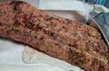 Example of fungal infection in wound. Image used with kind permission of Diane Merwarth PT.